
Rabbit Anti-Actin antibody
muscle Actin; ACTS_HUMAN; Actin, alpha skeletal muscle; Alpha-actin-1; ACTA1; ACTA; ASMA; CFTD; CFTD1; CFTDM; MPFD; NEM1; NEM2; NEM3; Actin alpha skeletal muscle; actin, alpha 1, skeletal muscle 1; actin, alpha 1, skeletal muscle; actina; aktin; alpha Act
View History [Clear]
Details
Product Name Actin Chinese Name 肌动蛋白α1抗体 Alias muscle Actin; ACTS_HUMAN; Actin, alpha skeletal muscle; Alpha-actin-1; ACTA1; ACTA; ASMA; CFTD; CFTD1; CFTDM; MPFD; NEM1; NEM2; NEM3; Actin alpha skeletal muscle; actin, alpha 1, skeletal muscle 1; actin, alpha 1, skeletal muscle; actina; aktin; alpha Actin 1; alpha skeletal muscle Actin; alpha skeletal muscle; alpha-actin; Beta cytoskeletal actin; nemaline myopathy type 3. α-skeletal muscle actin; α Skeletal Muscle Actin. literatures Immunogen Species Rabbit Clonality Polyclonal React Species Mouse, Rat, (predicted: Human, Dog, Cow, Rabbit, ) Applications WB=1:500-2000 ELISA=1:5000-10000 IHC-P=1:100-500 IHC-F=1:100-500 ICC=1:100-500 IF=1:100-500 (Paraffin sections need antigen repair)
not yet tested in other applications.
optimal dilutions/concentrations should be determined by the end user.Theoretical molecular weight 42kDa Cellular localization cytoplasmic Form Liquid Concentration 1mg/ml immunogen KLH conjugated synthetic peptide derived from human ACTA1: 3-100/377 Lsotype IgG Purification affinity purified by Protein A Buffer Solution 0.01M TBS(pH7.4) with 1% BSA, 0.03% Proclin300 and 50% Glycerol. Storage Shipped at 4℃. Store at -20 °C for one year. Avoid repeated freeze/thaw cycles. Attention This product as supplied is intended for research use only, not for use in human, therapeutic or diagnostic applications. PubMed PubMed Product Detail The product encoded by this gene belongs to the actin family of proteins, which are highly conserved proteins that play a role in cell motility, structure and integrity. Alpha, beta and gamma actin isoforms have been identified, with alpha actins being a major constituent of the contractile apparatus, while beta and gamma actins are involved in the regulation of cell motility. This actin is an alpha actin that is found in skeletal muscle. Mutations in this gene cause nemaline myopathy type 3, congenital myopathy with excess of thin myofilaments, congenital myopathy with cores, and congenital myopathy with fiber-type disproportion, diseases that lead to muscle fiber defects. [provided by RefSeq, Jul 2008]
Function:
Actins are highly conserved proteins that are involved in various types of cell motility and are ubiquitously expressed in all eukaryotic cells.
Subunit:
Polymerization of globular actin (G-actin) leads to a structural filament (F-actin) in the form of a two-stranded helix. Each actin can bind to 4 others. Identified in a complex composed of ACTA1, COBL, GSN AND TMSB4X. Interacts with TTID. Interacts (via its C-terminus) with USP25; the interaction occurs for all USP25 isoforms but is strongest for isoform USP25min muscle differentiating cells.
Subcellular Location:
Cytoplasm, cytoskeleton.
Post-translational modifications:
Oxidation of Met-46 and Met-49 by MICALs (MICAL1, MICAL2 or MICAL3) to form methionine sulfoxide promotes actin filament depolymerization. MICAL1 and MICAL2 produce the (R)-S-oxide form. The (R)-S-oxide form is reverted by MSRB1 and MSRB2, which promote actin repolymerization.
Monomethylation at Lys-86 (K84me1) regulates actin-myosin interaction and actomyosin-dependent processes. Demethylation by ALKBH4 is required for maintaining actomyosin dynamics supporting normal cleavage furrow ingression during cytokinesis and cell migration.
DISEASE:
Nemaline myopathy 3 (NEM3) [MIM:161800]: A form of nemaline myopathy. Nemaline myopathies are muscular disorders characterized by muscle weakness of varying severity and onset, and abnormal thread-like or rod-shaped structures in muscle fibers on histologic examination. Note=The disease is caused by mutations affecting the gene represented in this entry.
Myopathy, actin, congenital, with excess of thin myofilaments (MPCETM) [MIM:161800]: A congenital muscular disorder characterized at histological level by areas of sarcoplasm devoid of normal myofibrils and mitochondria, and replaced with dense masses of thin filaments. Central cores, rods, ragged red fibers and necrosis are absent. Note=The disease is caused by mutations affecting the gene represented in this entry.
Myopathy, congenital, with fiber-type disproportion (CFTD) [MIM:255310]: A genetically heterogeneous disorder in which there is relative hypotrophy of type 1 muscle fibers compared to type 2 fibers on skeletal muscle biopsy. However, these findings are not specific and can be found in many different myopathic and neuropathic conditions. Note=The disease is caused by mutations affecting the gene represented in this entry.
Similarity:
Belongs to the actin family.
SWISS:
P68133
Gene ID:
58
Database links:Entrez Gene: 421534 Chicken
Entrez Gene: 58 Human
Entrez Gene: 11459 Mouse
Omim: 102610 Human
SwissProt: P68139 Chicken
SwissProt: P68133 Human
SwissProt: P68134 Mouse
SwissProt: P68135 Rabbit
Unigene: 1288 Human
Unigene: 214950 Mouse
Unigene: 82732 Rat
Product Picture 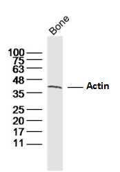 Sample:
Bone (Mouse) Lysate at 40 ug
Primary: Anti-Actin (SL10966R) at 1/300 dilution
Secondary: IRDye800CW Goat Anti-Rabbit IgG at 1/20000 dilution
Predicted band size: 42 kD
Observed band size: 42 kD
 Sample:
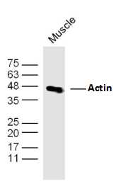
Muscle (Mouse) Lysate at 40 ug
Primary: Anti-Actin (SL10966R) at 1/300 dilution
Secondary: IRDye800CW Goat Anti-Rabbit IgG at 1/20000 dilution
Predicted band size: 42 kD
Observed band size: 42 kD
 Sample:
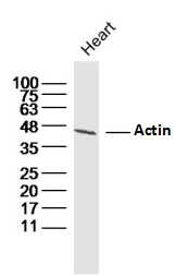
Heart (Mouse) Lysate at 40 ug
Primary: Anti-Actin (SL10966R) at 1/300 dilution
Secondary: IRDye800CW Goat Anti-Rabbit IgG at 1/20000 dilution
Predicted band size: 42 kD
Observed band size: 42 kD
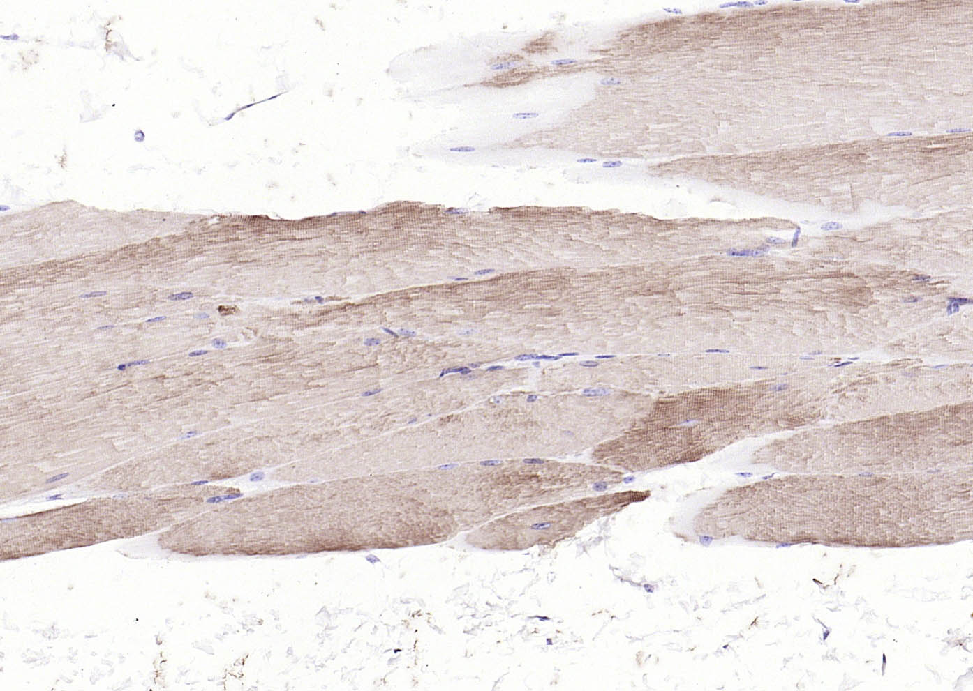 Paraformaldehyde-fixed, paraffin embedded (mouse skeletal muscle); Antigen retrieval by boiling in sodium citrate buffer (pH6.0) for 15min; Block endogenous peroxidase by 3% hydrogen peroxide for 20 minutes; Blocking buffer (normal goat serum) at 37°C for 30min; Antibody incubation with (Actin) Polyclonal Antibody, Unconjugated (SL10966R) at 1:200 overnight at 4°C, followed by operating according to SP Kit(Rabbit) (sp-0023) instructionsand DAB staining.
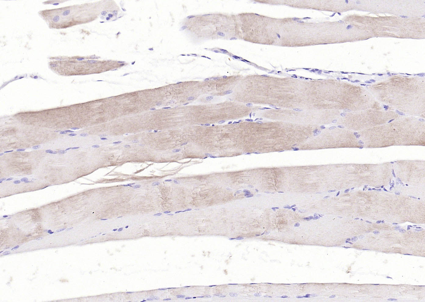 Paraformaldehyde-fixed, paraffin embedded (rat skeletal muscle); Antigen retrieval by boiling in sodium citrate buffer (pH6.0) for 15min; Block endogenous peroxidase by 3% hydrogen peroxide for 20 minutes; Blocking buffer (normal goat serum) at 37°C for 30min; Antibody incubation with (Actin) Polyclonal Antibody, Unconjugated (SL10966R) at 1:200 overnight at 4°C, followed by operating according to SP Kit(Rabbit) (sp-0023) instructionsand DAB staining.
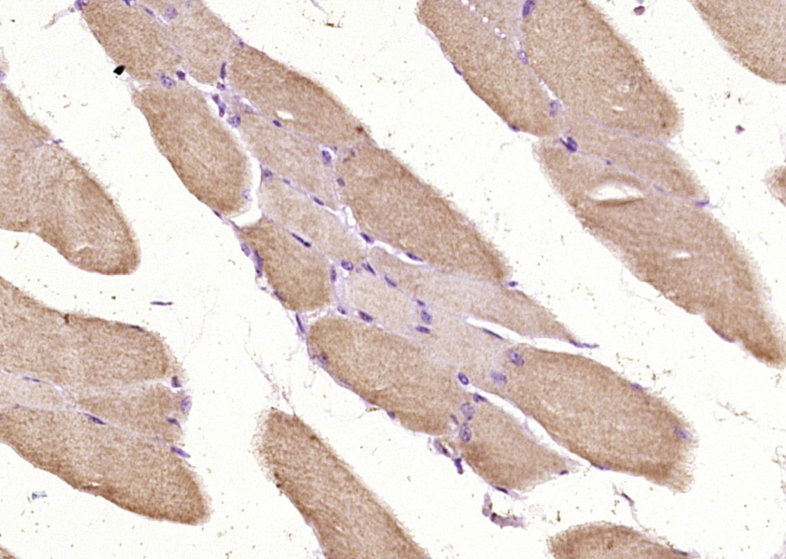 Paraformaldehyde-fixed, paraffin embedded (mouse skeletal muscle); Antigen retrieval by boiling in sodium citrate buffer (pH6.0) for 15min; Block endogenous peroxidase by 3% hydrogen peroxide for 20 minutes; Blocking buffer (normal goat serum) at 37°C for 30min; Antibody incubation with (Actin) Polyclonal Antibody, Unconjugated (SL10966R) at 1:200 overnight at 4°C, followed by operating according to SP Kit(Rabbit) (sp-0023) instructionsand DAB staining.
Cartpieces
Totalgoods,subtotals:¥Checkout
Bought notes(bought amounts latest0)
No one bought this product
User Comment(Total0User Comment Num)
- No comment


 +86 571 56623320
 +86 18668110335

