
Rabbit Anti-GFAP antibody
Astrocyte; FLJ45472; GFAP; Glial Fibrillary Acidic Protein; Intermediate filament protein; GFAP_HUMAN.
View History [Clear]
Details
Product Name GFAP Chinese Name 胶质纤维酸性蛋白抗体 Alias Astrocyte; FLJ45472; GFAP; Glial Fibrillary Acidic Protein; Intermediate filament protein; GFAP_HUMAN. Immunogen Species Rabbit Clonality Polyclonal React Species Human, Mouse, Rat, (predicted: Chicken, Pig, Cow, Rabbit, Sheep, ) Applications WB=1:500-2000 ELISA=1:5000-10000 IP=1:20-100 IHC-P=1:100-500 Flow-Cyt=3μg/Test ICC=1:100 (Paraffin sections need antigen repair)
not yet tested in other applications.
optimal dilutions/concentrations should be determined by the end user.Theoretical molecular weight 48kDa Cellular localization cytoplasmic Form Liquid Concentration 1mg/ml immunogen KLH conjugated synthetic peptide derived from human GFAP: 341-432/432 Lsotype IgG Purification affinity purified by Protein A Buffer Solution 0.01M TBS(pH7.4) with 1% BSA, 0.03% Proclin300 and 50% Glycerol. Storage Shipped at 4℃. Store at -20 °C for one year. Avoid repeated freeze/thaw cycles. Attention This product as supplied is intended for research use only, not for use in human, therapeutic or diagnostic applications. PubMed PubMed Product Detail This gene encodes one of the major intermediate filament proteins of mature astrocytes. It is used as a marker to distinguish astrocytes from other glial cells during development. Mutations in this gene cause Alexander disease, a rare disorder of astrocytes in the central nervous system. Alternative splicing results in multiple transcript variants encoding distinct isoforms. [provided by RefSeq, Oct 2008]
Function:
GFAP, a class-III intermediate filament, is a cell-specific marker that, during the development of the central nervous system, distinguishes astrocytes from other glial cells.
Subunit:
Interacts with SYNM. Isoform 3 interacts with PSEN1 (via N-terminus).
Subcellular Location:
Cytoplasm. Note=Associated with intermediate filaments.
Tissue Specificity:
Expressed in cells lacking fibronectin.
Post-translational modifications:
Phosphorylated by PKN1.
DISEASE:
Defects in GFAP are a cause of Alexander disease (ALEXD) [MIM:203450]. Alexander disease is a rare disorder of the central nervous system. It is a progressive leukoencephalopathy whose hallmark is the widespread accumulation of Rosenthal fibers which are cytoplasmic inclusions in astrocytes. The most common form affects infants and young children, and is characterized by progressive failure of central myelination, usually leading to death usually within the first decade. Infants with Alexander disease develop a leukoencephalopathy with macrocephaly, seizures, and psychomotor retardation. Patients with juvenile or adult forms typically experience ataxia, bulbar signs and spasticity, and a more slowly progressive course.
Similarity:
Belongs to the intermediate filament family.
SWISS:
P14136
Gene ID:
2670
Database links:
Entrez Gene: 2670 Human
Entrez Gene: 14580 Mouse
Omim: 137780 Human
SwissProt: P14136 Human
SwissProt: P03995 Mouse
Product Picture 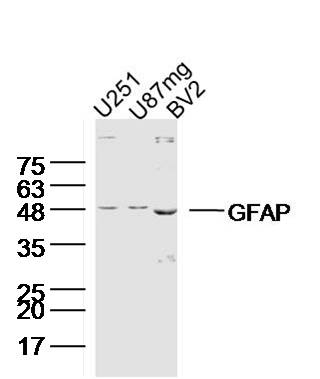 Sample:
U251(human) Cell Lysate at 40 ug
U87MG(human) Cell Lysate at 40 ug
BV2(mouse) Cell Lysate at 40 ug
Primary: Anti-GFAP (SL10950R) at 1/300 dilution
Secondary: IRDye800CW Goat Anti-Rabbit IgG at 1/20000 dilution
Predicted band size: 48 kD
Observed band size: 48 kD
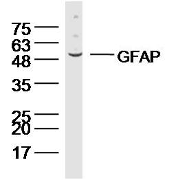 Sample:Cerebellum (Mouse) Lysate at 40 ug
Primary: Anti-GFAP (SL10950R) at 1/300 dilution
Secondary: IRDye800CW Goat Anti-Rabbit IgG at 1/20000 dilution
Predicted band size: 48 kD
Observed band size: 48 kD
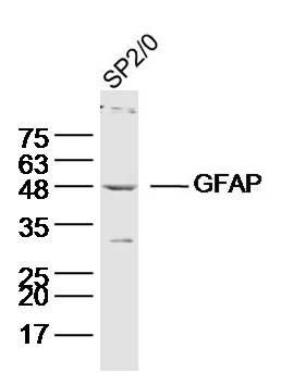 Sample:SP2/0(human) Cell Lysate at 40 ug
Primary: Anti-GFAP (SL10950R) at 1/300 dilution
Secondary: IRDye800CW Goat Anti-Rabbit IgG at 1/20000 dilution
Predicted band size: 48 kD
Observed band size: 48 kD
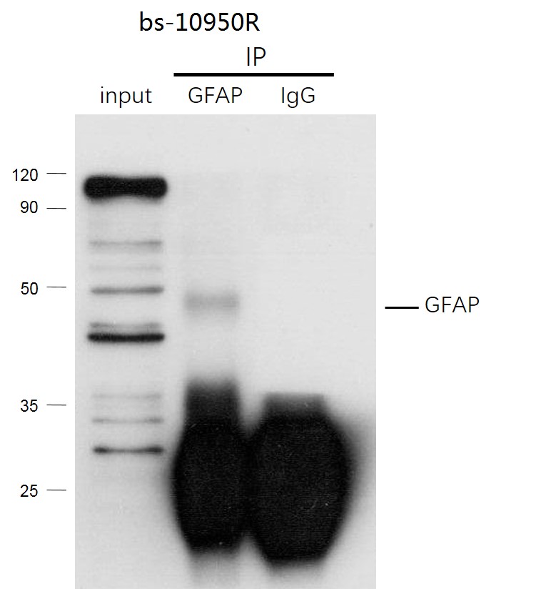 GFAP was immunoprecipitated from human hela cells lysate with SL10950R at 1/150 dilution. Western blot was performed from the immunoprecipitate using protein A/G beads. HRP Conjugated Mouse anti-Rabbit IgG (Light Chain specific) was used as secondary antibody at 1:5000 dilution.
Lane 1: human hela cells lysate 10 µg (Input).
Lane 2: SL10950R IP in human hela cells lysate.
Lane 3: native rabbit IgG IP in human hela cells lysate (negative control).
Secondary
All lanes : Mouse anti-Rabbit IgG (Light Chain specific), HRP Conjugated, 1:5000
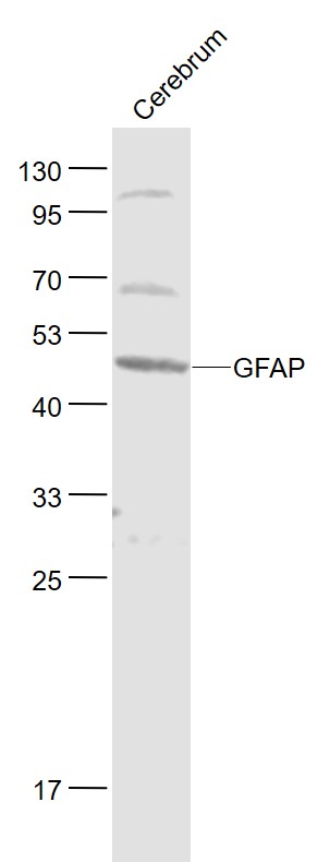 Sample:
Cerebrum (Rat) Lysate at 40 ug
Primary: Anti- GFAP (SL10950R) at 1/1000 dilution
Secondary: IRDye800CW Goat Anti-Rabbit IgG at 1/20000 dilution
Predicted band size: 48 kD
Observed band size: 48 kD
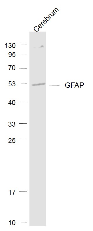 Sample:
Cerebrum (Mouse) Lysate at 40 ug
Primary: Anti-GFAP (SL10950R) at 1/1000 dilution
Secondary: IRDye800CW Goat Anti-Rabbit IgG at 1/20000 dilution
Predicted band size: 48 kD
Observed band size: 48 kD
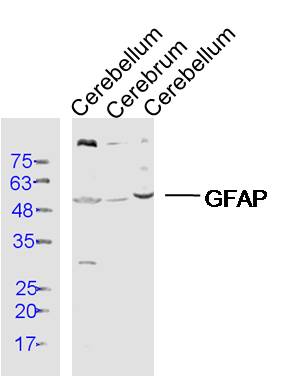 Sample:
Cerebellum (Rat) Lysate at 40 ug
Cerebrum (Mouse) Lysate at 40 ug
Cerebellum (Mouse) Lysate at 40 ug
Primary: Anti- GFAP (SL10950R) at 1/300 dilution
Secondary: IRDye800CW Goat Anti-Rabbit IgG at 1/20000 dilution
Predicted band size: 48 kD
Observed band size: 50 kD
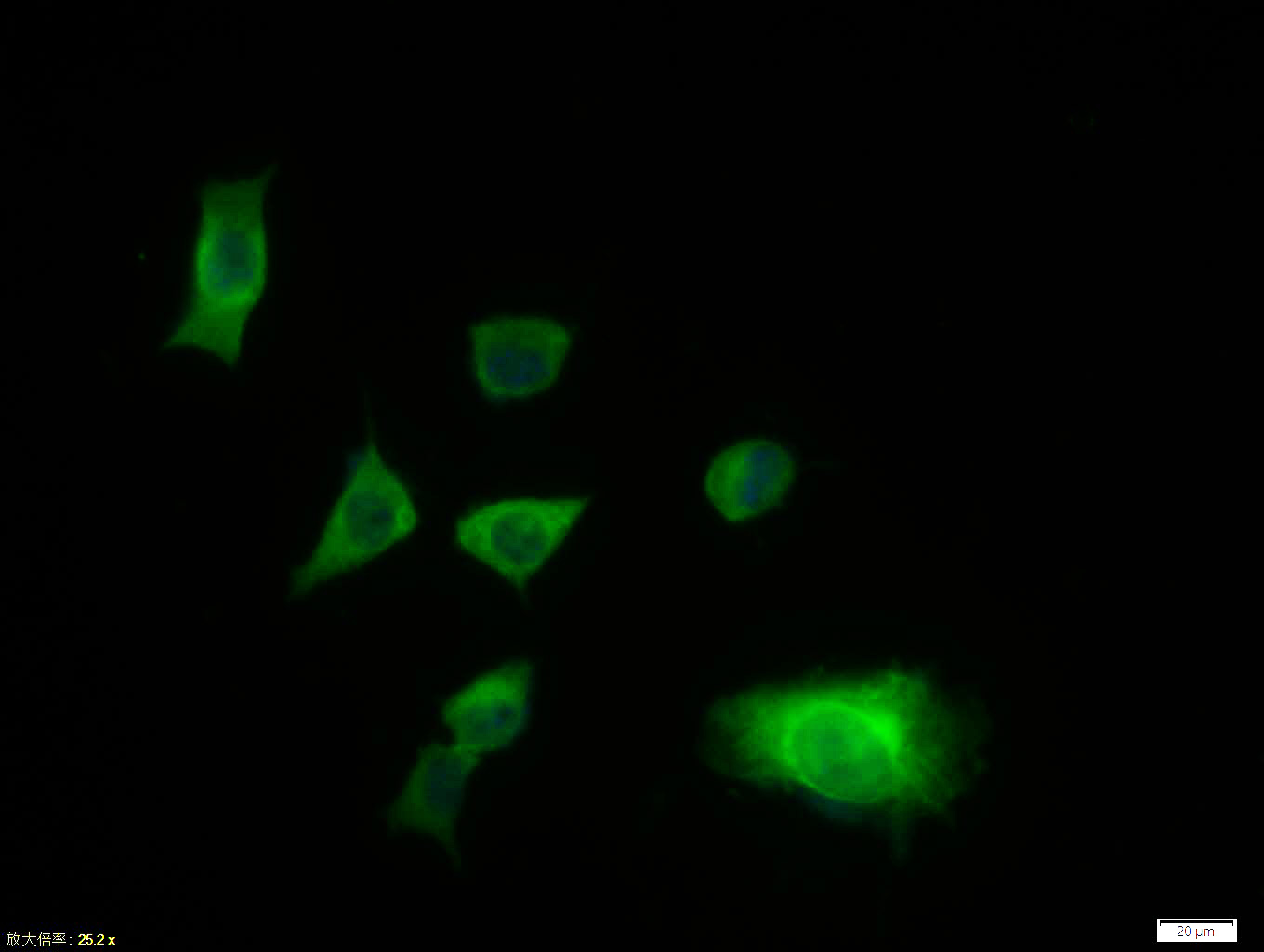 Tissue/cell:SH-SY5Y cell; 4% Paraformaldehyde-fixed; Triton X-100 at room temperature for 20 min; Blocking buffer (normal goat serum, C-0005) at 37°C for 20 min; Antibody incubation with (GFAP) polyclonal Antibody, Unconjugated (SL10950R) 1:100, 90 minutes at 37°C; followed by a FITC conjugated Goat Anti-Rabbit IgG antibody at 37°C for 90 minutes, DAPI (blue, C02-04002) was used to stain the cell nuclei.
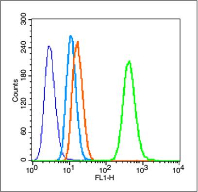 Blank control (blue line): Hela (fixed with 80% methanol (5 min at -20℃) and then permeabilized with 0.1% PBS-Tween for 20 min at room temperature ).
Primary Antibody (green line): Rabbit Anti-GFAP antibody (SL10950R),dilution: 3μg /10^6 cells;
Isotype Control Antibody (orange line): Rabbit IgG .
Secondary Antibody (white blue line): Goat anti-rabbit IgG-PE,Dilution: 1μg /test.
Cartpieces
Totalgoods,subtotals:¥Checkout
Bought notes(bought amounts latest0)
No one bought this product
User Comment(Total0User Comment Num)
- No comment


 +86 571 56623320
 +86 18668110335

