
Rabbit Anti-MRP2/ABCC2 antibody
multidrug resistance-associated protein2; ABC30; ABCC2; ATP binding cassette sub family C (CFTR/MRP) member 2; ATP binding cassette subfamily C member 2; Canalicular multidrug resistance protein; Canalicular multispecific organic anion transporter 1; CMOA
View History [Clear]
Details
Product Name MRP2/ABCC2 Chinese Name 多药耐药相关蛋白2抗体 Alias multidrug resistance-associated protein2; ABC30; ABCC2; ATP binding cassette sub family C (CFTR/MRP) member 2; ATP binding cassette subfamily C member 2; Canalicular multidrug resistance protein; Canalicular multispecific organic anion transporter 1; CMOAT; CMOAT1; cMRP; DJS; KIAA1010; MRP 2; MRP-2; MRP2; Multidrug resistance associated protein 2; MRP2_HUMAN; ATP-binding cassette sub-family C member 2; Multidrug resistance-associated protein 2. literatures Research Area Tumour Immunogen Species Rabbit Clonality Polyclonal React Species Human, Mouse, Rat, Applications WB=1:500-2000 ELISA=1:5000-10000 IHC-P=1:100-500 IHC-F=1:100-500 IF=1:100-500 (Paraffin sections need antigen repair)
not yet tested in other applications.
optimal dilutions/concentrations should be determined by the end user.Theoretical molecular weight 174kDa Cellular localization The cell membrane Form Liquid Concentration 1mg/ml immunogen KLH conjugated synthetic peptide derived from human MRP2: 485-615/1545 <Cytoplasmic> Lsotype IgG Purification affinity purified by Protein A Buffer Solution 0.01M TBS(pH7.4) with 1% BSA, 0.03% Proclin300 and 50% Glycerol. Storage Shipped at 4℃. Store at -20 °C for one year. Avoid repeated freeze/thaw cycles. Attention This product as supplied is intended for research use only, not for use in human, therapeutic or diagnostic applications. PubMed PubMed Product Detail multidrug resistance-associated protein 2 is a member of the superfamily of ATP-binding cassette (ABC) transporters. ABC proteins transport various molecules across extra- and intra-cellular membranes. ABC genes are divided into seven distinct subfamilies (ABC1, MDR/TAP, MRP, ALD, OABP, GCN20, White). This protein is a member of the MRP subfamily which is involved in multi-drug resistance. This protein is expressed in the canalicular (apical) part of the hepatocyte and functions in biliary transport. Substrates include anticancer drugs such as vinblastine; therefore, this protein appears to contribute to drug resistance in mammalian cells. Several different mutations in this gene have been observed in patients with Dubin-Johnson syndrome (DJS), an autosomal recessive disorder characterized by conjugated hyperbilirubinemia. Belongs to the ABC transporter family.
Function:
Mediates hepatobiliary excretion of numerous organic anions. May function as a cellular cisplatin transporter.
Subcellular Location:
Membrane; Multi-pass membrane protein.
Tissue Specificity:
Found on the apical membrane of polarized cells in liver, kidney and intestine. The highest expression is found in liver.
DISEASE:
Defects in ABCC2 are the cause of Dubin-Johnson syndrome (DJS) [MIM:237500]. DJS is an autosomal recessive disorder characterized by conjugated hyperbilirubinemia, an increase in the urinary excretion of coproporphyrin isomer I, deposition of melanin-like pigment in hepatocytes, and prolonged retention of sulfobromophthalein, but otherwise normal liver function.
Similarity:
Belongs to the ABC transporter superfamily. ABCC family. Conjugate transporter (TC 3.A.1.208) subfamily.
Contains 2 ABC transmembrane type-1 domains.
Contains 2 ABC transporter domains.
SWISS:
Q92887
Gene ID:
1244
Database links:
Entrez Gene: 1244 Human
Entrez Gene: 12780 Mouse
Omim: 601107 Human
SwissProt: Q92887 Human
SwissProt: Q8VI47 Mouse
Unigene: 368243 Human
Unigene: 39054 Mouse
Unigene: 10265 Rat
MRP-2作为一种结合输出泵,转运许多不同的药物结合物的蛋白。
MRP2蛋白的染色阳性产物分布于cytoplasmic/膜中。Product Picture 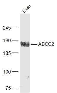 Sample:
Liver (Mouse) Lysate at 40 ug
Primary: Anti-ABCC2 (SL1092R) at 1/1000 dilution
Secondary: IRDye800CW Goat Anti-Rabbit IgG at 1/20000 dilution
Predicted band size: 174 kD
Observed band size: 174 kD
 Sample:
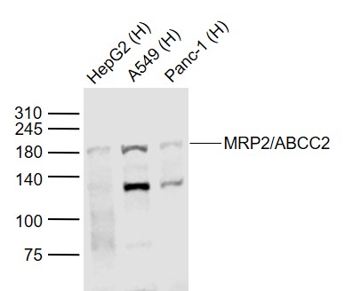
Lane 1: HepG2 (Human) Cell Lysate at 30 ug
Lane 2: A549 (Human) Cell Lysate at 30 ug
Lane 3: Panc-1 (Human) Cell Lysate at 30 ug
Primary: Anti-MRP2/ABCC2 (SL1092R) at 1/1000 dilution
Secondary: IRDye800CW Goat Anti-Rabbit IgG at 1/20000 dilution
Predicted band size: 180 kD
Observed band size: 180 kD
 Sample:
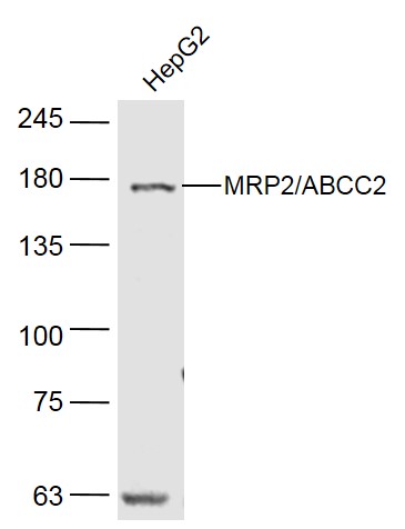
HepG2(Human) Cell Lysate at 40 ug
Primary: Anti- MRP2/ABCC2 (SL1092R) at 1/300 dilution
Secondary: IRDye800CW Goat Anti-Rabbit IgG at 1/20000 dilution
Predicted band size: 174 kD
Observed band size: 174 kD
 Paraformaldehyde-fixed, paraffin embedded (human lung carcinoma); Antigen retrieval by boiling in sodium citrate buffer (pH6.0) for 15min; Block endogenous peroxidase by 3% hydrogen peroxide for 20 minutes; Blocking buffer (normal goat serum) at 37°C for 30min; Antibody incubation with (MRP2/ABCC2) Polyclonal Antibody, Unconjugated (SL1092R) at 1:200 overnight at 4°C, followed by operating according to SP Kit(Rabbit) (sp-0023) instructionsand DAB staining.
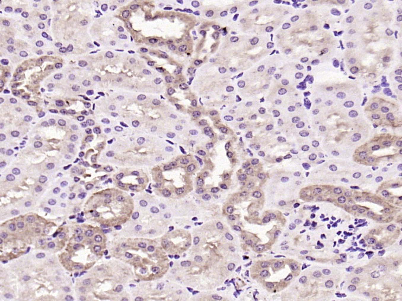 Paraformaldehyde-fixed, paraffin embedded (rat kidney); Antigen retrieval by boiling in sodium citrate buffer (pH6.0) for 15min; Block endogenous peroxidase by 3% hydrogen peroxide for 20 minutes; Blocking buffer (normal goat serum) at 37°C for 30min; Antibody incubation with (ABCC2) Polyclonal Antibody, Unconjugated (rat kidney) at 1:2000 overnight at 4°C, followed by operating according to SP Kit(Rabbit) (sp-0023) instructionsand DAB staining.
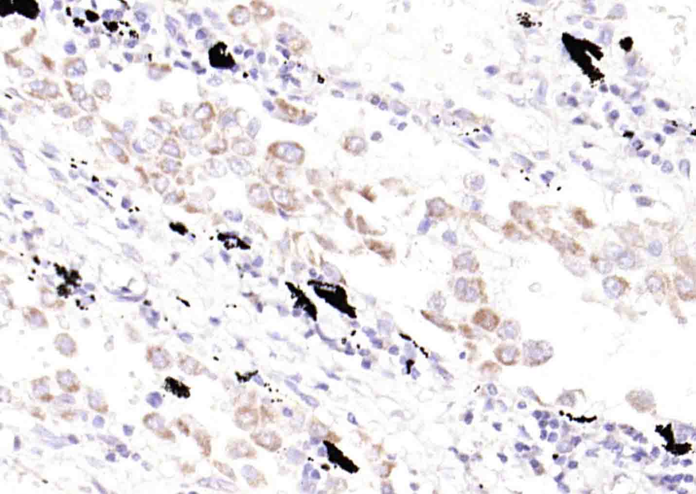
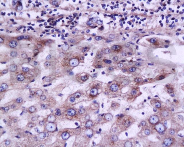 Tissue/cell: human liver carcinoma; 4% Paraformaldehyde-fixed and paraffin-embedded;
Antigen retrieval: citrate buffer ( 0.01M, pH 6.0 ), Boiling bathing for 15min; Block endogenous peroxidase by 3% Hydrogen peroxide for 30min; Blocking buffer (normal goat serum,C-0005) at 37℃ for 20 min;
Incubation: Anti-MRP2 Polyclonal Antibody, Unconjugated(SL1092R) 1:200, overnight at 4°C, followed by conjugation to the secondary antibody(SP-0023) and DAB(C-0010) staining
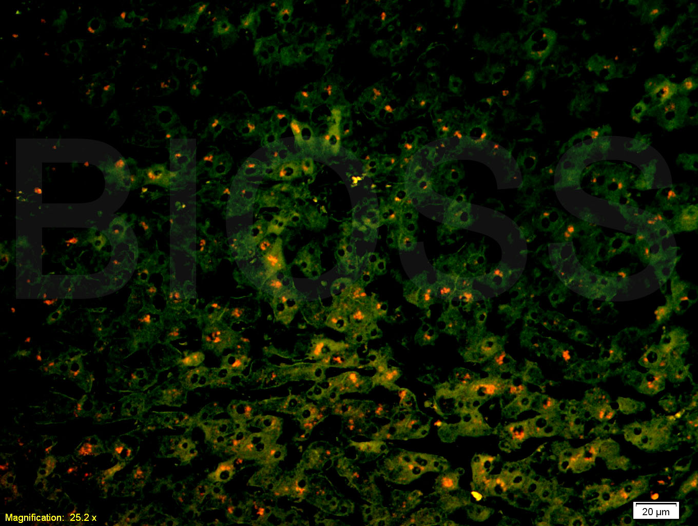 Tissue/cell: human liver carcinoma;4% Paraformaldehyde-fixed and paraffin-embedded;
Antigen retrieval: citrate buffer ( 0.01M, pH 6.0 ), Boiling bathing for 15min; Blocking buffer (normal goat serum,C-0005) at 37℃ for 20 min;
Incubation: Anti-MRP2 Polyclonal Antibody, Unconjugated(SL1092R) 1:200, overnight at 4°C; The secondary antibody was Goat Anti-Rabbit IgG, PE conjugated(SL0295G-PE)used at 1:200 dilution for 40 minutes at 37°C.
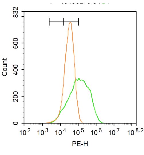 Blank control: A549.
Primary Antibody (green line): Rabbit Anti-ABCC2 antibody (SL1092R)
Dilution: 3μg /10^6 cells;
Isotype Control Antibody (orange line): Rabbit IgG .
Secondary Antibody : Goat anti-rabbit IgG-PE
Dilution: 1μg /test.
Protocol
The cells were incubated in 5%BSA to block non-specific protein-protein interactions for 30 min at at room temperature .Cells stained with Primary Antibody for 30 min at room temperature. The secondary antibody used for 40 min at room temperature. Acquisition of 20,000 events was performed.
Cartpieces
Totalgoods,subtotals:¥Checkout
Bought notes(bought amounts latest0)
No one bought this product
User Comment(Total0User Comment Num)
- No comment


 +86 571 56623320
 +86 18668110335

