
Rabbit Anti-Egr1 antibody
Egr-1; Early growth response 1; Krox-24; Ngf1; Ngfi; NGFI-A; zif-268; Egr1; A530045N19Rik; egr; Egr-1; ETR103; Krox-1; Krox-24; Krox24; NGF1-A; NGFI-A; NGFIA; TIS8; Zenk; Zfp-6; AT 225; AT225; Early growth response protein 1; G0S 30; G0S30; KROX 24; Krox
View History [Clear]
Details
Product Name Egr1 Chinese Name 早期生长应答蛋白1抗体 Alias Egr-1; Early growth response 1; Krox-24; Ngf1; Ngfi; NGFI-A; zif-268; Egr1; A530045N19Rik; egr; Egr-1; ETR103; Krox-1; Krox-24; Krox24; NGF1-A; NGFI-A; NGFIA; TIS8; Zenk; Zfp-6; AT 225; AT225; Early growth response protein 1; G0S 30; G0S30; KROX 24; Krox 24 protein; KROX24; TIS 8; TIS8; Transcription factor ETR 103; Transcription factor ETR103; Transcription factor Zif 268; Transcription factor Zif268; Zfp 6; ZIF268; Zinc finger protein 225; ZNF 225; ZNF225; EGR1_HUMAN. literatures Research Area Cell biology immunology Signal transduction Growth factors and hormones Cyclin transcriptional regulatory factor Immunogen Species Rabbit Clonality Polyclonal React Species Human, Mouse, Rat, Applications WB=1:500-2000 ELISA=1:5000-10000 IHC-P=1:100-500 IHC-F=1:100-500 Flow-Cyt=2ug/test ICC=1:100 IF=1:100-500 (Paraffin sections need antigen repair)
not yet tested in other applications.
optimal dilutions/concentrations should be determined by the end user.Theoretical molecular weight 60kDa Cellular localization The nucleus Form Liquid Concentration 1mg/ml immunogen KLH conjugated synthetic peptide derived from human Egr-1: 401-500/453 Lsotype IgG Purification affinity purified by Protein A Buffer Solution 0.01M TBS(pH7.4) with 1% BSA, 0.03% Proclin300 and 50% Glycerol. Storage Shipped at 4℃. Store at -20 °C for one year. Avoid repeated freeze/thaw cycles. Attention This product as supplied is intended for research use only, not for use in human, therapeutic or diagnostic applications. PubMed PubMed Product Detail The protein encoded by this gene belongs to the EGR family of C2H2-type zinc-finger proteins. It is a nuclear protein and functions as a transcriptional regulator. The products of target genes it activates are required for differentitation and mitogenesis. Studies suggest this is a cancer suppresor gene. [provided by RefSeq].
Function:
Transcriptional regulator. Recognizes and binds to the DNA sequence 5'-CGCCCCCGC-3'(EGR-site). Activates the transcription of target genes whose products are required for mitogenesis and differentiation.
Subcellular Location:
Nucleus.
Similarity:
Belongs to the EGR C2H2-type zinc-finger protein family.
Contains 3 C2H2-type zinc fingers.
SWISS:
P18146
Gene ID:
1958
Database links:Entrez Gene: 1958 Human
Entrez Gene: 13653 Mouse
Omim: 128990 Human
SwissProt: P18146 Human
SwissProt: P08046 Mouse
Unigene: 326035 Human
Unigene: 708393 Human
Unigene: 181959 Mouse
Egr-1为即刻早期基因家族中的一个成员,是一种含3个锌指结构的转录因子。有研究表明EGR-1在细胞生长分化中发挥重要作用,如在细胞周期G1-S期的转换过程中Egr-1被诱导表达。Product Picture 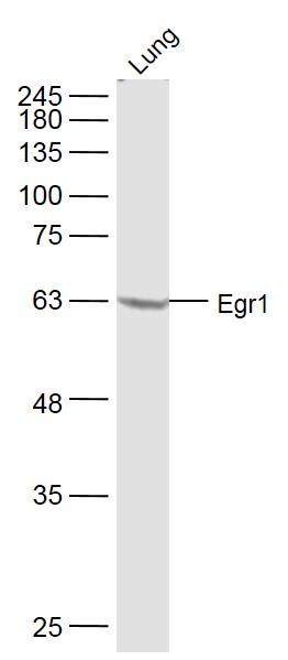 Sample:
Lung (Mouse) Lysate at 40 ug
Primary: Anti-Egr1 (SL1076R) at 1/300 dilution
Secondary: IRDye800CW Goat Anti-Rabbit IgG at 1/20000 dilution
Predicted band size: 60 kD
Observed band size: 60 kD
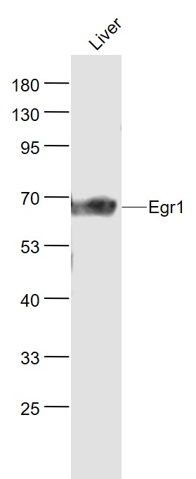 Sample:
Liver (Rat) Lysate at 40 ug
Primary: Anti- Egr1 (SL1076R) at 1/1000 dilution
Secondary: IRDye800CW Goat Anti-Rabbit IgG at 1/20000 dilution
Predicted band size: 60 kD
Observed band size: 60 kD
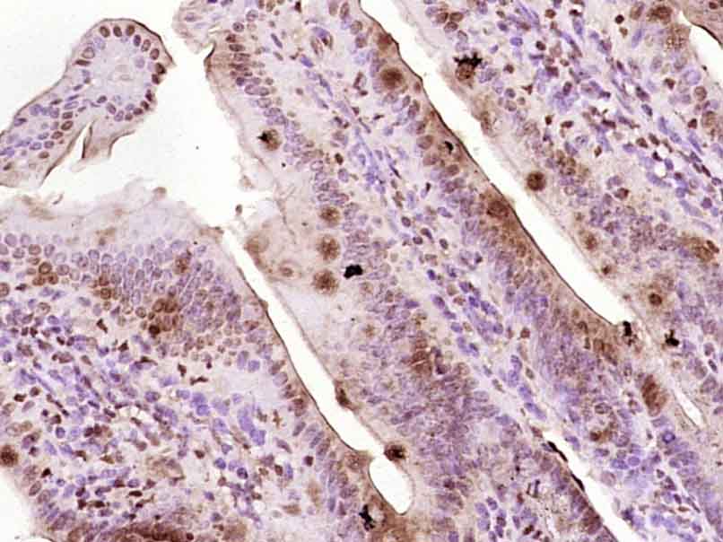 Paraformaldehyde-fixed, paraffin embedded (Mouse small intestine); Antigen retrieval by boiling in sodium citrate buffer (pH6.0) for 15min; Block endogenous peroxidase by 3% hydrogen peroxide for 20 minutes; Blocking buffer (normal goat serum) at 37°C for 30min; Antibody incubation with (Egr1) Polyclonal Antibody, Unconjugated (SL1076R) at 1:400 overnight at 4°C, followed by operating according to SP Kit(Rabbit) (sp-0023) instructionsand DAB staining.
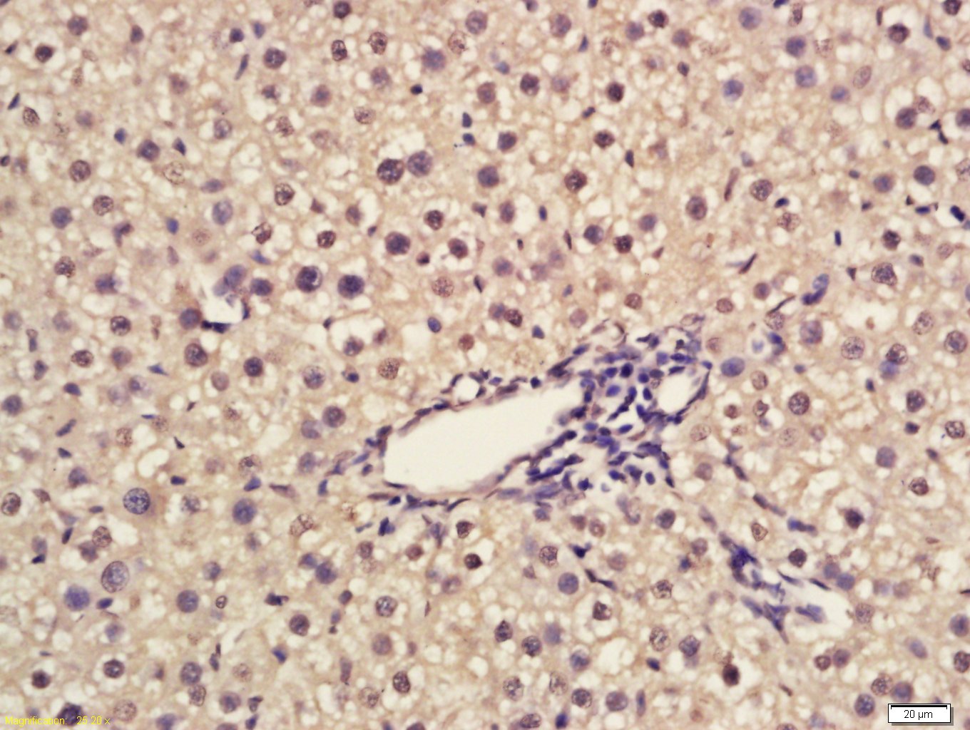 Tissue/cell: rat liver tissue; 4% Paraformaldehyde-fixed and paraffin-embedded;
Antigen retrieval: citrate buffer ( 0.01M, pH 6.0 ), Boiling bathing for 15min; Block endogenous peroxidase by 3% Hydrogen peroxide for 30min; Blocking buffer (normal goat serum,C-0005) at 37℃ for 20 min;
Incubation: Anti-Egr1 Polyclonal Antibody, Unconjugated(SL1076R) 1:200, overnight at 4°C, followed by conjugation to the secondary antibody(SP-0023) and DAB(C-0010) staining
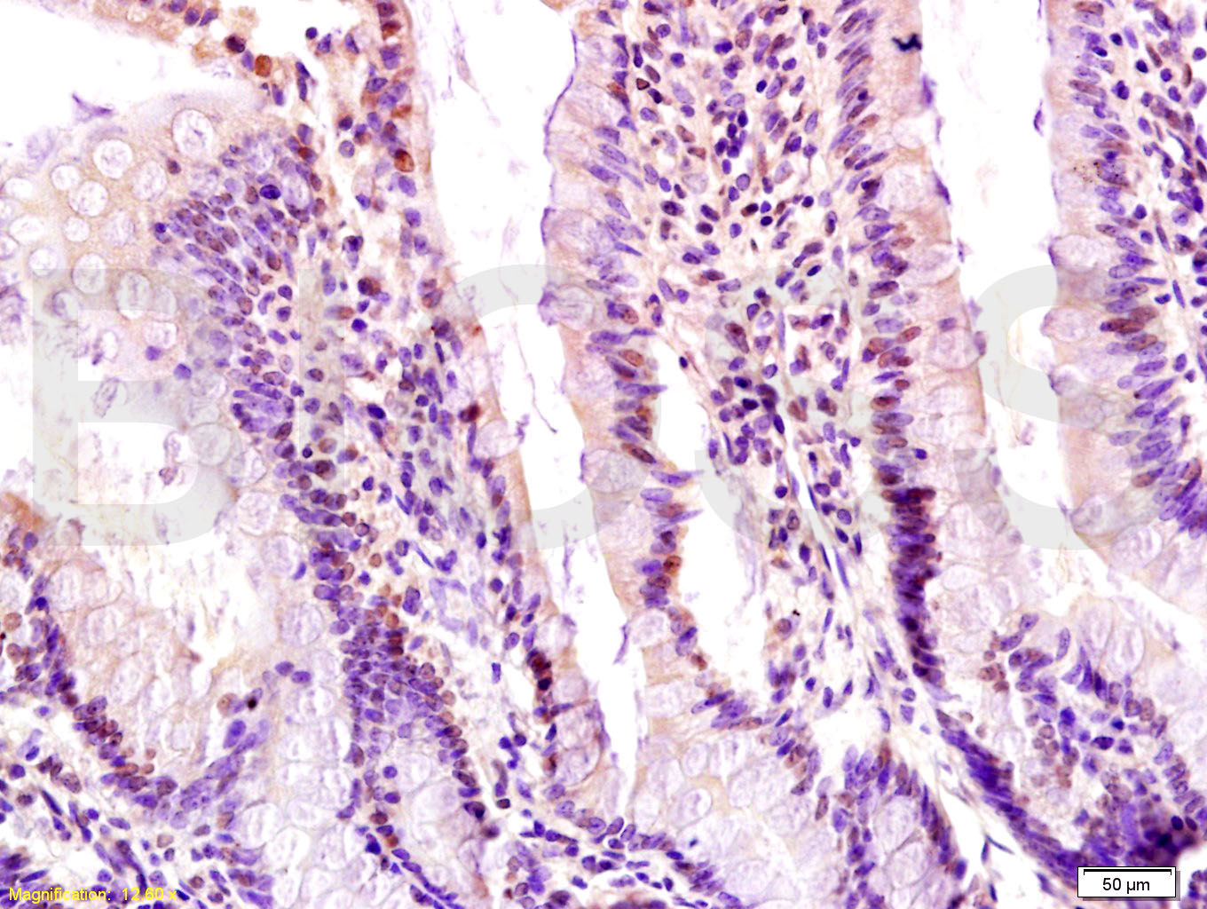 Tissue/cell: rat small intestine tissue; 4% Paraformaldehyde-fixed and paraffin-embedded;
Antigen retrieval: citrate buffer ( 0.01M, pH 6.0 ), Boiling bathing for 15min; Block endogenous peroxidase by 3% Hydrogen peroxide for 30min; Blocking buffer (normal goat serum,C-0005) at 37℃ for 20 min;
Incubation: Anti-Egr1 Polyclonal Antibody, Unconjugated(SL1076R) 1:200, overnight at 4°C, followed by conjugation to the secondary antibody(SP-0023) and DAB(C-0010) staining
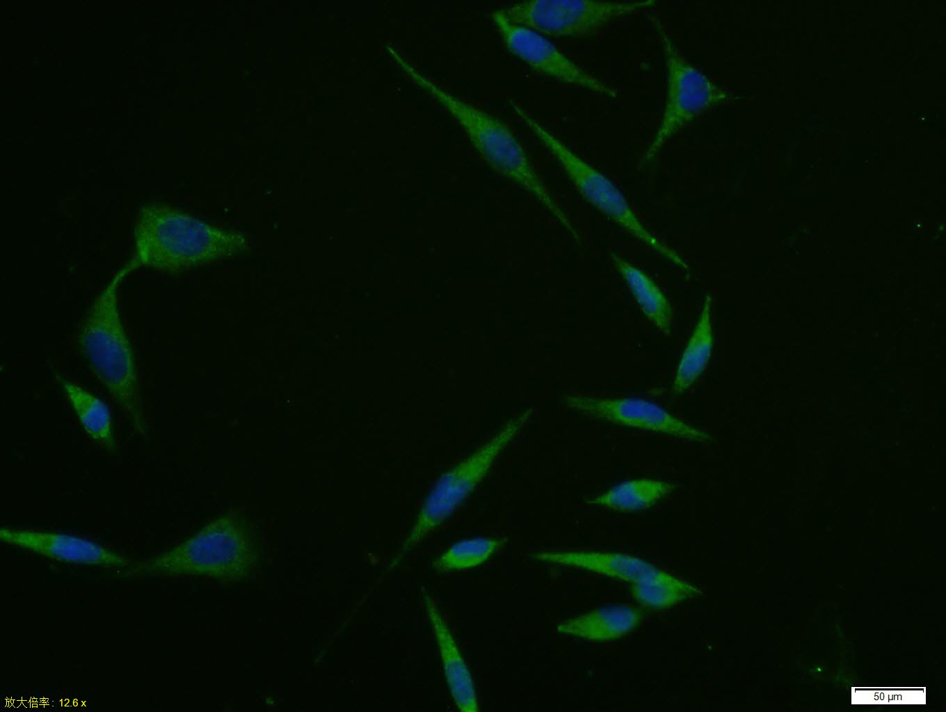 A431 cell; 4% Paraformaldehyde-fixed; Triton X-100 at room temperature for 20 min; Blocking buffer (normal goat serum, C-0005) at 37°C for 20 min; Antibody incubation with (Egr1) polyclonal Antibody, Unconjugated (SL1076R) 1:100, 90 minutes at 37°C; followed by a conjugated Goat Anti-Rabbit IgG antibody at 37°C for 90 minutes, DAPI (blue, C02-04002) was used to stain the cell nuclei.
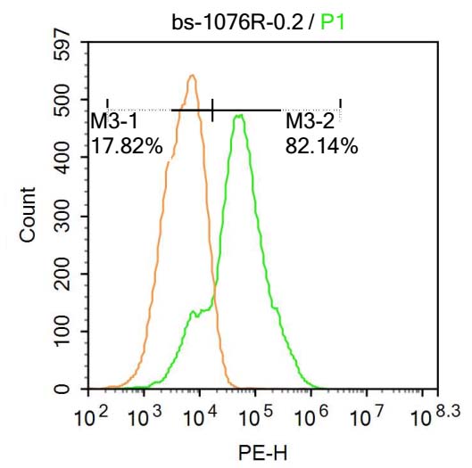 U-937 cells were fixed with 4% PFA for 10min at room temperature,permeabilized with 90% ice-cold methanol for 20 min at room temperature,and incubated in 5% BSA blocking buffer for 30 min at room temperature. Cells were then stained with Egr1 Antibody(SL1076R) at 1:500 dilution in blocking buffer and incubated for 30 min at room temperature, washed twice with 2%BSA in PBS, followed by secondary antibody incubation for 40 min at room temperature. Acquisitions of 20,000 events were performed.Cells stained with primary antibody (green), and isotype control (orange).
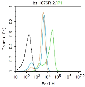 Blank control:K562.
Primary Antibody (green line): Rabbit Anti-Egr1 antibody (SL1076R)
Dilution: 2μg /10^6 cells;
Isotype Control Antibody (orange line): Rabbit IgG .
Secondary Antibody : Goat anti-rabbit IgG-FITC
Dilution: 0.5μg /test.
Protocol
The cells were fixed with 4% PFA (10min at room temperature)and then permeabilized with 0.1% PBST for 20 min at room temperature. The cells were then incubated in 5%BSA to block non-specific protein-protein interactions for 30 min at room temperature .Cells stained with Primary Antibody for 30 min at room temperature. The secondary antibody used for 40 min at room temperature. Acquisition of 20,000 events was performed.
Cartpieces
Totalgoods,subtotals:¥Checkout
Bought notes(bought amounts latest0)
No one bought this product
User Comment(Total0User Comment Num)
- No comment


 +86 571 56623320
 +86 18668110335

