
Rabbit Anti-CD101 antibody
IGSF2; Immunoglobulin superfamily member 2; Leukocyte surface protein; V7; V7 LSB; IGSF2_HUMAN; IgSF2; Cell surface glycoprotein V7; Glu-Trp-Ile EWI motif-containing protein 101; EWI-101; CD101.
View History [Clear]
Details
Product Name CD101 Chinese Name CD101抗体 Alias IGSF2; Immunoglobulin superfamily member 2; Leukocyte surface protein; V7; V7 LSB; IGSF2_HUMAN; IgSF2; Cell surface glycoprotein V7; Glu-Trp-Ile EWI motif-containing protein 101; EWI-101; CD101. literatures Research Area Cell biology t-lymphocyte Immunogen Species Rabbit Clonality Polyclonal React Species Human, (predicted: Mouse, Rat, ) Applications WB=1:500-2000 ELISA=1:5000-10000 IHC-P=1:100-500 IHC-F=1:100-500 Flow-Cyt=1μg/Test ICC=1:100-500 IF=1:10-100 (Paraffin sections need antigen repair)
not yet tested in other applications.
optimal dilutions/concentrations should be determined by the end user.Theoretical molecular weight 113kDa Cellular localization The cell membrane Form Liquid Concentration 1mg/ml immunogen KLH conjugated synthetic peptide derived from human CD101: 51-150/1021 <Extracellular> Lsotype IgG Purification affinity purified by Protein A Buffer Solution 0.01M TBS(pH7.4) with 1% BSA, 0.03% Proclin300 and 50% Glycerol. Storage Shipped at 4℃. Store at -20 °C for one year. Avoid repeated freeze/thaw cycles. Attention This product as supplied is intended for research use only, not for use in human, therapeutic or diagnostic applications. PubMed PubMed Product Detail CD101 is a disulfide-linked homodimeric type 1 glycoprotein. The peptide is comprised of 7 extracellular V-type IgSF domains. CD101 is highly expressed on monocytes, granulocytes, mucosal T cells, and on activated peripheral blood T cells. Expression is weak on resting T and B and NK cells, absent from platelets and weak or absent from most hematopoietic cell lines. CD101 is thought to play a co-stimulatory role in TCR/CD3-mediated T cell activation. Monoclonal antibodies against CD101 inhibit allogeneic T cell responses. Studies suggest that CD101 plays a major role in the activation of T cells by skin dendritic cells.
Function:
Plays a role as inhibitor of T-cells proliferation induced by CD3. Inhibits expression of IL2RA on activated T-cells and secretion of IL2. Inhibits tyrosine kinases that are required for IL2 production and cellular proliferation. Inhibits phospholipase C-gamma-1/PLCG1 phosphorylation and subsequent CD3-induced changes in intracellular free calcium. Prevents nuclear translocation of nuclear factor of activated T-cell to the nucleus. Plays a role in the inhibition of T-cell proliferation via IL10 secretion by cutaneous dendritic cells. May be a marker of CD4(+) CD56(+) leukemic tumor cells.
Subcellular Location:
Membrane; Single-pass type I membrane protein (Potential).
Tissue Specificity:
Expressed in lung, thymus and small intestine. Detected in cutaneous dendritic cells, activated T-cells, monocytes and granulocytes as well as with epithelial cells with dendritic morphology. Expressed in some leukemic cells, the CD4(+) CD56(+) blastic tumor cells, as well as in Langerhans cells from LCH (Langerhans cell histiocytosis) patients.
Post-translational modifications:
N-glycosylated.
Similarity:
Contains 7 Ig-like C2-type (immunoglobulin-like) domains.
SWISS:
Q93033
Gene ID:
9398
Database links:Entrez Gene: 9398 Human
Omim: 604516 Human
SwissProt: Q93033 Human
Unigene: 74115 Human
Product Picture 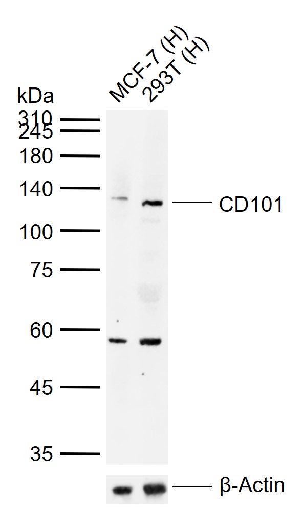 Sample:
Lane 1: Human MCF-7 cell lysates
Lane 2: Human 293T cell lysates
Primary: Anti-CD101 (SL10727R) at 1/1000 dilution
Secondary: IRDye800CW Goat Anti-Rabbit IgG at 1/20000 dilution
Predicted band size: 113 kDa
Observed band size: 125 kDa
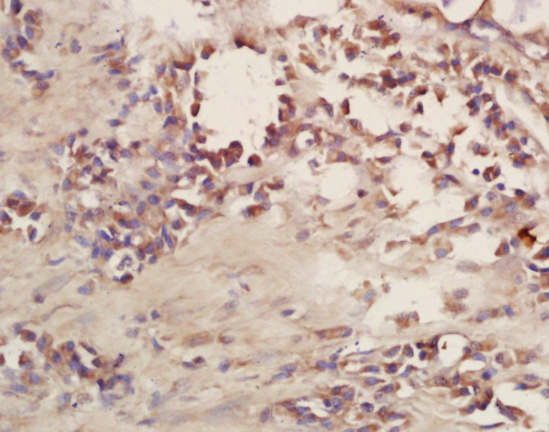 Tissue/cell: human lung carcinoma; 4% Paraformaldehyde-fixed and paraffin-embedded;
Antigen retrieval: citrate buffer ( 0.01M, pH 6.0 ), Boiling bathing for 15min; Block endogenous peroxidase by 3% Hydrogen peroxide for 30min; Blocking buffer (normal goat serum,C-0005) at 37℃ for 20 min;
Incubation: Anti-CD101 Polyclonal Antibody, Unconjugated(SL10727R) 1:200, overnight at 4°C, followed by conjugation to the secondary antibody(SP-0023) and DAB(C-0010) staining
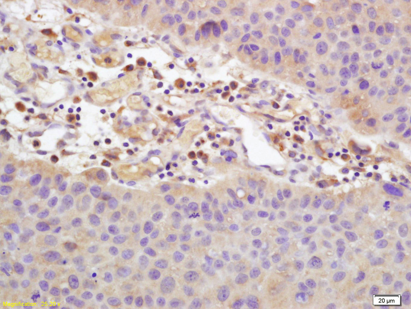 Tissue/cell: human bladder carcinoma; 4% Paraformaldehyde-fixed and paraffin-embedded;
Antigen retrieval: citrate buffer ( 0.01M, pH 6.0 ), Boiling bathing for 15min; Block endogenous peroxidase by 3% Hydrogen peroxide for 30min; Blocking buffer (normal goat serum,C-0005) at 37℃ for 20 min;
Incubation: Anti-CD101 Polyclonal Antibody, Unconjugated(SL10727R) 1:200, overnight at 4°C, followed by conjugation to the secondary antibody(SP-0023) and DAB(C-0010) staining
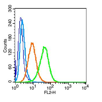 Blank control(blue): U937(fixed with 2% paraformaldehyde (10 min)).
Primary Antibody:Rabbit Anti-CD101 antibody(SL10727R), Dilution: 1μg in 100 μL 1X PBS containing 0.5% BSA;
Isotype Control Antibody: Rabbit IgG(orange) ,used under the same conditions );
Secondary Antibody: Goat anti-rabbit IgG-PE(white blue), Dilution: 1:200 in 1 X PBS containing 0.5% BSA.
Cartpieces
Totalgoods,subtotals:¥Checkout
Bought notes(bought amounts latest0)
No one bought this product
User Comment(Total0User Comment Num)
- No comment


 +86 571 56623320
 +86 18668110335

