
Rabbit Anti-NMDAR1 antibody
NMDA-NR1; N-Methyl-d-Asprtate receptor 1; GRIN1; NMDA1; NR1; Glutamate [NMDA] receptor subunit zeta 1; Glutamate receptor ionotropic N methyl D aspartate 1; Grin 1; Grin1; N methyl D aspartate receptor channel; N-methyl-D-aspartate receptor; N-methyl-D-as
View History [Clear]
Details
Product Name NMDAR1 Chinese Name 离子型谷氨酸受体1抗体 Alias NMDA-NR1; N-Methyl-d-Asprtate receptor 1; GRIN1; NMDA1; NR1; Glutamate [NMDA] receptor subunit zeta 1; Glutamate receptor ionotropic N methyl D aspartate 1; Grin 1; Grin1; N methyl D aspartate receptor channel; N-methyl-D-aspartate receptor; N-methyl-D-aspartate receptor subunit NR1; NMD-R1; NMDA 1; NMDA NR1; NMDA R1; NMDA receptor 1; NMDA1; NMDAR 1; NMDAR; NR 1; NMDZ1_HUMAN. Research Area Cell biology immunology Neurobiology Signal transduction Apoptosis transcriptional regulatory factor The cell membrane受体 Immunogen Species Rabbit Clonality Polyclonal React Species Human, Rat, (predicted: Mouse, Chicken, Dog, Cow, Horse, ) Applications WB=1:500-2000 ELISA=1:5000-10000 IHC-P=1:100-500 IHC-F=1:100-500 Flow-Cyt=2ug/test IF=1:100-500 (Paraffin sections need antigen repair)
not yet tested in other applications.
optimal dilutions/concentrations should be determined by the end user.Theoretical molecular weight 103kDa Cellular localization The cell membrane Form Liquid Concentration 1mg/ml immunogen KLH conjugated synthetic peptide derived from human NMDAR1: 801-900/938 Lsotype IgG Purification affinity purified by Protein A Buffer Solution 0.01M TBS(pH7.4) with 1% BSA, 0.03% Proclin300 and 50% Glycerol. Storage Shipped at 4℃. Store at -20 °C for one year. Avoid repeated freeze/thaw cycles. Attention This product as supplied is intended for research use only, not for use in human, therapeutic or diagnostic applications. PubMed PubMed Product Detail Neuronal Marker
The protein encoded by this gene is a critical subunit of N-methyl-D-aspartate receptors, members of the glutamate receptor channel superfamily which are heteromeric protein complexes with multiple subunits arranged to form a ligand-gated ion channel. These subunits play a key role in the plasticity of synapses, which is believed to underlie memory and learning. Cell-specific factors are thought to control expression of different isoforms, possibly contributing to the functional diversity of the subunits. Alternatively spliced transcript variants have been described. [provided by RefSeq, Jul 2008]
Function:
NMDA receptor subtype of glutamate-gated ion channels with high calcium permeability and voltage-dependent sensitivity to magnesium. Mediated by glycine. This protein plays a key role in synaptic plasticity, synaptogenesis, excitotoxicity, memory acquisition and learning. It mediates neuronal functions in glutamate neurotransmission. Is involved in the cell surface targeting of NMDA receptors.
Subunit:
Forms heteromeric channel of a zeta subunit (GRIN1), a epsilon subunit (GRIN2A, GRIN2B, GRIN2C or GRIN2D) and a third subunit (GRIN3A or GRIN3B); disulfide-linked. Found in a complex with GRIN2A or GRIN2B, GRIN3A or GRIN3B and PPP2CB. Interacts with DLG4 and MPDZ. Interacts with LRFN1 and LRFN2. Interacts with MYZAP.
Subcellular Location:
Cell membrane; Multi-pass membrane protein. Cell junction, synapse, postsynaptic cell membrane. Cell junction, synapse, postsynaptic cell membrane, postsynaptic density. Note=Enriched in post-synaptic plasma membrane and post-synaptic densities.
Tissue Specificity:
Post-translational modifications:
NMDA is probably regulated by C-terminal phosphorylation of an isoform of NR1 by PKC. Dephosphorylated on Ser-897 probably by protein phosphatase 2A (PPP2CB). Its phosphorylated state is influenced by the formation of the NMDAR-PPP2CB complex and the NMDAR channel activity.
DISEASE:
Defects in GRIN1 are the cause of mental retardation autosomal dominant type 8 (MRD8) [MIM:614254]. Mental retardation is characterized by significantly below average general intellectual functioning associated with impairments in adaptative behavior and manifested during the developmental period.
Similarity:
Belongs to the glutamate-gated ion channel (TC 1.A.10.1) family. NR1/GRIN1 subfamily.
SWISS:
Q05586
Gene ID:
2902
Database links:Entrez Gene: 2902 Human
Entrez Gene: 14810 Mouse
Omim: 138249 Human
SwissProt: Q05586 Human
SwissProt: P35438 Mouse
Unigene: 558334 Human
Unigene: 278672 Mouse
Unigene: 9840 Rat
神经细胞Maker
(NMDAR1)N-甲基-D-天门冬氨酸受体(NMDAR)是兴奋性氨基酸受体亚型之一,是由NMDAR1与不同的NMDAR2亚基组成的异聚体。
NMDAR1又称GluR1 (Glutamate Receptor 1)近年实验研究发现,许多NMDAR拮抗药均具有镇痛活性,表明NMDAR在痛觉传递中具有重要作用,这为新型镇痛药的研究开发提供了新的作用靶点。Product Picture 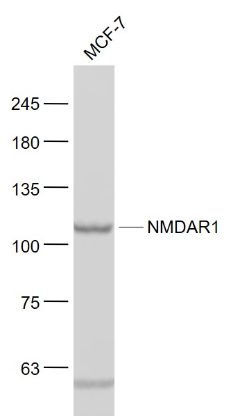 Sample:
MCF-7(Human) Cell Lysate at 30 ug
Primary: Anti- NMDAR1 (SL1068R) at 1/1000 dilution
Secondary: IRDye800CW Goat Anti-Rabbit IgG at 1/20000 dilution
Predicted band size: 103 kD
Observed band size: 105 kD
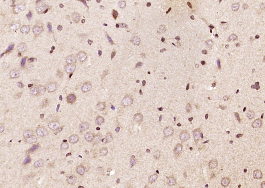 Paraformaldehyde-fixed, paraffin embedded (rat brain tissue); Antigen retrieval by boiling in sodium citrate buffer (pH6.0) for 15min; Block endogenous peroxidase by 3% hydrogen peroxide for 20 minutes; Blocking buffer (normal goat serum) at 37°C for 30min; Antibody incubation with (NMDAR1) Polyclonal Antibody, Unconjugated (SL1068R ) at 1:200 overnight at 4°C, followed by operating according to SP Kit(Rabbit) (sp-0023) instructionsand DAB staining.
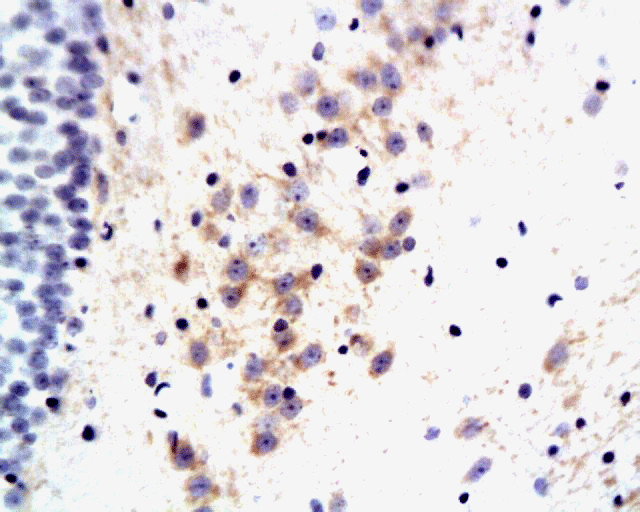 Tissue/cell: rat brain tissue; 4% Paraformaldehyde-fixed and paraffin-embedded;
Antigen retrieval: citrate buffer ( 0.01M, pH 6.0 ), Boiling bathing for 15min; Block endogenous peroxidase by 3% Hydrogen peroxide for 30min; Blocking buffer (normal goat serum,C-0005) at 37鈩� for 20 min;
Incubation: Anti-GluR1/AMPA Polyclonal Antibody, Unconjugated(SL1068R) 1:200, overnight at 4掳C, followed by conjugation to the secondary antibody(SP-0023) and DAB(C-0010) staining
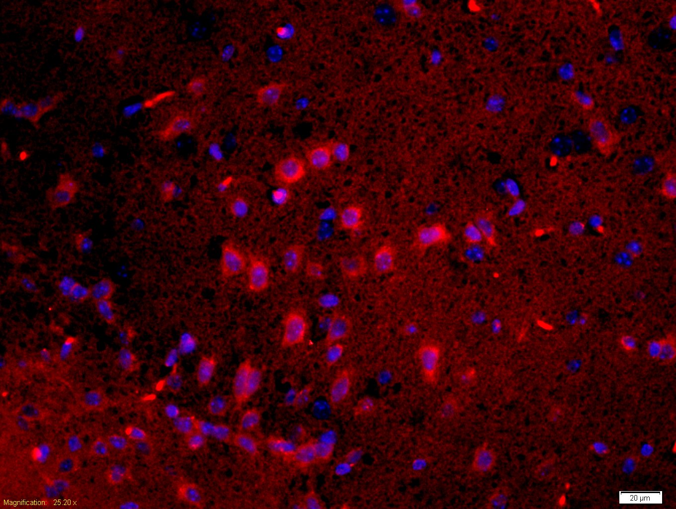 Tissue/cell: rat brain tissue;4% Paraformaldehyde-fixed and paraffin-embedded;
Antigen retrieval: citrate buffer ( 0.01M, pH 6.0 ), Boiling bathing for 15min; Blocking buffer (normal goat serum,C-0005) at 37℃ for 20 min;
Incubation: Anti-GluR1/AMPA Polyclonal Antibody, Unconjugated(SL1068R) 1:200, overnight at 4°C; The secondary antibody was Goat Anti-Rabbit IgG, Cy3 conjugated(SL0295G-Cy3)used at 1:200 dilution for 40 minutes at 37°C. DAPI(5ug/ml,blue,C-0033) was used to stain the cell nuclei
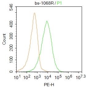 Blank control:MCF7.
Primary Antibody (green line): Rabbit Anti-NMDAR1 antibody (SL1068R)
Dilution: 1μg /10^6 cells;
Isotype Control Antibody (orange line): Rabbit IgG .
Secondary Antibody : Goat anti-rabbit IgG-PE
Dilution: 2μg /test.
Protocol
The cells were incubated in 5% BSA to block non-specific protein-protein interactions for 30 min at at room temperature .Cells stained with Primary Antibody for 30 min at room temperature. The secondary antibody used for 40 min at room temperature. Acquisition of 20,000 events was performed.
Cartpieces
Totalgoods,subtotals:¥Checkout
Bought notes(bought amounts latest0)
No one bought this product
User Comment(Total0User Comment Num)
- No comment


 +86 571 56623320
 +86 18668110335

