
Rabbit Anti-NME1/NM23A antibody
NDKA_HUMAN; Nucleoside diphosphate kinase A; EC:2.7.4.6; NDK A; NDP kinase A; Granzyme A-activated DNase (GAAD); Metastasis inhibition factor nm23; NM23-H1; Tumor metastatic process-associated protein; NME/NM23 nucleoside diphosphate kinase 1; NDPKA; NM23
View History [Clear]
Details
Product Name NME1/NM23A Chinese Name Tumour抑制基因抗体 Alias NDKA_HUMAN; Nucleoside diphosphate kinase A; EC:2.7.4.6; NDK A; NDP kinase A; Granzyme A-activated DNase (GAAD); Metastasis inhibition factor nm23; NM23-H1; Tumor metastatic process-associated protein; NME/NM23 nucleoside diphosphate kinase 1; NDPKA; NM23; NB; AWD; NBS; GAAD; NDKA; NDPK-A; Research Area Tumour Neurobiology Signal transduction transcriptional regulatory factor Immunogen Species Rabbit Clonality Polyclonal React Species Human, Mouse, Rat, (predicted: Dog, Pig, Cow, Horse, Rabbit, ) Applications WB=1:500-2000 ELISA=1:5000-10000 IHC-P=1:100-500 IHC-F=1:100-500 Flow-Cyt=1μg/Test IF=1:100-500 (Paraffin sections need antigen repair)
not yet tested in other applications.
optimal dilutions/concentrations should be determined by the end user.Theoretical molecular weight 17kDa Cellular localization The nucleus Form Liquid Concentration 1mg/ml immunogen KLH conjugated synthetic peptide derived from human Nm23-H1: 41-152/152 Lsotype IgG Purification affinity purified by Protein A Buffer Solution 0.01M TBS(pH7.4) with 1% BSA, 0.03% Proclin300 and 50% Glycerol. Storage Shipped at 4℃. Store at -20 °C for one year. Avoid repeated freeze/thaw cycles. Attention This product as supplied is intended for research use only, not for use in human, therapeutic or diagnostic applications. PubMed PubMed Product Detail NM23A plays a major role in the synthesis of nucleoside triphosphates other than ATP. Possesses nucleoside-diphosphate kinase, serine/threonine-specific protein kinase, geranyl and farnesyl pyrophosphate kinase, histidine protein kinase and 3'-5' exonuclease activities. Involved in cell proliferation, differentiation and development, signal transduction, G protein-coupled receptor endocytosis, and gene expression. Required for neural development including neural patterning and cell fate determination. Has tumor metastasis-suppressive capacity.
Function:
Major role in the synthesis of nucleoside triphosphates other than ATP. Possesses nucleoside-diphosphate kinase, serine/threonine-specific protein kinase, geranyl and farnesyl pyrophosphate kinase, histidine protein kinase and 3'-5' exonuclease activities. Involved in cell proliferation, differentiation and development, signal transduction, G protein-coupled receptor endocytosis, and gene expression. Required for neural development including neural patterning and cell fate determination.
Subunit:
Hexamer of two different chains: A and B (A6, A5B, A4B2, A3B3, A2B4, AB5, B6). Interacts with SET and PRUNE.
Subcellular Location:
Cytoplasm. Nucleus. Note=Cell-cycle dependent nuclear localization which can be induced by interaction with Epstein-barr viral proteins or by degradation of the SET complex by GzmA.
Tissue Specificity:
Isoform 1 is expressed in heart, brain, placenta, lung, liver, skeletal muscle, pancreas, spleen and thymus. Expressed in lung carcinoma cell lines but not in normal lung tissues. Isoform 2 is ubiquitously expressed and its expression is also related to tumor differentiation. Isoform 3 is ubiquitously expressed.
Similarity:
Belongs to the NDK family.
SWISS:
P15531
Gene ID:
4830
Database links:Entrez Gene: 4830 Human
Entrez Gene: 18102 Mouse
Omim: 156490 Human
SwissProt: P15531 Human
SwissProt: P15532 Mouse
Unigene: 463456 Human
Unigene: 439702 Mouse
Unigene: 6236 Rat
Product Picture 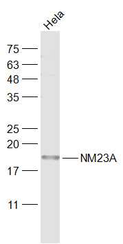 Sample:
Hela(Human) Cell Lysate at 30 ug
Primary: Anti-NM23A (SL1066R) at 1/1000 dilution
Secondary: IRDye800CW Goat Anti-Rabbit IgG at 1/20000 dilution
Predicted band size: 17 kD
Observed band size: 18 kD
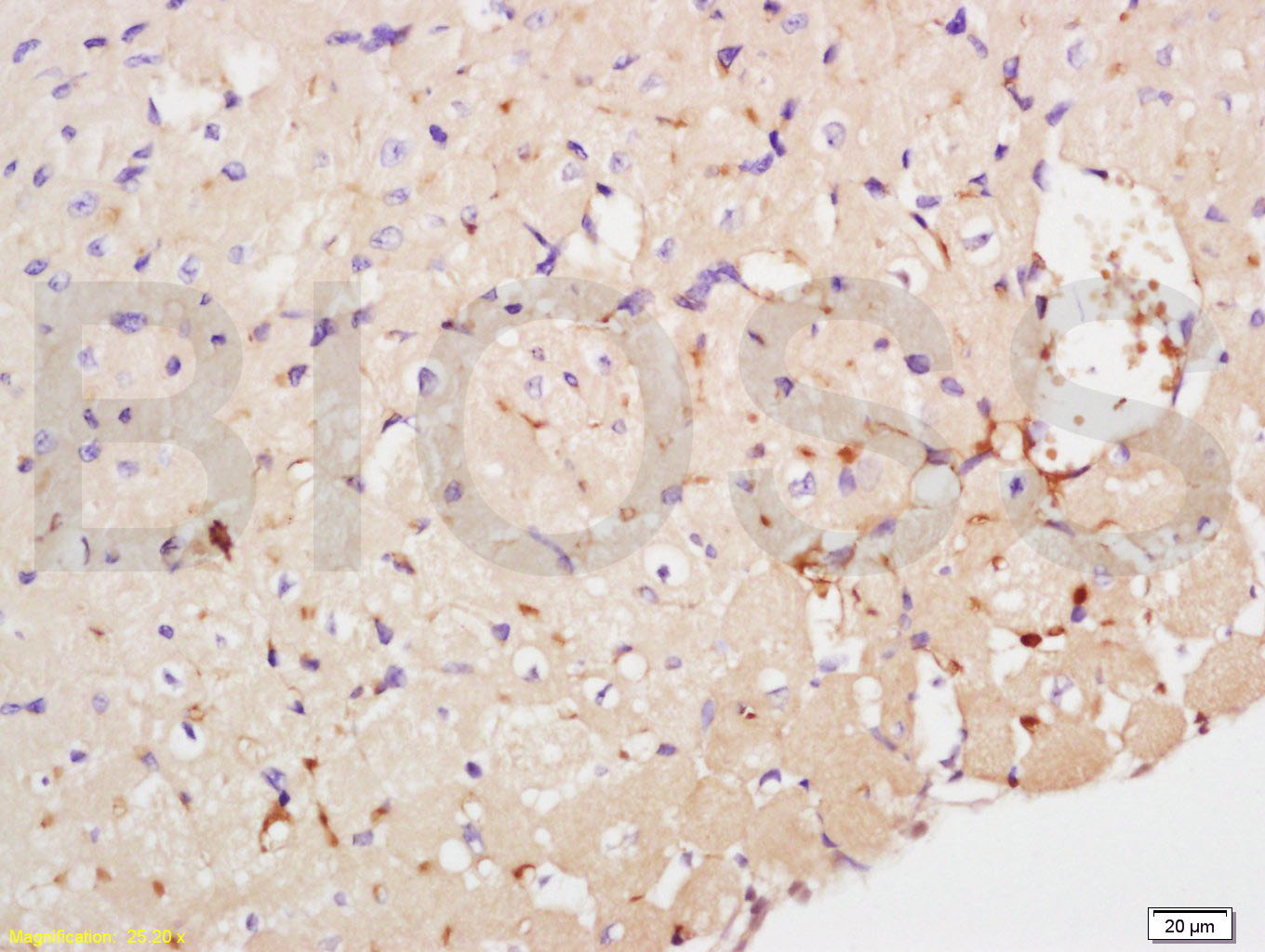 Tissue/cell: mouse heart tissue; 4% Paraformaldehyde-fixed and paraffin-embedded;
Antigen retrieval: citrate buffer ( 0.01M, pH 6.0 ), Boiling bathing for 15min; Block endogenous peroxidase by 3% Hydrogen peroxide for 30min; Blocking buffer (normal goat serum,C-0005) at 37℃ for 20 min;
Incubation: Anti-NME1/Nm23-H1/NDKA Polyclonal Antibody, Unconjugated(SL1066R) 1:200, overnight at 4°C, followed by conjugation to the secondary antibody(SP-0023) and DAB(C-0010) staining
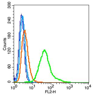 Blank control: RSC96(blue).
Primary Antibody:Rabbit Anti-NME1 antibody(SL1066R), Dilution: 1μg in 100 μL 1X PBS containing 0.5% BSA;
Isotype Control Antibody: Rabbit IgG(orange) ,used under the same conditions );
Secondary Antibody: Goat anti-rabbit IgG-PE(white blue), Dilution: 1:200 in 1 X PBS containing 0.5% BSA.
Protocol
The cells were fixed with 2% paraformaldehyde (10 min) , then permeabilized with 90% ice-cold methanol for 30 min on ice. Antibody (SL1066R, 1μg /1x10^6 cells) were incubated for 30 min on the ice, followed by 1 X PBS containing 0.5% BSA + 1 0% goat serum (15 min) to block non-specific protein-protein interactions. Then the Goat Anti-rabbit IgG/PE antibody was added into the blocking buffer mentioned above to react with the primary antibody of SL1066R at 1/200 dilution for 30 min on ice. Acquisition of 20,000 events was performed.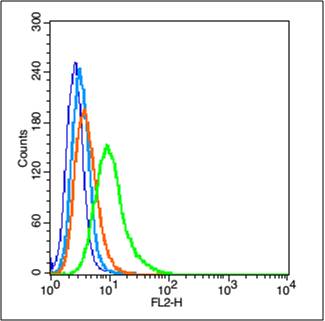 Blank control (blue line): A549 (blue).
Primary Antibody (green line): Rabbit Anti-NME1 antibody (SL1066R)
Dilution: 1μg /10^6 cells;
Isotype Control Antibody (orange line): Rabbit IgG .
Secondary Antibody (white blue line): Goat anti-rabbit IgG-PE
Dilution: 1μg /test.
Protocol
The cells were fixed with 2% paraformaldehyde (10 min) , then permeabilized with 90% ice-cold methanol for 30 min on ice. Cells stained with Primary Antibody for 30 min at room temperature. The cells were then incubated in 1 X PBS/2%BSA/10% goat serum to block non-specific protein-protein interactions followed by the antibody for 15 min at room temperature. The secondary antibody used for 40 min at room temperature. Acquisition of 20,000 events was performed.
Cartpieces
Totalgoods,subtotals:¥Checkout
Bought notes(bought amounts latest0)
No one bought this product
User Comment(Total0User Comment Num)
- No comment


 +86 571 56623320
 +86 18668110335

