
Rabbit Anti-Cardiac Troponin T antibody
Cardiac muscle troponin T; Cardiomyopathy dilated 1D (autosomal dominant); Cardiomyopathy hypertrophic 2; CMD1D; CMH2; CMPD2; cTnT; LVNC6; MGC3889; OTTHUMP00000033864; OTTHUMP00000033865; OTTHUMP00000033866; OTTHUMP00000033867; OTTHUMP00000033870; OTTHUMP
View History [Clear]
Details
Product Name Cardiac Troponin T Chinese Name 心肌特异性肌钙蛋白T抗体 Alias Cardiac muscle troponin T; Cardiomyopathy dilated 1D (autosomal dominant); Cardiomyopathy hypertrophic 2; CMD1D; CMH2; CMPD2; cTnT; LVNC6; MGC3889; OTTHUMP00000033864; OTTHUMP00000033865; OTTHUMP00000033866; OTTHUMP00000033867; OTTHUMP00000033870; OTTHUMP00000218095; RCM3; TNNT 2; TNNT2; TNNT2_HUMAN; TnTC; Troponin T cardiac muscle; Troponin T type 2 (cardiac); Troponin T type 2 cardiac; Troponin T, cardiac muscle; Troponin T2; Troponin T2 cardiac. literatures Research Area Cardiovascular immunology Immunogen Species Rabbit Clonality Polyclonal React Species Human, Mouse, Rat, (predicted: Dog, Pig, Cow, ) Applications WB=1:500-2000 ELISA=1:5000-10000 IHC-P=1:100-500 IHC-F=1:100-500 Flow-Cyt=1μg/Test ICC=1:100-500 IF=1:100-500 (Paraffin sections need antigen repair)
not yet tested in other applications.
optimal dilutions/concentrations should be determined by the end user.Theoretical molecular weight 36kDa Cellular localization cytoplasmic Form Liquid Concentration 1mg/ml immunogen KLH conjugated synthetic peptide derived from human Cardiac Troponin T: 201-298/298 Lsotype IgG Purification affinity purified by Protein A Buffer Solution 0.01M TBS(pH7.4) with 1% BSA, 0.03% Proclin300 and 50% Glycerol. Storage Shipped at 4℃. Store at -20 °C for one year. Avoid repeated freeze/thaw cycles. Attention This product as supplied is intended for research use only, not for use in human, therapeutic or diagnostic applications. PubMed PubMed Product Detail The protein encoded by this gene is the tropomyosin-binding subunit of the troponin complex, which is located on the thin filament of striated muscles and regulates muscle contraction in response to alterations in intracellular calcium ion concentration. Mutations in this gene have been associated with familial hypertrophic cardiomyopathy as well as with dilated cardiomyopathy. Transcripts for this gene undergo alternative splicing that results in many tissue-specific isoforms, however, the full-length nature of some of these variants has not yet been determined. [provided by RefSeq].
Function:
Troponin T is the tropomyosin-binding subunit of troponin, the thin filament regulatory complex which confers calcium-sensitivity to striated muscle actomyosin ATPase activity.
Subcellular Location:
Cytoplasm.
Tissue Specificity:
Heart. The fetal heart shows a greater expression in the atrium than in the ventricle, while the adult heart shows a greater expression in the ventricle than in the atrium. Isoform 6 predominates in normal adult heart. Isoforms 1, 7 and 8 are expressed in fetal heart. Isoform 7 is also expressed in failing adult heart.
DISEASE:
Cardiomyopathy, familial hypertrophic 2 (CMH2) [MIM:115195]: A hereditary heart disorder characterized by ventricular hypertrophy, which is usually asymmetric and often involves the interventricular septum. The symptoms include dyspnea, syncope, collapse, palpitations, and chest pain. They can be readily provoked by exercise. The disorder has inter- and intrafamilial variability ranging from benign to malignant forms with high risk of cardiac failure and sudden cardiac death. Note=The disease is caused by mutations affecting the gene represented in this entry.
Cardiomyopathy, dilated 1D (CMD1D) [MIM:601494]: A disorder characterized by ventricular dilation and impaired systolic function, resulting in congestive heart failure and arrhythmia. Patients are at risk of premature death. Note=The disease is caused by mutations affecting the gene represented in this entry.
Cardiomyopathy, familial restrictive 3 (RCM3) [MIM:612422]: A heart disorder characterized by impaired filling of the ventricles with reduced diastolic volume, in the presence of normal or near normal wall thickness and systolic function. Note=The disease is caused by mutations affecting the gene represented in this entry.
Similarity:
Belongs to the troponin T family.
SWISS:
P45379
Gene ID:
7139
Database links:
Entrez Gene: 7139 Human
Entrez Gene: 21956 Mouse
Entrez Gene: 100009428 Rabbit
Omim: 191045 Human
SwissProt: P45379 Human
SwissProt: P50752 Mouse
SwissProt: P09741 Rabbit
Unigene: 533613 Human
Unigene: 247470 Mouse
Unigene: 9965 Rat
Product Picture 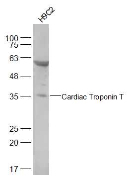 Sample:
H9C2(Rat) Cell Lysate at 30 ug
Primary: Anti-alpha smooth muscle Actin (SL10648R) at 1/500 dilution
Secondary: IRDye800CW Goat Anti-Rabbit IgG at 1/20000 dilution
Predicted band size: 36 kD
Observed band size: 36 kD
 Paraformaldehyde-fixed, paraffin embedded (mouse heart); Antigen retrieval by boiling in sodium citrate buffer (pH6.0) for 15min; Block endogenous peroxidase by 3% hydrogen peroxide for 20 minutes; Blocking buffer (normal goat serum) at 37°C for 30min; Antibody incubation with (Cardiac Troponin T) Polyclonal Antibody, Unconjugated (SL10648R) at 1:200 overnight at 4°C, followed by operating according to SP Kit(Rabbit) (sp-0023) instructionsand DAB staining.
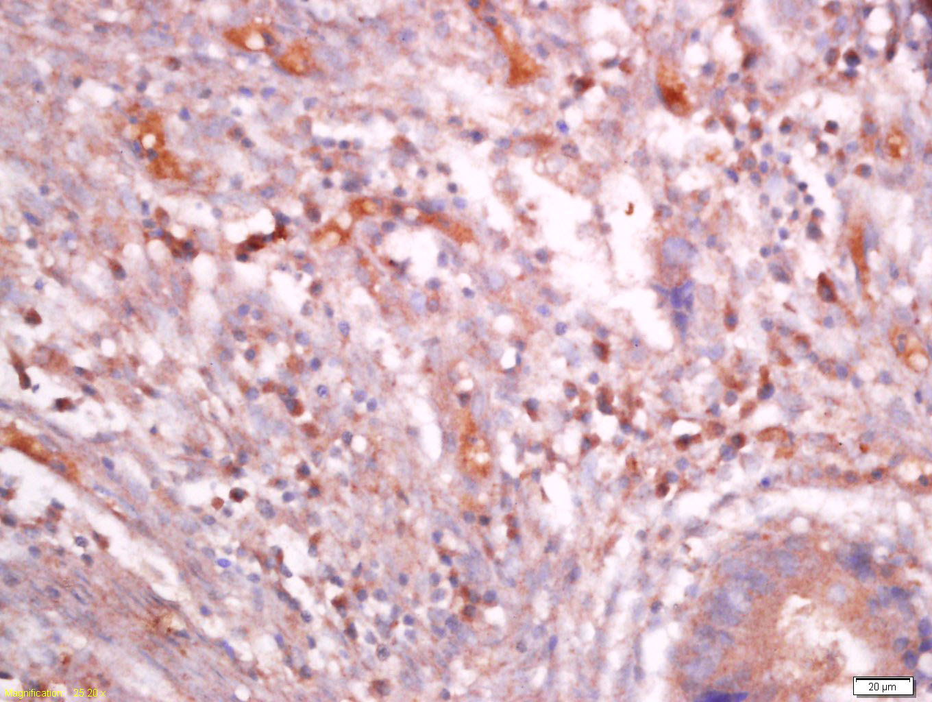 Tissue/cell: human colon cancer; 4% Paraformaldehyde-fixed and paraffin-embedded;
Antigen retrieval: citrate buffer ( 0.01M, pH 6.0 ), Boiling bathing for 15min; Block endogenous peroxidase by 3% Hydrogen peroxide for 30min; Blocking buffer (normal goat serum,C-0005) at 37℃ for 20 min;
Incubation: Anti-TNNT2 Polyclonal Antibody, Unconjugated(SL10648R) 1:400, overnight at 4°C, followed by conjugation to the secondary antibody(SP-0023) and DAB(C-0010) staining
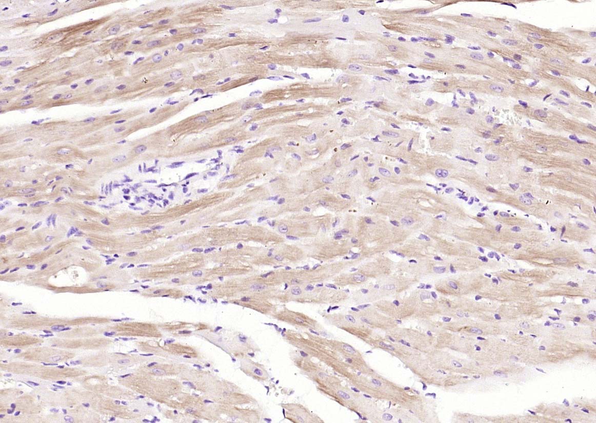
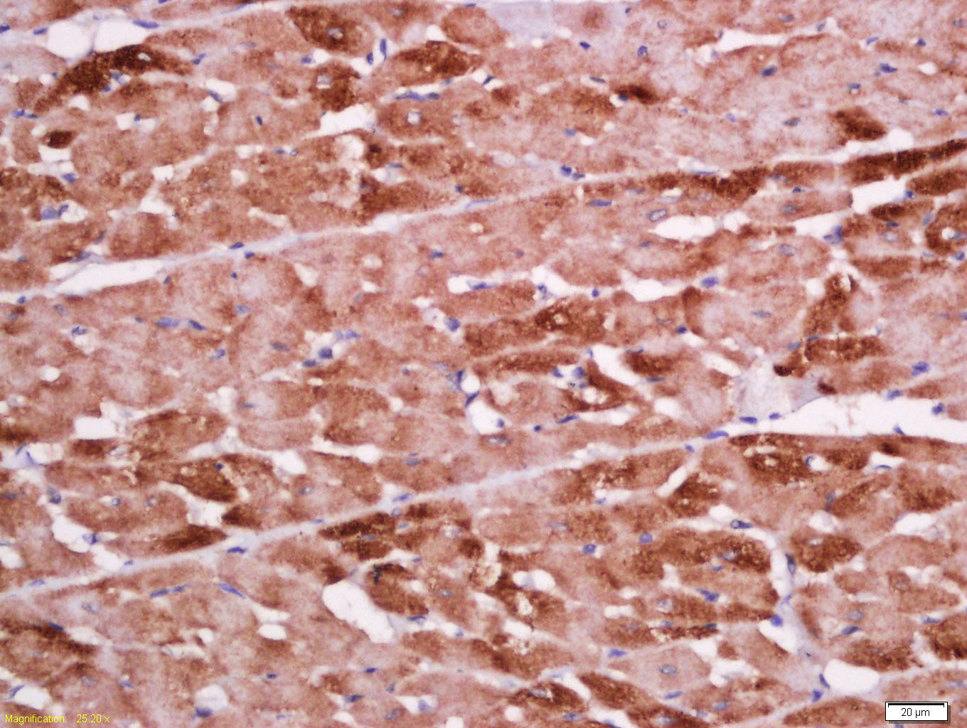 Tissue/cell: rat heart tissue; 4% Paraformaldehyde-fixed and paraffin-embedded;
Antigen retrieval: citrate buffer ( 0.01M, pH 6.0 ), Boiling bathing for 15min; Block endogenous peroxidase by 3% Hydrogen peroxide for 30min; Blocking buffer (normal goat serum,C-0005) at 37℃ for 20 min;
Incubation: Anti-TNNT2 Polyclonal Antibody, Unconjugated(SL10648R) 1:500, overnight at 4°C, followed by conjugation to the secondary antibody(SP-0023) and DAB(C-0010) staining
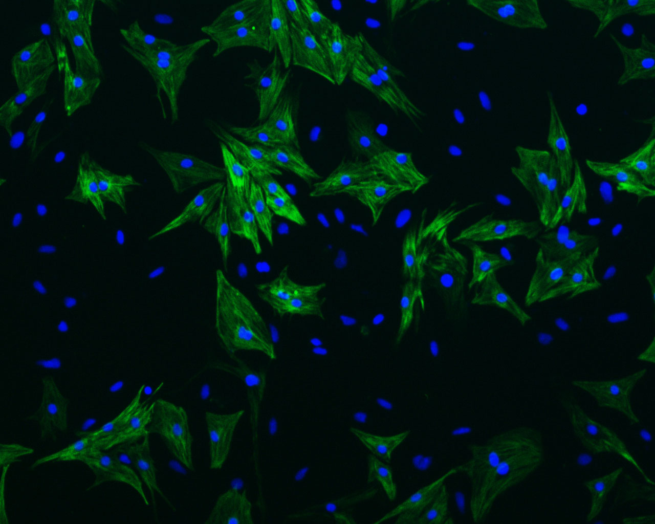 Cell: Neonatal rat ventricular cardiomyocytes;
Dilution: 1:400;
Incubation: Anti-Cardiac Troponin T Antibody, unconjugated (SL10648R);
DAPI was used to stain the cell nuclei.
The image is provided by Tongji University.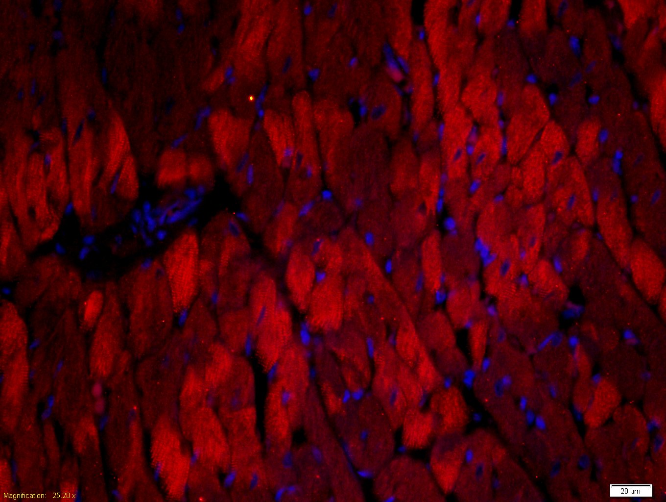 Tissue/cell: rat heart tissue;4% Paraformaldehyde-fixed and paraffin-embedded;
Antigen retrieval: citrate buffer ( 0.01M, pH 6.0 ), Boiling bathing for 15min; Blocking buffer (normal goat serum,C-0005) at 37℃ for 20 min;
Incubation: Anti-TNNT2 Polyclonal Antibody, Unconjugated(SLR) 1:500, overnight at 4°C; The secondary antibody was Goat Anti-Rabbit IgG, Cy3 conjugated(SL0295G-Cy3)used at 1:200 dilution for 40 minutes at 37°C. DAPI(5ug/ml,blue,C-0033) was used to stain the cell nuclei
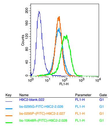 Positive control: H9C2 cells
Concebtration: 2μg/10^6 cells
Incubation conditions: Avoid light , 30 minutes on the ice.
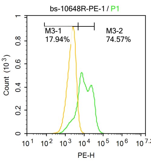 Blank control:U-2OS.
Primary Antibody (green line): Rabbit Anti-TNNT2 antibody (SL10648R)
Dilution: 1μg /10^6 cells;
Isotype Control Antibody (orange line): Rabbit IgG .
Secondary Antibody : Goat anti-rabbit IgG-AF647
Dilution: 1μg /test.
Protocol
The cells were fixed with 4% PFA (10min at room temperature)and then permeabilized with 0.1% PBST for 20 min at-20℃. The cells were then incubated in 5%BSA to block non-specific protein-protein interactions for 30 min at at room temperature .Cells stained with Primary Antibody for 30 min at room temperature. The secondary antibody used for 40 min at room temperature. Acquisition of 20,000 events was performed.
Cartpieces
Totalgoods,subtotals:¥Checkout
Bought notes(bought amounts latest0)
No one bought this product
User Comment(Total0User Comment Num)
- No comment


 +86 571 56623320
 +86 18668110335

