
Rabbit Anti-TNNI3 antibody
Cardiac Troponin I; cardiac muscle; Troponin I, cardiac muscle; Cardiomyopathy, familial hypertrophic, 7, included; CMD1FF; CMD2A; CMH7; cTnI; Familial hypertrophic cardiomyopathy 7; MGC116817; RCM1; Tn1; Tni; TNN I3; TNNC 1; TNNC1; TNNI3; TNNI3; TNNI3_HU
View History [Clear]
Details
Product Name TNNI3 Chinese Name 心肌肌钙蛋白I抗体 Alias Cardiac Troponin I; cardiac muscle; Troponin I, cardiac muscle; Cardiomyopathy, familial hypertrophic, 7, included; CMD1FF; CMD2A; CMH7; cTnI; Familial hypertrophic cardiomyopathy 7; MGC116817; RCM1; Tn1; Tni; TNN I3; TNNC 1; TNNC1; TNNI3; TNNI3; TNNI3_HUMAN; Troponin I; Troponin I cardiac; Troponin I cardiac muscle; Troponin I cardiac muscle isoform; Troponin I type 3 cardiac; troponin I, cardiac 3; TroponinI; Ttroponin I type 3 (cardiac). Research Area Cardiovascular Developmental biology Signal transduction Stem cells Cytoskeleton Immunogen Species Rabbit Clonality Polyclonal React Species Human, Mouse, Rat, Applications WB=1:500-2000 ELISA=1:5000-10000 IHC-P=1:100-500 IHC-F=1:100-500 Flow-Cyt=2ug/Test ICC=1:100-500 IF=1:100-500 (Paraffin sections need antigen repair)
not yet tested in other applications.
optimal dilutions/concentrations should be determined by the end user.Theoretical molecular weight 24kDa Cellular localization cytoplasmic Form Liquid Concentration 1mg/ml immunogen KLH conjugated synthetic peptide derived from human TNNI3: 131-210/210 Lsotype IgG Purification affinity purified by Protein A Buffer Solution 0.01M TBS(pH7.4) with 1% BSA, 0.03% Proclin300 and 50% Glycerol. Storage Shipped at 4℃. Store at -20 °C for one year. Avoid repeated freeze/thaw cycles. Attention This product as supplied is intended for research use only, not for use in human, therapeutic or diagnostic applications. PubMed PubMed Product Detail Troponin I (TnI), along with troponin T (TnT) and troponin C (TnC), is one of 3 subunits that form the troponin complex of the thin filaments of striated muscle. TnI is the inhibitory subunit; blocking actin-myosin interactions and thereby mediating striated muscle relaxation. The TnI subfamily contains three genes: TnI-skeletal-fast-twitch, TnI-skeletal-slow-twitch, and TnI-cardiac. This gene encodes the TnI-cardiac protein and is exclusively expressed in cardiac muscle tissues. Mutations in this gene cause familial hypertrophic cardiomyopathy type 7 (CMH7) and familial restrictive cardiomyopathy (RCM). [provided by RefSeq].
Function:
Troponin I is the inhibitory subunit of troponin, the thin filament regulatory complex which confers calcium-sensitivity to striated muscle actomyosin ATPase activity.
Post-translational modifications:
Phosphorylated at Ser-42 and Ser-44 by PRKCE; phosphorylation increases myocardium contractile dysfunction. Phosphorylated at Ser-23 and Ser-24 by PRKD1; phosphorylation reduces myofilament calcium sensitivity. Phosphorylated preferentially at Thr-31. Phosphorylation by STK4/MST1 alters its binding affinity to TNNC1 (cardiac Tn-C) and TNNT2 (cardiac Tn-T).
DISEASE:
Defects in TNNI3 are the cause of cardiomyopathy familial hypertrophic type 7 (CMH7) [MIM:613690]. Familial hypertrophic cardiomyopathy is a hereditary heart disorder characterized by ventricular hypertrophy, which is usually asymmetric and often involves the interventricular septum. The symptoms include dyspnea, syncope, collapse, palpitations, and chest pain. They can be readily provoked by exercise. The disorder has inter- and intrafamilial variability ranging from benign to malignant forms with high risk of cardiac failure and sudden cardiac death.
Defects in TNNI3 are the cause of cardiomyopathy familial restrictive type 1 (RCM1) [MIM:115210]. RCM1 is an heart muscle disorder characterized by impaired filling of the ventricles with reduced diastolic volume, in the presence of normal or near normal wall thickness and systolic function.
Defects in TNNI3 are the cause of cardiomyopathy dilated type 2A (CMD2A) [MIM:611880]. Dilated cardiomyopathy is a disorder characterized by ventricular dilation and impaired systolic function, resulting in congestive heart failure and arrhythmia. Patients are at risk of premature death.
Defects in TNNI3 are the cause of cardiomyopathy dilated type 1FF (CMD1FF) [MIM:613286]. Dilated cardiomyopathy is a disorder characterized by ventricular dilation and impaired systolic function, resulting in congestive heart failure and arrhythmia. Patients are at risk of premature death.
Similarity:
Belongs to the troponin I family.
SWISS:
P19429
Gene ID:
7137
Database links:
Entrez Gene: 7137 Human
Entrez Gene: 21954 Mouse
Omim: 191044 Human
SwissProt: P19429 Human
SwissProt: P48787 Mouse
SwissProt: P02646 Rabbit
Unigene: 709179 Human
Unigene: 27674 Mouse
Unigene: 64141 Rat
Product Picture 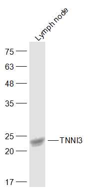 Sample:
Lymph node(Rat) Cell Lysate at 40 ug
Primary: Anti-TNNI3 (SL10614R) at 1/300 dilution
Secondary: IRDye800CW Goat Anti-Rabbit IgG at 1/20000 dilution
Predicted band size: 24 kD
Observed band size: 24 kD
 Sample:
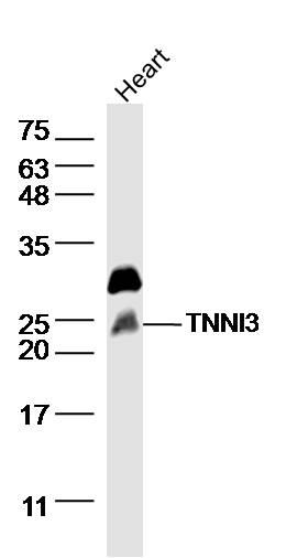
heart(rat)Lysate at 40 ug
Primary: Anti- TNNI3 (SL10614R)at 1/300 dilution
Secondary: IRDye800CW Goat Anti-Rabbit IgG at 1/20000 dilution
Predicted band size: 24kD
Observed band size: 24 kD
 Sample:
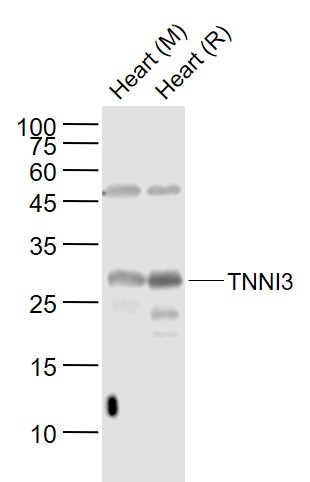
Lane 1: Heart (Mouse) Lysate at 40 ug
Lane 2: Heart (Rat) Lysate at 40 ug
Primary: Anti-TNNI3 (SL10614R ) at 1/1000 dilution
Secondary: IRDye800CW Goat Anti-Rabbit IgG at 1/20000 dilution
Predicted band size: 28 kD
Observed band size: 28 kD
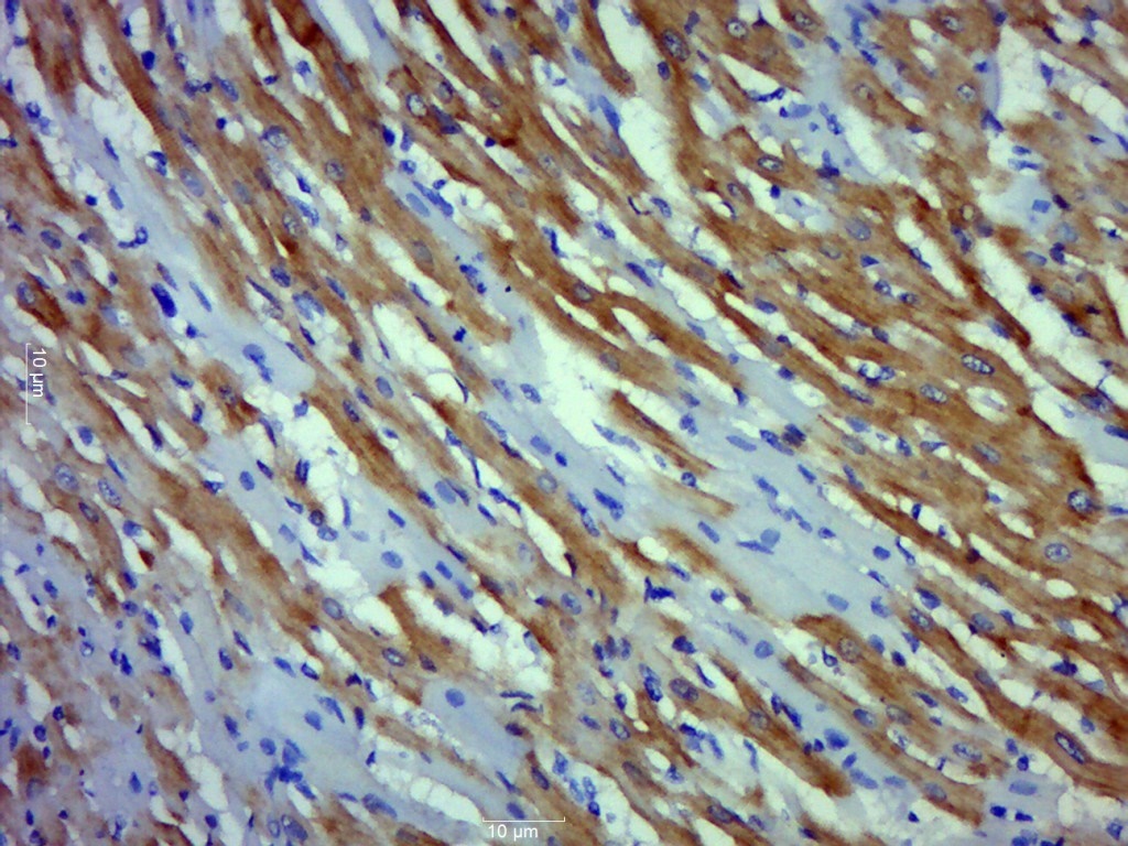 Paraformaldehyde-fixed, paraffin embedded (Rat heart); Antigen retrieval by boiling in sodium citrate buffer (pH6.0) for 15min; Block endogenous peroxidase by 3% hydrogen peroxide for 20 minutes; Blocking buffer (normal goat serum) at 37°C for 30min; Antibody incubation with (TNNI3) Polyclonal Antibody, Unconjugated (SL10614R) at 1:500 overnight at 4°C, followed by a conjugated secondary (sp-0023) for 20 minutes and DAB staining.
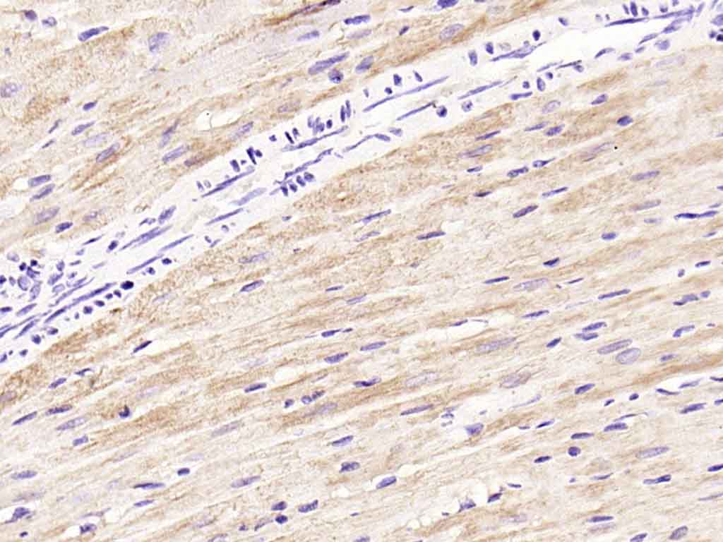 Paraformaldehyde-fixed, paraffin embedded (mouse heart); Antigen retrieval by boiling in sodium citrate buffer (pH6.0) for 15min; Block endogenous peroxidase by 3% hydrogen peroxide for 20 minutes; Blocking buffer (normal goat serum) at 37°C for 30min; Antibody incubation with (TNNI3) Polyclonal Antibody, Unconjugated (SL10614R) at 1:200 overnight at 4°C, followed by operating according to SP Kit(Rabbit) (sp-0023) instructionsand DAB staining.
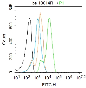 Blank control: K562.
Primary Antibody (green line): Rabbit Anti-TNNI3 antibody (SL10614R)
Dilution: 1μg /10^6 cells;
Isotype Control Antibody (orange line): Rabbit IgG .
Secondary Antibody : Goat anti-rabbit IgG-FITC
Dilution: 1μg /test.
Protocol
The cells were fixed with 4% PFA (10min at room temperature)and then permeabilized with 0.1% PBST for 20 min at room temperature. The cells were then incubated in 5%BSA to block non-specific protein-protein interactions for 30 min at room temperature .Cells stained with Primary Antibody for 30 min at room temperature. The secondary antibody used for 40 min at room temperature. Acquisition of 20,000 events was performed.
Cartpieces
Totalgoods,subtotals:¥Checkout
References (0)
No References
Bought notes(bought amounts latest0)
No one bought this product
User Comment(Total0User Comment Num)
- No comment


 +86 571 56623320
 +86 18668110335

