
Rabbit Anti-Cytokeratin 5 antibody
Cytokeratin 5; Keratin, type II cytoskeletal 5; CK-5; CK 5; CK5; Keratin-5; K5; 58 kDa cytokeratin; KRT5; DDD; EBS 2; EBS2; K5; Keratin 5 (epidermolysis bullosa simplex Dowling-Meara/Kobner/Weber-Cockayne types); Keratin 5; Keratin Type II Cytoskeletal 5;
View History [Clear]
Details
Product Name Cytokeratin 5 Chinese Name 细胞角蛋白5抗体 Alias Cytokeratin 5; Keratin, type II cytoskeletal 5; CK-5; CK 5; CK5; Keratin-5; K5; 58 kDa cytokeratin; KRT5; DDD; EBS 2; EBS2; K5; Keratin 5 (epidermolysis bullosa simplex Dowling-Meara/Kobner/Weber-Cockayne types); Keratin 5; Keratin Type II Cytoskeletal 5; Keratin5; KRT 5; KRT 5A; KRT5; Cytokeratin-5; Cytokeratin5; DDD; epidermolysis bullosa simplex 2 Dowling-Meara/Kobner/Weber-Cockayne types; K2C5_HUMAN; Keratin 5; Keratin; Keratin Type II Cytoskeletal 5; Keratin5; KRT5A; type II cytoskeletal 5; Type-II keratin Kb5. literatures Research Area Tumour Signal transduction Immunogen Species Rabbit Clonality Polyclonal React Species Human, Mouse, Rat, (predicted: Dog, Pig, Cow, Horse, Rabbit, ) Applications WB=1:500-2000 ELISA=1:5000-10000 IHC-P=1:100-500 IHC-F=1:100-500 IF=1:100-500 (Paraffin sections need antigen repair)
not yet tested in other applications.
optimal dilutions/concentrations should be determined by the end user.Theoretical molecular weight 64kDa Cellular localization The nucleus cytoplasmic Extracellular matrix Form Liquid Concentration 1mg/ml immunogen KLH conjugated synthetic peptide derived from human CK5: 331-430/590 Lsotype IgG Purification affinity purified by Protein A Buffer Solution 0.01M TBS(pH7.4) with 1% BSA, 0.03% Proclin300 and 50% Glycerol. Storage Shipped at 4℃. Store at -20 °C for one year. Avoid repeated freeze/thaw cycles. Attention This product as supplied is intended for research use only, not for use in human, therapeutic or diagnostic applications. PubMed PubMed Product Detail Cytokeratins (CK) are intermediate filaments of epithelial cells, both in keratinising tissue (ie., skin) and non keratinising cells (ie., mesothelial cells). Although not a traditional marker for endothelial cells, cytokeratins have also been found in some microvascular endothelial cells. At least 20 different cytokeratins (CK) in the molecular range of 40 to 70 kDa and isoelectric points of 5 to 8.5 can be identified using two dimensional gel electrophoresis. Biochemically, most members of the CK family fall into one of two classes, type I (acidic polypeptides) and type II (basic polypeptides). At least one member of the acidic family and one member of the basic family is expressed in all epithelial cells. Defects in KRT5 are a cause of epidermolysis bullosa simplex.
Subunit:
Heterotetramer of two type I and two type II keratins. Keratin-5 associates with keratin-14. Interacts with TCHP.
DISEASE:
Defects in KRT5 are a cause of epidermolysis bullosa simplex Dowling-Meara type (DM-EBS) [MIM:131760]. DM-EBS is a severe form of intraepidermal epidermolysis bullosa characterized by generalized herpetiform blistering, milia formation, dystrophic nails, and mucous membrane involvement.
Defects in KRT5 are the cause of epidermolysis bullosa simplex with migratory circinate erythema (EBSMCE) [MIM:609352]. EBSMCE is a form of intraepidermal epidermolysis bullosa characterized by unusual migratory circinate erythema. Skin lesions appear from birth primarily on the hands, feet, and legs but spare nails, ocular epithelia and mucosae. Lesions heal with brown pigmentation but no scarring. Electron microscopy findings are distinct from those seen in the DM-EBS, with no evidence of tonofilament clumping.
Defects in KRT5 are a cause of epidermolysis bullosa simplex Weber-Cockayne type (WC-EBS) [MIM:131800]. WC-EBS is a form of intraepidermal epidermolysis bullosa characterized by blistering limited to palmar and plantar areas of the skin.
Defects in KRT5 are a cause of epidermolysis bullosa simplex Koebner type (K-EBS) [MIM:131900]. K-EBS is a form of intraepidermal epidermolysis bullosa characterized by generalized skin blistering. The phenotype is not fundamentally distinct from the Dowling-Meara type, althought it is less severe.
Defects in KRT5 are the cause of epidermolysis bullosa simplex with mottled pigmentation (MP-EBS) [MIM:131960]. MP-EBS is a form of intraepidermal epidermolysis bullosa characterized by blistering at acral sites and 'mottled' pigmentation of the trunk and proximal extremities with hyper- and hypopigmentation macules.
Defects in KRT5 are the cause of Dowling-Degos disease (DDD) [MIM:179850]; also known as Dowling-Degos-Kitamura disease or reticulate acropigmentation of Kitamura. DDD is an autosomal dominant genodermatosis. Affected individuals develop a postpubertal reticulate hyperpigmentation that is progressive and disfiguring, and small hyperkeratotic dark brown papules that affect mainly the flexures and great skin folds. Patients usually show no abnormalities of the hair or nails.
Similarity:
Belongs to the intermediate filament family.
SWISS:
P13647
Gene ID:
3852
Database links:Entrez Gene: 3852 Human
Entrez Gene: 110308 Mouse
Omim: 148040 Human
SwissProt: P13647 Human
SwissProt: Q922U2 Mouse
Unigene: 433845 Human
Unigene: 451847 Mouse
Unigene: 129725 Rat
Unigene: 195318 Rat
结构蛋白(Structural Proteins)
细胞角蛋白5,为高分子量细胞角蛋白 58 kDa ,表达在皮肤的基底细胞和棘层细胞,部分前列腺基底细胞,与其它单层腺上皮不表达。主要用于间皮瘤与腺癌的鉴别诊断。
细胞角蛋白是一类与结构相关的蛋白家族,其在epithelial cells中形成Cytoskeleton中间丝。CK5是在表皮角质化细胞中大量表达的4种角蛋白之一。CK5可用以区分正常细胞和Tumour细胞。在基底细胞上皮瘤、多种鳞状细胞癌(皮肤和舌)、多种epithelial cells和间皮瘤都有CK5的表达。Product Picture 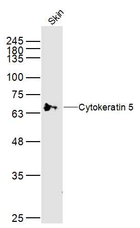 Sample:
Skin (Mouse) Lysate at 40 ug
Primary: Anti-Cytokeratin 5 (SL1060R) at 1/300 dilution
Secondary: IRDye800CW Goat Anti-Rabbit IgG at 1/20000 dilution
Predicted band size: 64 kD
Observed band size: 64 kD
 Sample:
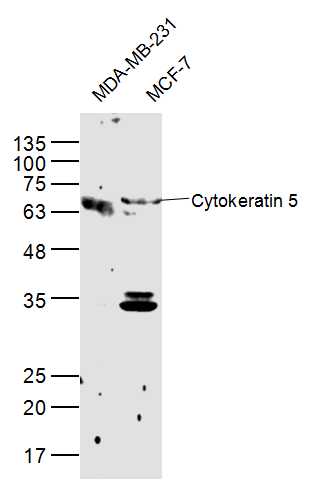
MDA-MB-231(Human) Cell Lysate at 40 ug
MCF-7(Human) Cell Lysate at 40 ug
Primary: Anti-Cytokeratin 5 (SL1060R) at 1/300 dilution
Secondary: IRDye800CW Goat Anti-Rabbit IgG at 1/20000 dilution
Predicted band size: 64 kD
Observed band size: 64 kD
 Sample:
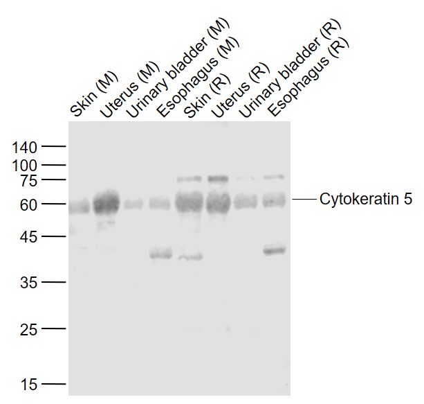
Lane 1: Skin (Mouse) Lysate at 40 ug
Lane 2: Uterus (Mouse) Lysate at 40 ug
Lane 3: Urinary bladder (Mouse) Lysate at 40 ug
Lane 4: Esophagus (Mouse) Lysate at 40 ug
Lane 5: Skin (Rat) Lysate at 40 ug
Lane 6: Uterus (Rat) Lysate at 40 ug
Lane 7: Urinary bladder (Rat) Lysate at 40 ug
Lane 8: Esophagus (Rat) Lysate at 40 ug
Primary: Anti-Cytokeratin 5 (SL1060R) at 1/1000 dilution
Secondary: IRDye800CW Goat Anti-Rabbit IgG at 1/20000 dilution
Predicted band size: 62 kD
Observed band size: 60 kD
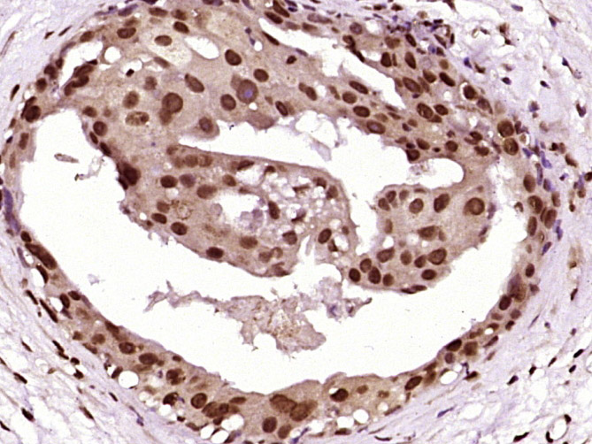 Paraformaldehyde-fixed, paraffin embedded (human breast carcinoma); Antigen retrieval by boiling in sodium citrate buffer (pH6.0) for 15min; Block endogenous peroxidase by 3% hydrogen peroxide for 20 minutes; Blocking buffer (normal goat serum) at 37°C for 30min; Antibody incubation with (Cytokeratin 5) Polyclonal Antibody, Unconjugated (SL1060R) at 1:400 overnight at 4°C, followed by operating according to SP Kit(Rabbit) (sp-0023) instructionsand DAB staining.
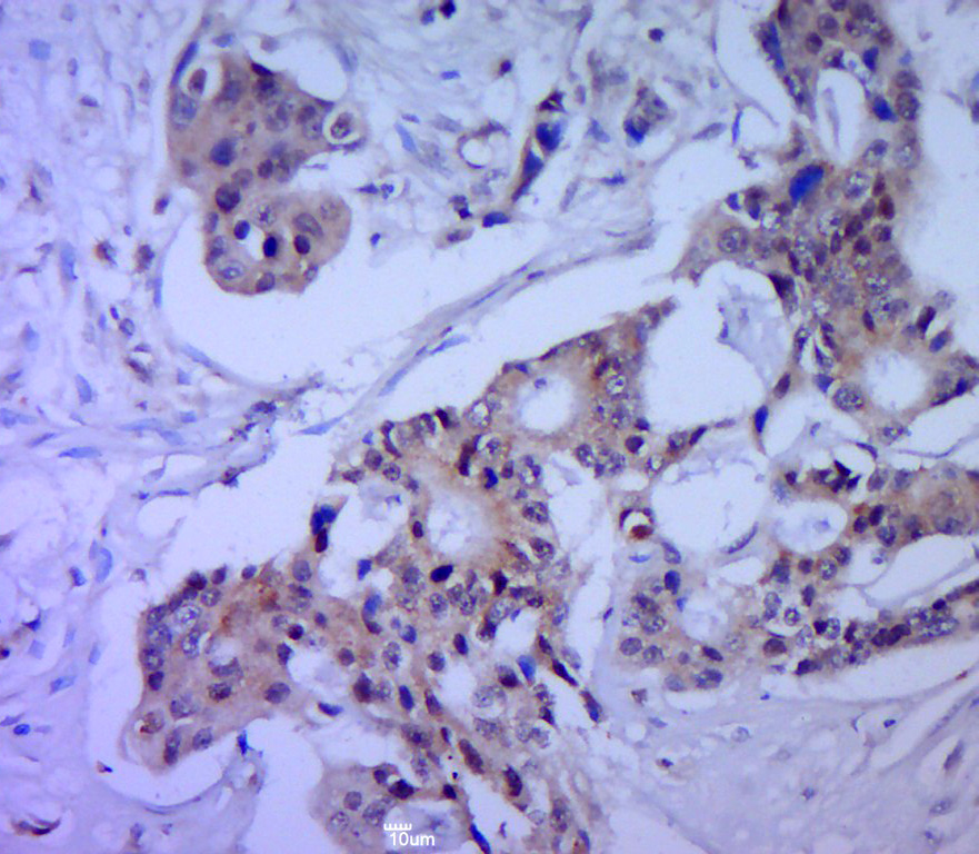 Paraformaldehyde-fixed, paraffin embedded (human cervical cancer); Antigen retrieval by boiling in sodium citrate buffer (pH6.0) for 15min; Block endogenous peroxidase by 3% hydrogen peroxide for 20 minutes; Blocking buffer (normal goat serum) at 37°C for 30min; Antibody incubation with (Cytokeratin 5) Polyclonal Antibody, Unconjugated (SL1060R) at 1:400 overnight at 4°C, followed by a conjugated secondary (sp-0023) for 20 minutes and DAB staining.
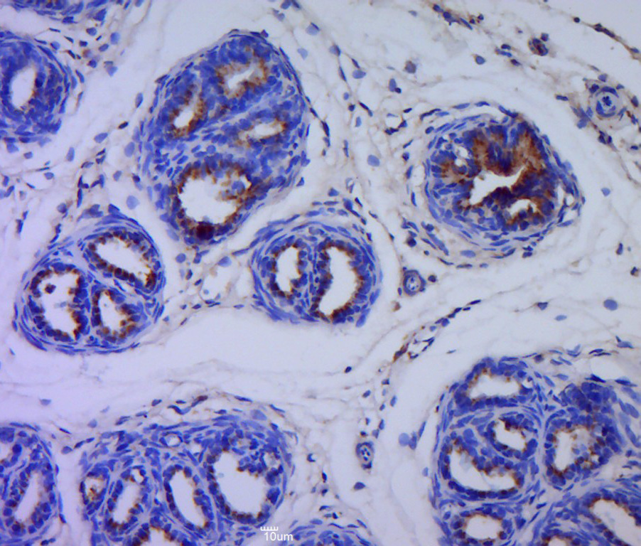 Paraformaldehyde-fixed, paraffin embedded (rat bladder tissue); Antigen retrieval by boiling in sodium citrate buffer (pH6.0) for 15min; Block endogenous peroxidase by 3% hydrogen peroxide for 20 minutes; Blocking buffer (normal goat serum) at 37°C for 30min; Antibody incubation with (Cytokeratin 5) Polyclonal Antibody, Unconjugated (SL1060R) at 1:400 overnight at 4°C, followed by a conjugated secondary (sp-0023) for 20 minutes and DAB staining.
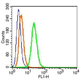 Blank control: Hela Cells(blue).
Primary Antibody: Rabbit Anti-Cytokeratin 5/AF488 Conjugated antibody (SL1060R/AF488), Dilution: 1μg in 100 μL 1X PBS containing 0.5% BSA;
Isotype Control Antibody: Rabbit IgG/AF488 (orange) ,used under the same conditions.
Protocol
The cells were fixed with 2% paraformaldehyde (10 min) and then permeabilized with ice-cold 90% methanol for 30 min on ice. The cells were washed twice with 1 X PBS. The cells were incubated in 1 X PBS containing 0.5% BSA + 1 0% goat serum (15 min) to block non-specific protein-protein interactions followed by the incubated with antibody (SL1060R/AF488, 1μg /1x10^6 cells) for 30 min on ice. Acquisition of 20,000 events was performed.
Cartpieces
Totalgoods,subtotals:¥Checkout
Bought notes(bought amounts latest0)
No one bought this product
User Comment(Total0User Comment Num)
- No comment


 +86 571 56623320
 +86 18668110335

