
Rabbit Anti-Cdc25C antibody
CDC 25; Cdc 25C; CDC25; Cell division cycle 25 homolog C; Cell division cycle 25C; Cell division cycle 25C protein; Dual specificity phosphatase Cdc25C; M phase inducer phosphatase 3; Mitosis inducer CDC25; MPIP3; Phosphotyrosine phosphatase; MPIP3_HUMAN.
View History [Clear]
Details
Product Name Cdc25C Chinese Name 细胞分裂周期蛋白25C抗体 Alias CDC 25; Cdc 25C; CDC25; Cell division cycle 25 homolog C; Cell division cycle 25C; Cell division cycle 25C protein; Dual specificity phosphatase Cdc25C; M phase inducer phosphatase 3; Mitosis inducer CDC25; MPIP3; Phosphotyrosine phosphatase; MPIP3_HUMAN. literatures Research Area Signal transduction Cyclin Kinases and Phosphatases Epigenetics Immunogen Species Rabbit Clonality Polyclonal React Species Human, Mouse, Rat, (predicted: Dog, Pig, Cow, ) Applications ELISA=1:5000-10000 IHC-P=1:100-500 IHC-F=1:100-500 ICC=1:100 IF=1:100-500 (Paraffin sections need antigen repair)
not yet tested in other applications.
optimal dilutions/concentrations should be determined by the end user.Theoretical molecular weight 53kDa Cellular localization The nucleus Form Liquid Concentration 1mg/ml immunogen KLH conjugated synthetic peptide derived from human Cdc25C: 321-420/473 Lsotype IgG Purification affinity purified by Protein A Buffer Solution 0.01M TBS(pH7.4) with 1% BSA, 0.03% Proclin300 and 50% Glycerol. Storage Shipped at 4℃. Store at -20 °C for one year. Avoid repeated freeze/thaw cycles. Attention This product as supplied is intended for research use only, not for use in human, therapeutic or diagnostic applications. PubMed PubMed Product Detail This gene is highly conserved during evolution and it plays a key role in the regulation of cell division. The encoded protein is a tyrosine phosphatase and belongs to the Cdc25 phosphatase family. It directs dephosphorylation of cyclin B-bound CDC2 and triggers entry into mitosis. It is also thought to suppress p53-induced growth arrest. Multiple alternatively spliced transcript variants of this gene have been described, however, the full-length nature of many of them is not known. [provided by RefSeq, Jul 2008]
Function:
Functions as a dosage-dependent inducer in mitotic control. It is a tyrosine protein phosphatase required for progression of the cell cycle. It directly dephosphorylates CDK1 and activate its kinase activity.
Subunit:
Interacts with HIV-1 Vpr, thereby inactivating CDC25C phosphatase activity. Interacts with MAPK14 and 14-3-3 proteins.
Subcellular Location:
Nucleus.
Post-translational modifications:
Phosphorylated by CHEK1 and MAPK14 at Ser-216. This phosphorylation creates a binding site for 14-3-3 protein and inhibits the phosphatase. Phosphorylated by PLK4. Phosphorylated by PLK1, leading to activate the phosphatase activity. Phosphorylation by PLK3 at Ser-191 promotes nuclear translocation. Ser-198 is a minor phosphorylation site. Was initially reported to be phosphorylated by PLK3 at Ser-216 (PubMed:10557092). However, such phosphorylation by PLK3 was not confirmed by other groups. Phosphorylation at Thr-48, Thr-67, Ser-122, Thr-130, Ser-168 and Ser-214 occurs at G2 and G2-M transition and is probably catalyzed by CDK1. Ser-168 phosphorylation levels are lower than those at the other 5 CDK1 sites. Phosphorylation by CDK1 leads to increased activity.
Similarity:
Belongs to the MPI phosphatase family.
Contains 1 rhodanese domain.
SWISS:
P30307
Gene ID:
995
Database links:Entrez Gene: 995 Human
Omim: 157680 Human
SwissProt: P30307 Human
Unigene: 656 Human
Product Picture 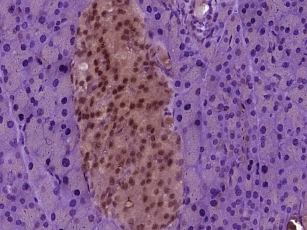 Paraformaldehyde-fixed, paraffin embedded (mouse pancreas tissue); Antigen retrieval by boiling in sodium citrate buffer (pH6.0) for 15min; Block endogenous peroxidase by 3% hydrogen peroxide for 20 minutes; Blocking buffer (normal goat serum) at 37°C for 30min; Antibody incubation with (Cdc25C) Polyclonal Antibody, Unconjugated (SL10579R) at 1:400 overnight at 4°C, followed by operating according to SP Kit(Rabbit) (sp-0023) instructionsand DAB staining.
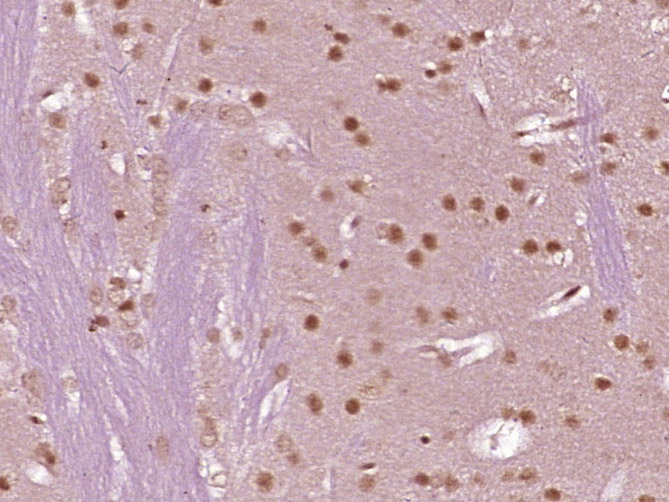 Paraformaldehyde-fixed, paraffin embedded (mouse brain tissue); Antigen retrieval by boiling in sodium citrate buffer (pH6.0) for 15min; Block endogenous peroxidase by 3% hydrogen peroxide for 20 minutes; Blocking buffer (normal goat serum) at 37°C for 30min; Antibody incubation with (Cdc25C) Polyclonal Antibody, Unconjugated (SL10579R) at 1:400 overnight at 4°C, followed by operating according to SP Kit(Rabbit) (sp-0023) instructionsand DAB staining.
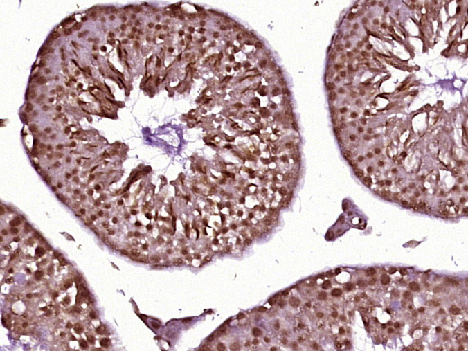 Paraformaldehyde-fixed, paraffin embedded (mouse testis tissue); Antigen retrieval by boiling in sodium citrate buffer (pH6.0) for 15min; Block endogenous peroxidase by 3% hydrogen peroxide for 20 minutes; Blocking buffer (normal goat serum) at 37°C for 30min; Antibody incubation with (Cdc25C) Polyclonal Antibody, Unconjugated (SL10579R) at 1:400 overnight at 4°C, followed by operating according to SP Kit(Rabbit) (sp-0023) instructionsand DAB staining.
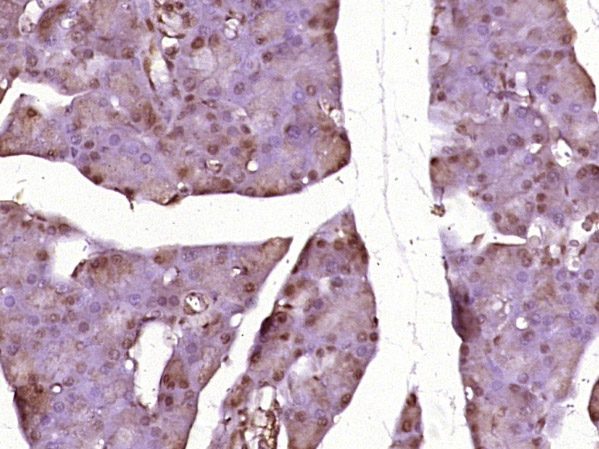 Paraformaldehyde-fixed, paraffin embedded (rat pancreas tissue); Antigen retrieval by boiling in sodium citrate buffer (pH6.0) for 15min; Block endogenous peroxidase by 3% hydrogen peroxide for 20 minutes; Blocking buffer (normal goat serum) at 37°C for 30min; Antibody incubation with (Cdc25C) Polyclonal Antibody, Unconjugated (SL10579R) at 1:400 overnight at 4°C, followed by operating according to SP Kit(Rabbit) (sp-0023) instructionsand DAB staining.
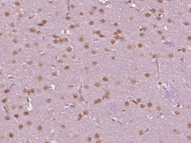 Paraformaldehyde-fixed, paraffin embedded (rat brain tissue); Antigen retrieval by boiling in sodium citrate buffer (pH6.0) for 15min; Block endogenous peroxidase by 3% hydrogen peroxide for 20 minutes; Blocking buffer (normal goat serum) at 37°C for 30min; Antibody incubation with (Cdc25C) Polyclonal Antibody, Unconjugated (SL10579R) at 1:400 overnight at 4°C, followed by operating according to SP Kit(Rabbit) (sp-0023) instructionsand DAB staining.
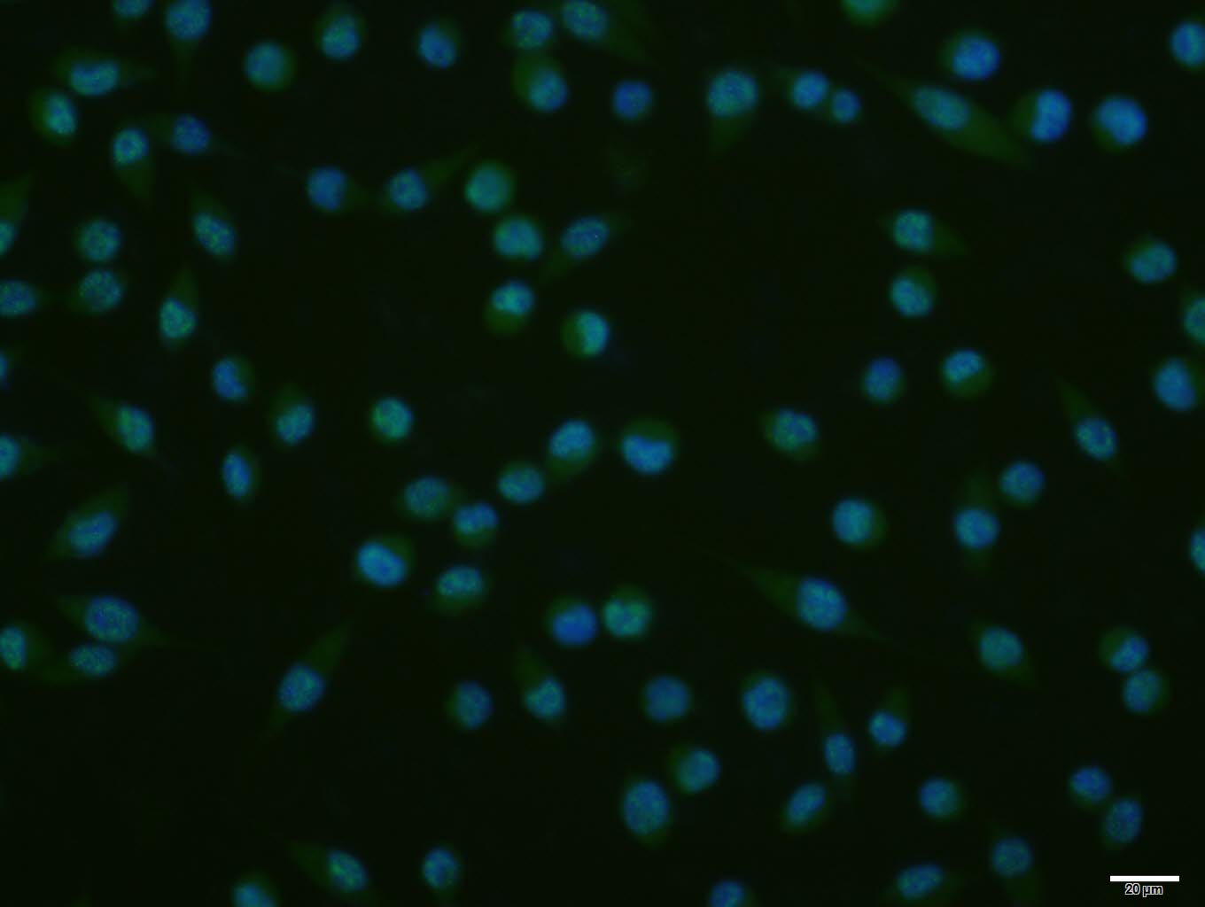 A431 cell; 4% Paraformaldehyde-fixed; Triton X-100 at room temperature for 20 min; Blocking buffer (normal goat serum, C-0005) at 37°C for 20 min; Antibody incubation with (Cdc25C) polyclonal Antibody, Unconjugated (SL10579R) 1:100, 90 minutes at 37°C; followed by a conjugated Goat Anti-Rabbit IgG antibody at 37°C for 90 minutes, DAPI (blue, C02-04002) was used to stain the cell nuclei.
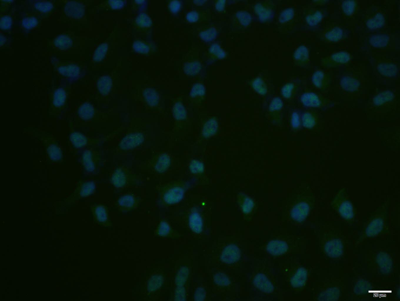 Hela cell; 4% Paraformaldehyde-fixed; Triton X-100 at room temperature for 20 min; Blocking buffer (normal goat serum, C-0005) at 37°C for 20 min; Antibody incubation with (Cdc25C) polyclonal Antibody, Unconjugated (SL10579R) 1:100, 90 minutes at 37°C; followed by a conjugated Goat Anti-Rabbit IgG antibody at 37°C for 90 minutes, DAPI (blue, C02-04002) was used to stain the cell nuclei.
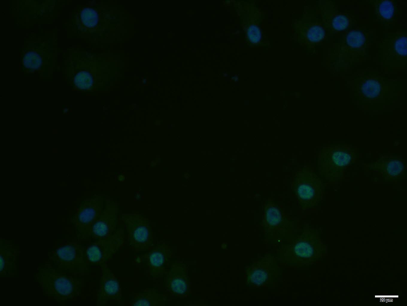 HepG2 cell; 4% Paraformaldehyde-fixed; Triton X-100 at room temperature for 20 min; Blocking buffer (normal goat serum, C-0005) at 37°C for 20 min; Antibody incubation with (Cdc25C) polyclonal Antibody, Unconjugated (SL10579R) 1:100, 90 minutes at 37°C; followed by a conjugated Goat Anti-Rabbit IgG antibody at 37°C for 90 minutes, DAPI (blue, C02-04002) was used to stain the cell nuclei.
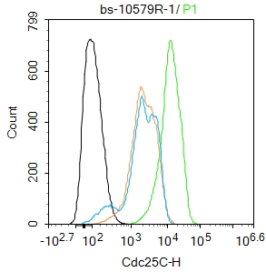 Blank control(black line):HepG2.
Primary Antibody (green line): Rabbit Anti-Cdc25C antibody (SL10579R)
Dilution:1ug/Test;
Secondary Antibody(white blue line): Goat anti-rabbit IgG-AF488
Dilution: 0.5ug/Test.
Isotype control(orange line): Normal Rabbit IgG
Protocol
The cells were fixed with 4% PFA (10min at room temperature)and then permeabilized with 90% ice-cold methanol for 20 min at -20℃, The cells were then incubated in 5%BSA to block non-specific protein-protein interactions for 30 min at room temperature .Cells stained with Primary Antibody for 30 min at room temperature. The secondary antibody used for 40 min at room temperature. Acquisition of 20,000 events was performed.
Cartpieces
Totalgoods,subtotals:¥Checkout
Bought notes(bought amounts latest0)
No one bought this product
User Comment(Total0User Comment Num)
- No comment


 +86 571 56623320
 +86 18668110335

