
Rabbit Anti-phospho-HSP70 (Tyr41)antibody
HSP70 (phospho 41); HSP70 (phospho Y41); p-HSP70 (Tyr41); p-HSP70 (Y41); HSP70; HSP-70; HSP 70; Heat shock 70 kDa protein 1; heat shock 70kDa protein 1A; Heat shock 70kDa protein 1B; Heat shock induced protein; heat shock protein 70; HSP70 1; HSP70 2; HSP
View History [Clear]
Details
Product Name phospho-HSP70 (Tyr41) Chinese Name 磷酸化热休克蛋白-70抗体 Alias HSP70 (phospho 41); HSP70 (phospho Y41); p-HSP70 (Tyr41); p-HSP70 (Y41); HSP70; HSP-70; HSP 70; Heat shock 70 kDa protein 1; heat shock 70kDa protein 1A; Heat shock 70kDa protein 1B; Heat shock induced protein; heat shock protein 70; HSP70 1; HSP70 2; HSP70.1; HSP72; HSPA1; HSPA1A; HSPA1B; XXbac BCX40G17.3 001; Heat shock 70 kDa protein 1A/1B; HSP71A_HUMAN; Hspa1a; Hsp70-1; HSP72; Hspa1; Hspa1b; Heat shock 70 kDa protein 1A/1B; shock 70 kDa protein 1/2; HSP70-1/HSP70-2; HSP70.1/HSP70.2. Product Type Phosphorylated anti Immunogen Species Rabbit Clonality Polyclonal React Species Human, Mouse, Rat, (predicted: Chicken, Cow, ) Applications WB=1:500-2000 ELISA=1:5000-10000 IHC-P=1:100-500 IHC-F=1:100-500 Flow-Cyt=1μg/Test ICC=1:100 IF=1:100-500 (Paraffin sections need antigen repair)
not yet tested in other applications.
optimal dilutions/concentrations should be determined by the end user.Theoretical molecular weight 70kDa Cellular localization cytoplasmic Form Liquid Concentration 1mg/ml immunogen KLH conjugated Synthesised phosphopeptide derived from human HSP70 around the phosphorylation site of Tyr41: PS(p-Y)VA Lsotype IgG Purification affinity purified by Protein A Buffer Solution 0.01M TBS(pH7.4) with 1% BSA, 0.03% Proclin300 and 50% Glycerol. Storage Shipped at 4℃. Store at -20 °C for one year. Avoid repeated freeze/thaw cycles. Attention This product as supplied is intended for research use only, not for use in human, therapeutic or diagnostic applications. PubMed PubMed Product Detail This intronless gene encodes a 70kDa heat shock protein which is a member of the heat shock protein 70 family. In conjuction with other heat shock proteins, this protein stabilizes existing proteins against aggregation and mediates the folding of newly translated proteins in the cytosol and in organelles. It is also involved in the ubiquitin-proteasome pathway through interaction with the AU-rich element RNA-binding protein 1. The gene is located in the major histocompatibility complex class III region, in a cluster with two closely related genes which encode similar proteins. [provided by RefSeq, Jul 2008].
Function:
In cooperation with other chaperones, Hsp70s stabilize preexistent proteins against aggregation and mediate the folding of newly translated polypeptides in the cytosol as well as within organelles. These chaperones participate in all these processes through their ability to recognize nonnative conformations of other proteins. They bind extended peptide segments with a net hydrophobic character exposed by polypeptides during translation and membrane translocation, or following stress-induced damage. In case of rotavirus A infection, serves as a post-attachment receptor for the virus to facilitate entry into the cell.
Subunit:
Component of the CatSper complex. Identified in a mRNP granule complex, at least composed of ACTB, ACTN4, DHX9, ERG, HNRNPA1, HNRNPA2B1, HNRNPAB, HNRNPD, HNRNPL, HNRNPR, HNRNPU, HSPA1, HSPA8, IGF2BP1, ILF2, ILF3, NCBP1, NCL, PABPC1, PABPC4, PABPN1, RPLP0, RPS3, RPS3A, RPS4X, RPS8, RPS9, SYNCRIP, TROVE2, YBX1 and untranslated mRNAs. Interacts with TSC2. Interacts with IRAK1BP1. Interacts with TERT; the interaction occurs in the absence of the RNA component, TERC, and dissociates once the TERT complex has formed. Interacts with DNAJC7. Interacts with CHCHD3.
Subcellular Location:
Cytoplasm. Note=Localized in cytoplasmic mRNP granules containing untranslated mRNAs.
Tissue Specificity:
HSPA1B is testis-specific.
Similarity:
Belongs to the heat shock protein 70 family.
SWISS:
P0DMV8
Gene ID:
3303
Database links:
Entrez Gene: 3303 Human
Entrez Gene: 3304 Human
Entrez Gene: 15511 Mouse
Entrez Gene: 193740 Mouse
Omim: 140550 Human
Omim: 603012 Human
SwissProt: P0DMV8 Human
SwissProt: P0DMV9 Human
SwissProt: P17879 Mouse
SwissProt: Q61696 Mouse
Unigene: 274402 Human
Unigene: 719966 Human
Unigene: 728810 Human
Unigene: 1950 Rat
Unigene: 228225 Rat
Product Picture 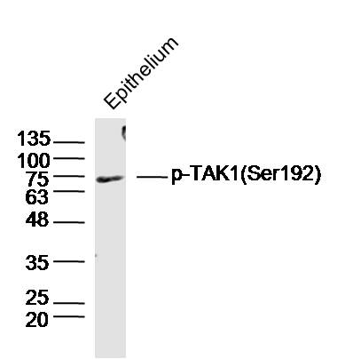 Sample: Epithelium (Mouse) Lysate at 40 ug
Primary: Anti-phospho-HSP70 (Tyr41) (SL10451R) at 1/300 dilution
Secondary: IRDye800CW Goat Anti-Rabbit IgG at 1/20000 dilution
Predicted band size: 70 kD
Observed band size: 70 kD
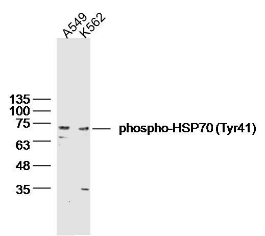 Sample:
A549(Human) Cell Lysate at 30 ug
K562(Human) Cell Lysate at 30 ug
Primary: Anti-phospho-HSP70 (Tyr41) (SL10451R) at 1/300 dilution
Secondary: IRDye800CW Goat Anti-Rabbit IgG at 1/20000 dilution
Predicted band size: 70 kD
Observed band size: 70 kD
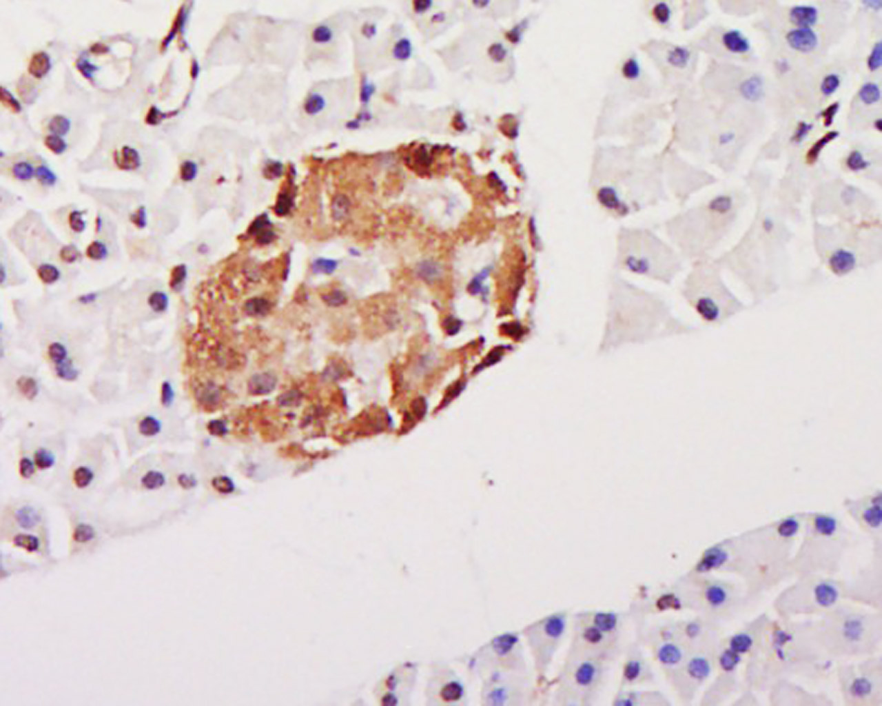 Tissue/cell: Rat pancreas tissue; 4% Paraformaldehyde-fixed and paraffin-embedded;
Antigen retrieval: citrate buffer ( 0.01M, pH 6.0 ), Boiling bathing for 15min; Block endogenous peroxidase by 3% Hydrogen peroxide for 30min; Blocking buffer (normal goat serum,C-0005) at 37℃ for 20 min;
Incubation: Anti-phospho-HSP70(Tyr41)Polyclonal Antibody, Unconjugated(SL10451R) 1:500, overnight at 4°C, followed by conjugation to the secondary antibody(SP-0023) and DAB(C-0010) staining
 Blank control:A549.
Primary Antibody (green line): Rabbit Anti-phospho-HSP70 (Tyr41) antibody (SL10451R)
Dilution: 1μg /10^6 cells;
Isotype Control Antibody (orange line): Rabbit IgG .
Secondary Antibody : Goat anti-rabbit IgG-AF488
Dilution: 1μg /test.
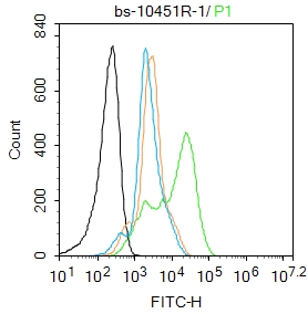
Protocol
The cells were fixed with 4% PFA (10min at room temperature)and then permeabilized with 90% ice-cold methanol for 20 min at -20℃. The cells were then incubated in 5%BSA to block non-specific protein-protein interactions for 30 min at room temperature .Cells stained with Primary Antibody for 30 min at room temperature. The secondary antibody used for 40 min at room temperature. Acquisition of 20,000 events was performed.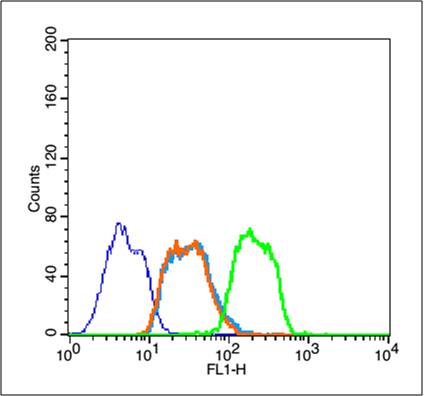 Blank control (blue line): Jurkat (fixed with 2% paraformaldehyde (10 min) , then permeabilized with 90% ice-cold methanol for 30 min on ice).
Primary Antibody (green line): Rabbit Anti-phospho-HSP70 (Tyr41) antibody (SL10451R),Dilution: 1μg /10^6 cells;
Isotype Control Antibody (orange line): Rabbit IgG .
Secondary Antibody (white blue line): Goat anti-rabbit IgG-FITC,Dilution: 1μg /test.
 Blank control: MCF7.
Primary Antibody (green line): Rabbit Anti- phospho-HSP70 (Tyr41) antibody (SL10451R)
Dilution: 2μg /10^6 cells;
Isotype Control Antibody (orange line): Rabbit IgG .
Secondary Antibody : Goat anti-rabbit IgG-FITC
Dilution: 1μg /test.
Protocol
The cells were fixed with 4% PFA (10min at room temperature)and then permeabilized with 0.1% PBST for 20 min at room temperature. The cells were then incubated in 5%BSA to block non-specific protein-protein interactions for 30 min at room temperature .Cells stained with Primary Antibody for 30 min at room temperature. The secondary antibody used for 40 min at room temperature. Acquisition of 20,000 events was performed.
Cartpieces
Totalgoods,subtotals:¥Checkout
Bought notes(bought amounts latest0)
No one bought this product
User Comment(Total0User Comment Num)
- No comment


 +86 571 56623320
 +86 18668110335

