
Rabbit Anti-VEGFR2 antibody
VEGF Receptor 2; CD309; CD309 antigen; Fetal liver kinase 1; FLK-1; FLK1; KDR; Kinase insert domain receptor (a type III receptor tyrosine kinase); Kinase insert domain receptor; KRD1; Ly73; Protein tyrosine kinase receptor FLK1; Protein-tyrosine kinase r
View History [Clear]
Details
Product Name VEGFR2 Chinese Name 血管内皮生长因子受体2抗体 Alias VEGF Receptor 2; CD309; CD309 antigen; Fetal liver kinase 1; FLK-1; FLK1; KDR; Kinase insert domain receptor (a type III receptor tyrosine kinase); Kinase insert domain receptor; KRD1; Ly73; Protein tyrosine kinase receptor FLK1; Protein-tyrosine kinase receptor flk-1; Tyrosine kinase growth factor receptor; Vascular endothelial growth factor receptor 2; VEGFR 2; VEGFR; VEGFR-2; VEGFR2; VGFR2_HUMAN. literatures Research Area Tumour Cell biology immunology Neurobiology Signal transduction Growth factors and hormones Kinases and Phosphatases The cell membrane受体 Cell type markers endothelial cells Immunogen Species Rabbit Clonality Polyclonal React Species Human, (predicted: Mouse, Rat, Dog, Pig, Cow, Horse, Sheep, ) Applications WB=1:500-2000 ELISA=1:5000-10000 IHC-P=1:100-500 IHC-F=1:100-500 Flow-Cyt=1μg/Test (Paraffin sections need antigen repair)
not yet tested in other applications.
optimal dilutions/concentrations should be determined by the end user.Theoretical molecular weight 147kDa Cellular localization The nucleus cytoplasmic The cell membrane Secretory protein Form Liquid Concentration 1mg/ml immunogen KLH conjugated synthetic peptide derived from human VEGFR2: 101-200/1356 <Extracellular> Lsotype IgG Purification affinity purified by Protein A Buffer Solution 0.01M TBS(pH7.4) with 1% BSA, 0.03% Proclin300 and 50% Glycerol. Storage Shipped at 4℃. Store at -20 °C for one year. Avoid repeated freeze/thaw cycles. Attention This product as supplied is intended for research use only, not for use in human, therapeutic or diagnostic applications. PubMed PubMed Product Detail Vascular endothelial growth factor (VEGF) is a major growth factor for endothelial cells. This gene encodes one of the two receptors of the VEGF. This receptor, known as kinase insert domain receptor, is a type III receptor tyrosine kinase. It functions as the main mediator of VEGF-induced endothelial proliferation, survival, migration, tubular morphogenesis and sprouting. The signalling and trafficking of this receptor are regulated by multiple factors, including Rab GTPase, P2Y purine nucleotide receptor, integrin alphaVbeta3, T-cell protein tyrosine phosphatase, etc.. Mutations of this gene are implicated in infantile capillary hemangiomas. [provided by RefSeq, May 2009].
Function:
Tyrosine-protein kinase that acts as a cell-surface receptor for VEGFA, VEGFC and VEGFD. Plays an essential role in the regulation of angiogenesis, vascular development, vascular permeability, and embryonic hematopoiesis. Promotes proliferation, survival, migration and differentiation of endothelial cells. Promotes reorganization of the actin cytoskeleton. Isoforms lacking a transmembrane domain, such as isoform 2 and isoform 3, may function as decoy receptors for VEGFA, VEGFC and/or VEGFD. Isoform 2 plays an important role as negative regulator of VEGFA-and VEGFC-mediated lymphangiogenesis by limiting the amount of free VEGFA and/or VEGFC and preventing their binding to FLT4. Modulates FLT1 and FLT4 signaling by forming heterodimers. Binding of vascular growth factors to isoform 1 leads to the activation of several signaling cascades. Activation of PLCG1 leads to the production of the cellular signaling molecules diacylglycerol and inositol 1,4,5-trisphosphate and the activation of protein kinase C. Mediates activation of MAPK1/ERK2, MAPK3/ERK1 and the MAP kinase signaling pathway, as well as of the AKT1 signaling pathway. Mediates phosphorylation of PIK3R1, the regulatory subunit of phosphatidylinositol 3-kinase, reorganization of the actin cytoskeleton and activation of PTK2/FAK1. Required for VEGFA-mediated induction of NOS2 and NOS3, leading to the production of the signaling molecule nitric oxide (NO) by endothelial cells. Phosphorylates PLCG1. Promotes phosphorylation of FYN, NCK1, NOS3, PIK3R1, PTK2/FAK1 and SRC.
Subunit:
Interacts with MYOF. Interacts with VEGFA, VEGFC and VEGFD. Monomer in the absence of bound VEGFA, VEGFC or VEGFD. Homodimer in the presence of bound dimeric VEGFA, VEGFC or VEGFD. Can also form heterodimers with FLT1 and FLT4. Interacts (tyrosine phosphorylated) with FYN, NCK1, PLCG1 and SHB. Interacts with HIV-1 Tat. Interacts with CBL. Interacts with SH2D2A/TSAD and GRB2.
Subcellular Location:
Isoform 1: Cell membrane; Single-pass type I membrane protein. Cytoplasm. Nucleus. Cytoplasmic vesicle. Early endosome. Note=Detected on caveolae-enriched lipid rafts at the cell surface. Is recycled from the plasma membrane to endosomes and back again. Phosphorylation triggered by VEGFA binding promotes internalization and subsequent degradation. VEGFA binding triggers internalization and translocation to the nucleus.
Isoform 2: Secreted (Probable).
Isoform 3: Secreted.
Tissue Specificity:
Detected in cornea (at protein level). Widely expressed.
Post-translational modifications:
N-glycosylated.
Ubiquitinated. Tyrosine phosphorylation of the receptor promotes its poly-ubiquitination, leading to its degradation via the proteasome or lysosomal proteases.
Autophosphorylated on tyrosine residues upon ligand binding. Autophosphorylation occurs in trans, i.e. one subunit of the dimeric receptor phosphorylates tyrosine residues on the other subunit. Phosphorylation at Tyr-951 is important for interaction with SH2D2A/TSAD and VEGFA-mediated reorganization of the actin cytoskeleton. Phosphorylation at Tyr-1175 is important for interaction with PLCG1 and SHB. Phosphorylation at Tyr-1214 is important for interaction with NCK1 and FYN. Dephosphorylated by PTPRB. Dephosphorylated by PTPRJ at Tyr-951, Tyr-996, Tyr-1054, Tyr-1059, Tyr-1175 and Tyr-1214.
DISEASE:
Defects in KDR are associated with susceptibility to hemangioma capillary infantile (HCI) [MIM:602089]. HCI are benign, highly proliferative lesions involving aberrant localized growth of capillary endothelium. They are the most common tumor of infancy, occurring in up to 10% of all births. Hemangiomas tend to appear shortly after birth and show rapid neonatal growth for up to 12 months characterized by endothelial hypercellularity and increased numbers of mast cells. This phase is followed by slow involution at a rate of about 10% per year and replacement by fibrofatty stroma.
Note=Plays a major role in tumor angiogenesis. In case of HIV-1 infection, the interaction with extracellular viral Tat protein seems to enhance angiogenesis in Kaposi's sarcoma lesions.
Similarity:
Belongs to the protein kinase superfamily. Tyr protein kinase family. CSF-1/PDGF receptor subfamily.
Contains 7 Ig-like C2-type (immunoglobulin-like) domains.
Contains 1 protein kinase domain.
SWISS:
P35968
Gene ID:
3791
Database links:Entrez Gene: 3791 Human
Entrez Gene: 16542 Mouse
Omim: 191306 Human
SwissProt: P35968 Human
SwissProt: P35918 Mouse
Product Picture 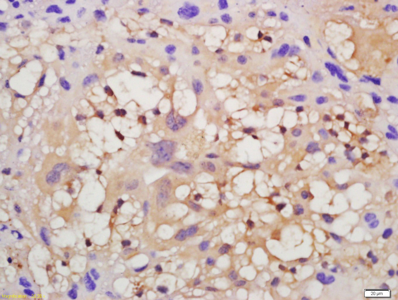 Tissue/cell: mouse placenta tissue; 4% Paraformaldehyde-fixed and paraffin-embedded;
Antigen retrieval: citrate buffer ( 0.01M, pH 6.0 ), Boiling bathing for 15min; Block endogenous peroxidase by 3% Hydrogen peroxide for 30min; Blocking buffer (normal goat serum,C-0005) at 37℃ for 20 min;
Incubation: Anti-VEGFR2 Polyclonal Antibody, Unconjugated(SL10412R) 1:200, overnight at 4°C, followed by conjugation to the secondary antibody(SP-0023) and DAB(C-0010) staining
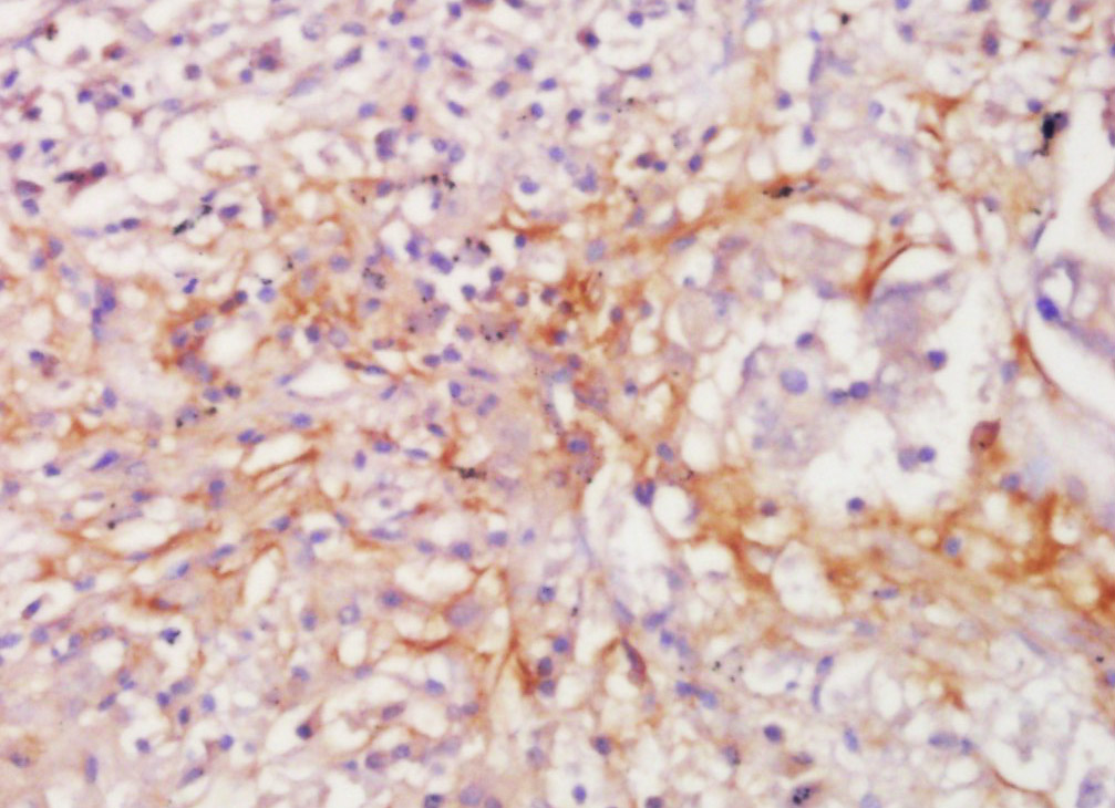 Tissue/cell: human lung carcinoma; 4% Paraformaldehyde-fixed and paraffin-embedded;
Antigen retrieval: citrate buffer ( 0.01M, pH 6.0 ), Boiling bathing for 15min; Block endogenous peroxidase by 3% Hydrogen peroxide for 30min; Blocking buffer (normal goat serum,C-0005) at 37℃ for 20 min;
Incubation: Anti-VEGFR2 Polyclonal Antibody, Unconjugated(SL10412R) 1:200, overnight at 4°C, followed by conjugation to the secondary antibody(SP-0023) and DAB(C-0010) staining
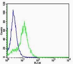 Cell: Hela
Concentration:1:100
Host/Isotype:Rabbit/IgG
Flow cytometric analysis of Rabbit IgG isotype control (Cat#: SL10412R) on Hela(green) compared with control in the absence of primary antibody (blue) followed by Alexa Fluor 488-conjugated goat anti-rabbit IgG(H+L) secondary antibody .
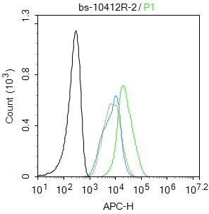 Blank control: MCF7.
Primary Antibody (green line): Rabbit Anti-VEGFR2 antibody (SL10412R)
Dilution: 2μg /10^6 cells;
Isotype Control Antibody (orange line): Rabbit IgG .
Secondary Antibody : Goat anti-rabbit IgG-AF647
Dilution: 1μg /test.
Protocol
The cells were fixed with 4% PFA (10min at room temperature)and then permeabilized with 90% ice-cold methanol for 20 min at-20℃.The cells were then incubated in 5%BSA to block non-specific protein-protein interactions for 30 min at at room temperature .Cells stained with Primary Antibody for 30 min at room temperature. The secondary antibody used for 40 min at room temperature. Acquisition of 20,000 events was performed.
Cartpieces
Totalgoods,subtotals:¥Checkout
Bought notes(bought amounts latest0)
No one bought this product
User Comment(Total0User Comment Num)
- No comment


 +86 571 56623320
 +86 18668110335

