
Rabbit Anti-LPA2 antibody
Endothelial Cell Differentiation Gene 4; Endothelial differentiation lysophosphatidic acid G protein coupled receptor 4; IPA2; LPA receptor 2; LPA receptor EDG4; LPA2; LPAR2; Lysophosphatidic acid receptor Edg4; Edg4; LPAR2_HUMAN.
View History [Clear]
Details
Product Name LPA2 Chinese Name 溶血磷脂酸受体蛋白2抗体 Alias Endothelial Cell Differentiation Gene 4; Endothelial differentiation lysophosphatidic acid G protein coupled receptor 4; IPA2; LPA receptor 2; LPA receptor EDG4; LPA2; LPAR2; Lysophosphatidic acid receptor Edg4; Edg4; LPAR2_HUMAN. literatures Immunogen Species Rabbit Clonality Polyclonal React Species Human, Mouse, Rat, (predicted: Dog, Pig, Cow, Horse, Rabbit, Sheep, ) Applications ELISA=1:5000-10000 IHC-P=1:100-500 IHC-F=1:100-500 Flow-Cyt=1μg/Test ICC=1:100 IF=1:100-500 (Paraffin sections need antigen repair)
not yet tested in other applications.
optimal dilutions/concentrations should be determined by the end user.Theoretical molecular weight 39kDa Cellular localization The cell membrane Form Liquid Concentration 1mg/ml immunogen KLH conjugated synthetic peptide derived from human LPA2: 1-100/351 <Extracellular> Lsotype IgG Purification affinity purified by Protein A Buffer Solution 0.01M TBS(pH7.4) with 1% BSA, 0.03% Proclin300 and 50% Glycerol. Storage Shipped at 4℃. Store at -20 °C for one year. Avoid repeated freeze/thaw cycles. Attention This product as supplied is intended for research use only, not for use in human, therapeutic or diagnostic applications. PubMed PubMed Product Detail This gene encodes a member of family I of the G protein-coupled receptors, as well as the EDG family of proteins. This protein functions as a lysophosphatidic acid (LPA) receptor and contributes to Ca2+ mobilization, a critical cellular response to LPA in cells, through association with Gi and Gq proteins. An alternative splice variant has been described but its full length sequence has not been determined. [provided by RefSeq, Jul 2008]
Function:
Receptor for lysophosphatidic acid (LPA), a mediator of diverse cellular activities. Seems to be coupled to the G(i)/G(o), G(12)/G(13), and G(q) families of heteromeric G proteins. Plays a key role in phospholipase C-beta (PLC-beta) signaling pathway. Stimulates phospholipase C (PLC) activity in a manner that is independent of RALA activation.
Subunit:
Interacts with SLC9A3R2/NHERF2, MAGI3 and PLCB3. Interacts with RALA and ADRBK1.
Subcellular Location:
Cell surface. Cell membrane; Multi-pass membrane protein. Note=Prior to LPA treatment found predominantly at the cell surface but in the presence of LPA co-localizes with RALA in the endocytic vesicles.
Tissue Specificity:
Expressed most abundantly in testes and peripheral blood leukocytes with less expression in pancreas, spleen, thymus and prostate. Little or no expression in heart, brain, placenta, lung, liver, skeletal muscle, kidney, ovary, small intestine, or colon.
Similarity:
Belongs to the G-protein coupled receptor 1 family.
SWISS:
Q9HBW0
Gene ID:
9170
Database links:Entrez Gene: 9170 Human
Entrez Gene: 53978 Mouse
Omim: 605110 Human
SwissProt: Q9HBW0 Human
SwissProt: Q9JL06 Mouse
Unigene: 122575 Human
Unigene: 23253 Mouse
Product Picture  Paraformaldehyde-fixed, paraffin embedded (mouse stomach); Antigen retrieval by boiling in sodium citrate buffer (pH6.0) for 15min; Block endogenous peroxidase by 3% hydrogen peroxide for 20 minutes; Blocking buffer (normal goat serum) at 37°C for 30min; Antibody incubation with (LPA2) Polyclonal Antibody, Unconjugated (SL10368R) at 1:200 overnight at 4°C, followed by operating according to SP Kit(Rabbit) (sp-0023) instructionsand DAB staining.
 Tissue/cell: rat spleen tissue; 4% Paraformaldehyde-fixed and paraffin-embedded;
Antigen retrieval: citrate buffer ( 0.01M, pH 6.0 ), Boiling bathing for 15min; Block endogenous peroxidase by 3% Hydrogen peroxide for 30min; Blocking buffer (normal goat serum,C-0005) at 37℃ for 20 min;
Incubation: Anti-LPA2 Polyclonal Antibody, Unconjugated(SL10368R) 1:100, overnight at 4°C, followed by conjugation to the secondary antibody(SP-0023) and DAB(C-0010) staining
 Blank control: mouse thymouses(blue)
Isotype Control Antibody: Rabbit IgG(orange) ; Secondary Antibody: Goat anti-rabbit IgG-FITC(white blue), Dilution: 1:100 in 1 X PBS containing 0.5% BSA ; Primary Antibody Dilution: 1μl in 100 μL1X PBS containing 0.5% BSA(green). Positive control: (mo)Splenocytes(2% Paraformaldehyde-fixed )
Isotype Control Antibody: Rabbit IgG,Dilution: 1μg in 100 μl 1 X PBS containing 0.5% BSA
Secondary Antibody: Goat anti-rabbit IgG-FITC,Dilution: 1:200 in 1 X PBS containing 0.5% BSA
Primary Antibody: rabbit Anti-LPA2 (SL10368R),Dilution: 1μg in 100 μl 1X PBS containing 0.5% BSA
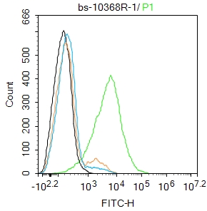 Blank control:THP-1.
Primary Antibody (green line): Rabbit Anti-LPA2 antibody (SL10368R)
Dilution: 1μg /10^6 cells;
Isotype Control Antibody (orange line): Rabbit IgG .
Secondary Antibody : Goat anti-rabbit IgG-FITC
Dilution: 0.5μg /test.
Protocol
The cells were incubated in 5%BSA to block non-specific protein-protein interactions for 30 min at room temperature .Cells stained with Primary Antibody for 30 min at room temperature. The secondary antibody used for 40 min at room temperature. Acquisition of 20,000 events was performed.
Cartpieces
Totalgoods,subtotals:¥Checkout
Bought notes(bought amounts latest0)
No one bought this product
User Comment(Total0User Comment Num)
- No comment


 +86 571 56623320
 +86 18668110335

