
Rabbit Anti-VNN1 antibody
HDLCQ8; High density lipoprotein cholesterol level quantitative trait locus 8, included; Pantetheinase; Pantetheine hydrolase; Tiff66; V-1 antibody Vanin 1; Vanin-1; Vannin 1; Vascular non inflammatory molecule 1; Vascular non-inflammatory molecule 1; VNN
View History [Clear]
Details
Product Name VNN1 Chinese Name 血管非炎症分子1抗体 Alias HDLCQ8; High density lipoprotein cholesterol level quantitative trait locus 8, included; Pantetheinase; Pantetheine hydrolase; Tiff66; V-1 antibody Vanin 1; Vanin-1; Vannin 1; Vascular non inflammatory molecule 1; Vascular non-inflammatory molecule 1; VNN 1; VNN1; VNN1_HUMAN. Research Area Cardiovascular Cell biology Signal transduction lymphocyte t-lymphocyte Immunogen Species Rabbit Clonality Polyclonal React Species Human, Mouse, Rat, (predicted: Cow, Rabbit, Sheep, ) Applications WB=1:500-2000 ELISA=1:5000-10000 IHC-P=1:100-500 IHC-F=1:100-500 Flow-Cyt=2μg/Test ICC=1:100-500 IF=1:100-500 (Paraffin sections need antigen repair)
not yet tested in other applications.
optimal dilutions/concentrations should be determined by the end user.Theoretical molecular weight 52kDa Cellular localization The cell membrane Form Liquid Concentration 1mg/ml immunogen KLH conjugated synthetic peptide derived from human VNN1: 151-250/513 Lsotype IgG Purification affinity purified by Protein A Buffer Solution 0.01M TBS(pH7.4) with 1% BSA, 0.03% Proclin300 and 50% Glycerol. Storage Shipped at 4℃. Store at -20 °C for one year. Avoid repeated freeze/thaw cycles. Attention This product as supplied is intended for research use only, not for use in human, therapeutic or diagnostic applications. PubMed PubMed Product Detail This gene encodes a member of the vanin family of proteins, which share extensive sequence similarity with each other, and also with biotinidase. The family includes secreted and membrane-associated proteins, a few of which have been reported to participate in hematopoietic cell trafficking. No biotinidase activity has been demonstrated for any of the vanin proteins, however, they possess pantetheinase activity, which may play a role in oxidative-stress response. This protein, like its mouse homolog, is likely a GPI-anchored cell surface molecule. The mouse protein is expressed by the perivascular thymic stromal cells and regulates migration of T-cell progenitors to the thymus. This gene lies in close proximity to, and in the same transcriptional orientation as, two other vanin genes on chromosome 6q23-q24. [provided by RefSeq, Feb 2009]
Function:
Amidohydrolase that hydrolyzes specifically one of the carboamide linkages in D-pantetheine thus recycling pantothenic acid (vitamin B5) and releasing cysteamine.
Subcellular Location:
Cell membrane; Lipid-anchor, GPI-anchor
Tissue Specificity:
Widely expressed with higher expression in spleen, kidney and blood. Overexpressed in lesional psoriatic skin.
Similarity:
Belongs to the CN hydrolase family. BTD/VNN subfamily.
Contains 1 CN hydrolase domain.
SWISS:
O95497
Gene ID:
8876
Database links:Entrez Gene: 8876 Human
Omim: 603570 Human
SwissProt: O95497 Human
Unigene: 12114 Human
Unigene: 720659 Human
Product Picture 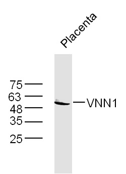 Sample: placenta(Mouse) Lysate at 40 ug
Primary: Anti-VNN1(SL10314R) at 1/300 dilution
Secondary: IRDye800CW Goat Anti-Rabbit IgG at 1/20000 dilution
Predicted band size: 52 kD
Observed band size: 52 kD
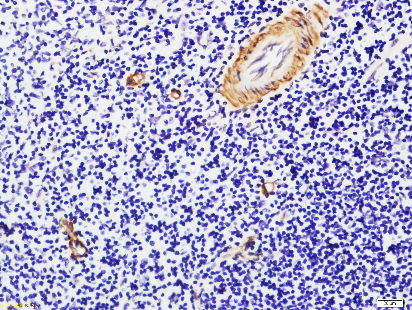 Tissue/cell: rat spleen tissue; 4% Paraformaldehyde-fixed and paraffin-embedded;
Antigen retrieval: citrate buffer ( 0.01M, pH 6.0 ), Boiling bathing for 15min; Block endogenous peroxidase by 3% Hydrogen peroxide for 30min; Blocking buffer (normal goat serum,C-0005) at 37℃ for 20 min;
Incubation: Anti-VNN1 Polyclonal Antibody, Unconjugated(SL10314R) 1:200, overnight at 4°C, followed by conjugation to the secondary antibody(SP-0023) and DAB(C-0010) staining
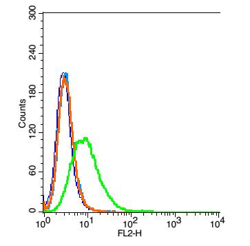 Blank control: 293T(blue).
Primary Antibody:Rabbit Anti-VNN1 antibody(SL10314R), Dilution: 5μg in 100 1μL 1X PBS containing 0.5% BSA;
Isotype Control Antibody: Rabbit IgG(orange) ,used under the same conditions );
Secondary Antibody: Goat anti-rabbit IgG-PE(white blue), Dilution: 1:200 in 1 X PBS containing 0.5% BSA.
Protocol
The cells were washed twice with phosphate-buffered saline (PBS).The cells were then incubated in 1 X PBS containing 0.5% BSA + 1 0% goat serum (15 min) to block non-specific protein-protein interactions followed by the antibody (SL10314R, 5μg /1x10^6 cells) for 30 min on ice. The secondary antibody used was Goat Anti-rabbit IgG/PE antibody at 1/200 dilution for 30 min on ice. Acquisition of 20,000 events was performed.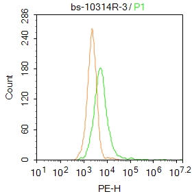 Blank control: 293T.
Primary Antibody (green line): Rabbit Anti-VNN1 antibody (SL10314R)
Dilution: 1μg /10^6 cells;
Isotype Control Antibody (orange line): Rabbit IgG .
Secondary Antibody : Goat anti-rabbit IgG-PE
Dilution: 1μg /test.
Protocol
The cells were incubated in 5%BSA to block non-specific protein-protein interactions for 30 min at at room temperature .Cells stained with Primary Antibody for 30 min at room temperature. The secondary antibody used for 40 min at room temperature. Acquisition of 20,000 events was performed.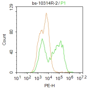 Blank control: A431.
Primary Antibody (green line): Rabbit Anti-VNN1 antibody (SL10314R)
Dilution: 2μg /10^6 cells;
Isotype Control Antibody (orange line): Rabbit IgG .
Secondary Antibody : Goat anti-rabbit IgG-PE
Dilution: 1μg /test.
Protocol
The cells were fixed with 4% PFA (10min at room temperature)and then permeabilized with 90% ice-cold methanol for 20 min at-20℃. The cells were then incubated in 5%BSA to block non-specific protein-protein interactions for 30 min at at room temperature .Cells stained with Primary Antibody for 30 min at room temperature. The secondary antibody used for 40 min at room temperature. Acquisition of 20,000 events was performed.
Cartpieces
Totalgoods,subtotals:¥Checkout
Bought notes(bought amounts latest0)
No one bought this product
User Comment(Total0User Comment Num)
- No comment


 +86 571 56623320
 +86 18668110335

