
Rabbit Anti-NSE antibody
Gamma-enolase; Gamma enolase; Neural enolase; Neuron specific enolase; neurone-specific enolase; 2 phospho D glycerate hydrolyase; Eno 2; Eno2; ENO2; ENOG; Enolase 2; Enolase 2 gamma neuronal; Enolase2; Neuron specific enolase; Neuron specific gamma enola
View History [Clear]
Details
Product Name NSE Chinese Name 神经元特异性烯醇化酶/γ 烯醇化酶抗体 Alias Gamma-enolase; Gamma enolase; Neural enolase; Neuron specific enolase; neurone-specific enolase; 2 phospho D glycerate hydrolyase; Eno 2; Eno2; ENO2; ENOG; Enolase 2; Enolase 2 gamma neuronal; Enolase2; Neuron specific enolase; Neuron specific gamma enolase; NSE; ENOG_HUMAN; Gamma-enolase; 2-phospho-D-glycerate hydro-lyase; enolase 2, gamma, neuronal. literatures Research Area Tumour Cell biology immunology Neurobiology TumourCell biologyMaker Immunogen Species Rabbit Clonality Polyclonal React Species Human, Mouse, Rat, Dog, (predicted: Chicken, Cow, ) Applications WB=1:500-2000 ELISA=1:5000-10000 IHC-P=1:100-500 IHC-F=1:100-500 Flow-Cyt=1μg/Test IF=1:100-500 (Paraffin sections need antigen repair)
not yet tested in other applications.
optimal dilutions/concentrations should be determined by the end user.Theoretical molecular weight 48kDa Cellular localization cytoplasmic The cell membrane Form Liquid Concentration 1mg/ml immunogen KLH conjugated synthetic peptide derived from human NSE: 201-300/434 Lsotype IgG Purification affinity purified by Protein A Buffer Solution 0.01M TBS(pH7.4) with 1% BSA, 0.03% Proclin300 and 50% Glycerol. Storage Shipped at 4℃. Store at -20 °C for one year. Avoid repeated freeze/thaw cycles. Attention This product as supplied is intended for research use only, not for use in human, therapeutic or diagnostic applications. PubMed PubMed Product Detail This gene encodes one of the three enolase isoenzymes found in mammals. This isoenzyme, a homodimer, is found in mature neurons and cells of neuronal origin. A switch from alpha enolase to gamma enolase occurs in neural tissue during development in rats and primates. [provided by RefSeq, Jul 2008].
Function:
Has neurotrophic and neuroprotective properties on a broad spectrum of central nervous system (CNS) neurons. Binds, in a calcium-dependent manner, to cultured neocortical neurons and promotes cell survival.
Subunit:
Mammalian enolase is composed of 3 isozyme subunits, alpha, beta and gamma, which can form homodimers or heterodimers which are cell-type and development-specific.
Subcellular Location:
Cytoplasm. Cell membrane. Can translocate to the plasma membrane in either the homodimeric (alpha/alpha) or heterodimeric (alpha/gamma) form.
Tissue Specificity:
The alpha/alpha homodimer is expressed in embryo and in most adult tissues. The alpha/beta heterodimer and the beta/beta homodimer are found in striated muscle, and the alpha/gamma heterodimer and the gamma/gamma homodimer in neurons.
Similarity:
Belongs to the enolase family.
SWISS:
P09104
Gene ID:
2026
Database links:Entrez Gene: 2026 Human
Entrez Gene: 13807 Mouse
Omim: 131360 Human
SwissProt: P09104 Human
SwissProt: P17183 Mouse
Unigene: 511915 Human
神经元特异性烯醇化酶(NSE)存在于神经元细胞和神经内分泌组织中,起源于神经内分泌细胞的Tumour可产生过量的NSE。NSE也是小细胞肺癌的检测指标,70%左右的小细胞肺癌患者血中NSE升高,而其他组织型肺癌NSE升高的患者仅为10%~20%。Product Picture 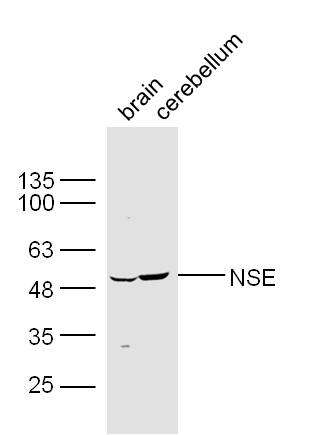 Sample:
Brain (Mouse) Lysate at 30 ug
Cerebellum (Mouse) Lysate at 30 ug
Primary: Anti-NSE (SL1027R) at 1/300 dilution
Secondary: IRDye800CW Goat Anti-Rabbit IgG at 1/20000 dilution
Predicted band size: 48 kD
Observed band size: 49kD
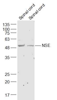 Sample:
Spinal cord (Mouse) Lysate at 40 ug
Spinal cord (Rat) Lysate at 40 ug
Primary: Anti-NSE (SL1027R) at 1/300 dilution
Secondary: IRDye800CW Goat Anti-Rabbit IgG at 1/20000 dilution
Predicted band size: 48 kD
Observed band size: 48 kD
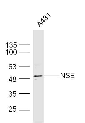 Sample: A431 Cell Lysate at 40 ug
Primary: Anti- NSE (SL1027R) at 1/300 dilution
Secondary: IRDye800CW Goat Anti-Rabbit IgG at 1/20000 dilution
Predicted band size: 48 kD
Observed band size: 48 kD
 Paraformaldehyde-fixed, paraffin embedded (rat brain); Antigen retrieval by boiling in sodium citrate buffer (pH6.0) for 15min; Block endogenous peroxidase by 3% hydrogen peroxide for 20 minutes; Blocking buffer (normal goat serum) at 37°C for 30min; Antibody incubation with (NSE) Polyclonal Antibody, Unconjugated (SL1027R) at 1:200 overnight at 4°C, followed by operating according to SP Kit(Rabbit) (sp-0023) instructionsand DAB staining.
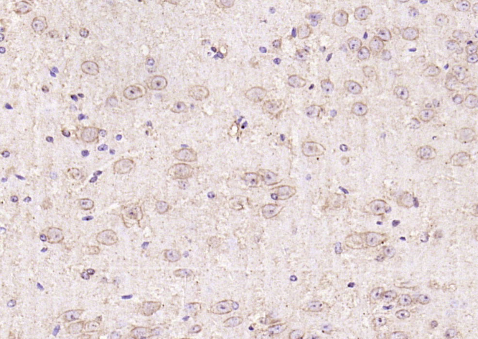 Paraformaldehyde-fixed, paraffin embedded (mouse brain); Antigen retrieval by boiling in sodium citrate buffer (pH6.0) for 15min; Block endogenous peroxidase by 3% hydrogen peroxide for 20 minutes; Blocking buffer (normal goat serum) at 37°C for 30min; Antibody incubation with (NSE) Polyclonal Antibody, Unconjugated (SL1027R) at 1:200 overnight at 4°C, followed by operating according to SP Kit(Rabbit) (sp-0023) instructionsand DAB staining.
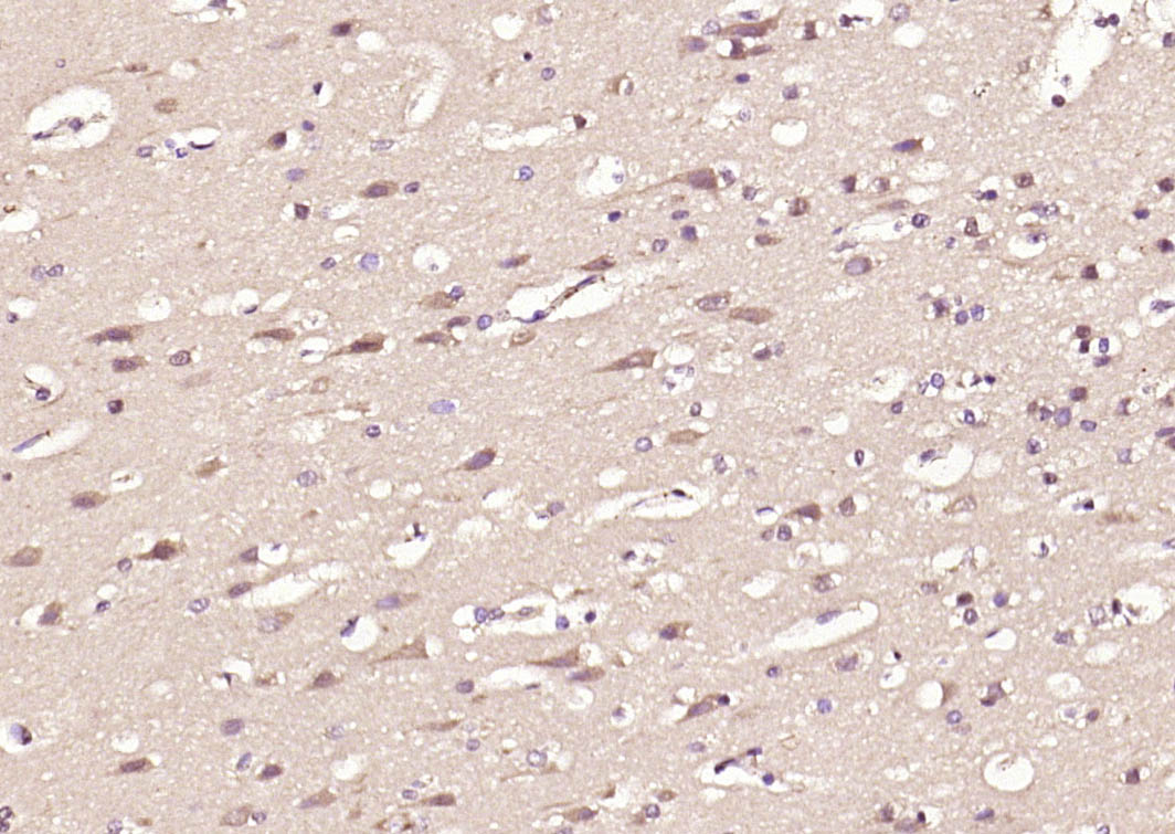 Paraformaldehyde-fixed, paraffin embedded (human brain); Antigen retrieval by boiling in sodium citrate buffer (pH6.0) for 15min; Block endogenous peroxidase by 3% hydrogen peroxide for 20 minutes; Blocking buffer (normal goat serum) at 37°C for 30min; Antibody incubation with (NSE) Polyclonal Antibody, Unconjugated (SL1027R) at 1:200 overnight at 4°C, followed by operating according to SP Kit(Rabbit) (sp-0023) instructionsand DAB staining.
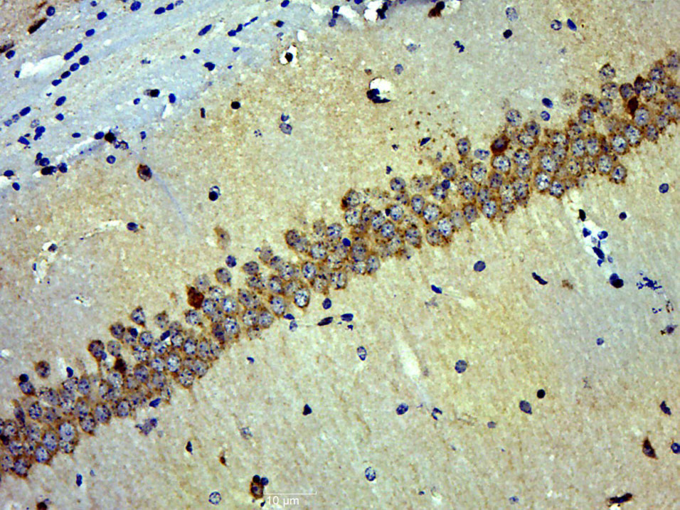 Paraformaldehyde-fixed, paraffin embedded (Rat brain); Antigen retrieval by boiling in sodium citrate buffer (pH6.0) for 15min; Block endogenous peroxidase by 3% hydrogen peroxide for 20 minutes; Blocking buffer (normal goat serum) at 37°C for 30min; Antibody incubation with (NSE) Polyclonal Antibody, Unconjugated (SL1027R) at 1:500 overnight at 4°C, followed by a conjugated secondary (sp-0023) for 20 minutes and DAB staining.
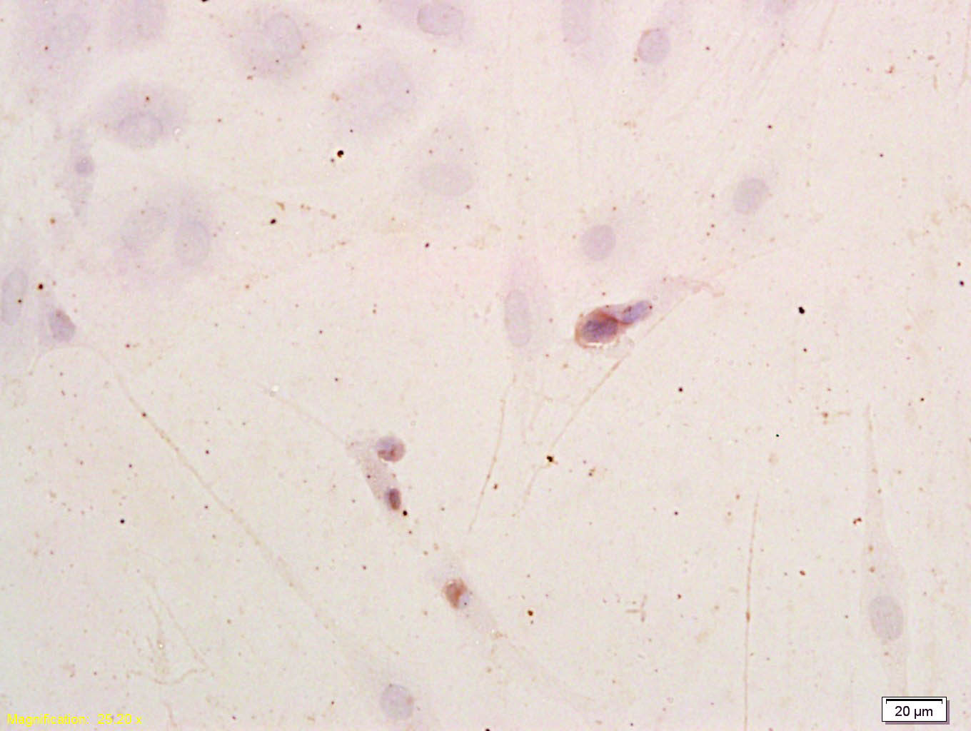 Tissue/cell: Neuroblastoma cells;
Block endogenous peroxidase by 3% Hydrogen peroxide for 30min; Blocking buffer (normal goat serum,C-0005) at 37℃ for 20 min;
Incubation: Anti-NSE/ENO2/γ Enolase Polyclonal Antibody, Unconjugated(SL1027R) 1:200, overnight at 4°C, followed by conjugation to the secondary antibody(SP-0023) and DAB(C-0010) staining
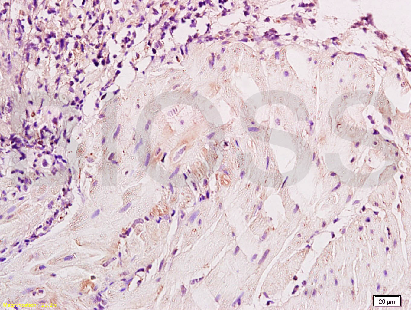 Tissue/cell: dog bladder tissue; 4% Paraformaldehyde-fixed and paraffin-embedded;
Antigen retrieval: citrate buffer ( 0.01M, pH 6.0 ), Boiling bathing for 15min; Block endogenous peroxidase by 3% Hydrogen peroxide for 30min; Blocking buffer (normal goat serum,C-0005) at 37℃ for 20 min;
Incubation: Anti-NSE/ENO2/γ Enolase Polyclonal Antibody, Unconjugated(SL1027R) 1:800, overnight at 4°C, followed by conjugation to the secondary antibody(SP-0023) and DAB(C-0010) staining
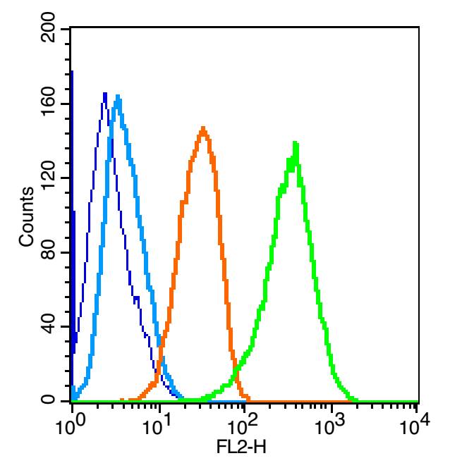 Blank control: U-87MG(blue).
Primary Antibody:Rabbit Anti-NSE antibody(SL1027R), Dilution: 1μg in 100 μL 1X PBS containing 0.5% BSA;
Isotype Control Antibody: Rabbit IgG(orange) ,used under the same conditions );
Secondary Antibody: Goat anti-rabbit IgG-PE(white blue), Dilution: 1:200 in 1 X PBS containing 0.5% BSA.
Protocol
The cells were fixed with 2% paraformaldehyde (10 min) , then permeabilized with 90% ice-cold methanol for 30 min on ice. Primary antibody (SL1027R,1μg /1x10^6 cells) were incubated for 30 min on the ice, followed by 1 X PBS containing 0.5% BSA + 1 0% goat serum (15 min) to block non-specific protein-protein interactions. Then the Goat Anti-rabbit IgG/PE antibody was added into the blocking buffer mentioned above to react with the primary antibody at 1/200 dilution for 30 min on ice. Acquisition of 20,000 events was performed.
Cartpieces
Totalgoods,subtotals:¥Checkout
Bought notes(bought amounts latest0)
No one bought this product
User Comment(Total0User Comment Num)
- No comment


 +86 571 56623320
 +86 18668110335

