
Rabbit Anti-Desmin antibody
DESM_HUMAN; DES;
View History [Clear]
Details
Product Name Desmin Chinese Name 结蛋白抗体 Alias DESM_HUMAN; DES; literatures Research Area Tumour Cardiovascular immunology Signal transduction Cell type markers Immunogen Species Rabbit Clonality Polyclonal React Species Human, Mouse, Rat, (predicted: Chicken, Dog, Pig, Cow, Horse, Rabbit, Sheep, Guinea Pig, ) Applications ELISA=1:5000-10000 IHC-P=1:100-500 IHC-F=1:100-500 Flow-Cyt=1μg/Test (Paraffin sections need antigen repair)
not yet tested in other applications.
optimal dilutions/concentrations should be determined by the end user.Theoretical molecular weight 52kDa Cellular localization cytoplasmic Form Liquid Concentration 1mg/ml immunogen KLH conjugated synthetic peptide derived from human Desmin: 261-360/470 Lsotype IgG Purification affinity purified by Protein A Buffer Solution 0.01M TBS(pH7.4) with 1% BSA, 0.03% Proclin300 and 50% Glycerol. Storage Shipped at 4℃. Store at -20 °C for one year. Avoid repeated freeze/thaw cycles. Attention This product as supplied is intended for research use only, not for use in human, therapeutic or diagnostic applications. PubMed PubMed Product Detail Desmin is a muscle-specific, type III intermediate filament that integrates the sarcolemma, Z disk, and nuclear membrane in sarcomeres and regulates sarcomere architecture. In adult striated muscle they form a fibrous network connecting myofibrils to each other and to the plasma membrane from the periphery of the Z line structures. Defects in Desmin are the cause of desmin related cardio skeletal myopathy (CSM) also known as desmin related myopathy (DRM). CSM is characterized by skeletal muscle weakness associated with cardiac conduction blocks, arrhythmias, restrictive heart failure, and by intracytoplasmic accumulation of desmin reactive deposits in cardiac and skeletal muscle cells. A desmin related myopathy can have a distal onset, it is then known as hereditary distal myopathy (HDM). Defects in Desmin are also the cause of dilated cardiomyopathy type 1I (CMD1I). CMD1I is an autosomal form of dilated cardiomyopathy characterized by ventricular dilatation and impaired systolic function. Antidesmin antibodies are useful in identification of tumours of myogenic origin.
Function:
Desmin are class-III intermediate filaments found in muscle cells. In adult striated muscle they form a fibrous network connecting myofibrils to each other and to the plasma membrane from the periphery of the Z-line structures.
Subunit:
Homopolymer. Interacts with DST. Interacts with MTM1.
Subcellular Location:
Cytoplasm.
Post-translational modifications:
ADP-ribosylation prevents ability to form intermediate filaments.
DISEASE:
Defects in DES are the cause of myopathy myofibrillar type 1 (MFM1) [MIM:601419]. A neuromuscular disorder characterized by skeletal muscle weakness associated with cardiac conduction blocks, arrhythmias, restrictive heart failure, and by myofibrillar destruction with intracytoplasmic accumulation of desmin-reactive deposits in cardiac and skeletal muscle cells. Note=Mutations in the DES gene are associated with a variable clinical phenotype which encompasses isolated myopathies, pure cardiac phenotypes (including dilated cardiomyopathy, restrictive cardiomyopathy and arrhythmogenic right ventricular cardiomyopathy), cardiac conduction disease, and combinations of these disorders. If both cardiologic and neurologic features occur, they can manifest in any order, as cardiologic features can precede, occur simultaneously with, or follow manifestation of generalized neuromuscular disease (PubMed:19879535).
Defects in DES are the cause of cardiomyopathy dilated type 1I (CMD1I) [MIM:604765]. Dilated cardiomyopathy is a disorder characterized by ventricular dilation and impaired systolic function, resulting in congestive heart failure and arrhythmia. Patients are at risk of premature death.
Defects in DES are the cause of neurogenic scapuloperoneal syndrome Kaeser type (Kaeser syndrome) [MIM:181400]. Kaeser syndrome is an autosomal dominant disorder with a peculiar scapuloperoneal distribution of weakness and atrophy. A large clinical variability is observed ranging from scapuloperoneal, limb grindle and distal phenotypes with variable cardiac or respiratory involvement. Facial weakness, dysphagia and gynaecomastia are frequent additional symptoms. Affected men seemingly bear a higher risk of sudden, cardiac death as compared to affected women. Histological and immunohistochemical examination of muscle biopsy specimens reveal a wide spectrum of findings ranging from near normal or unspecific pathology to typical, myofibrillar changes with accumulation of desmin.
Similarity:
Belongs to the intermediate filament family.
SWISS:
P17661
Gene ID:
1674
Database links:Entrez Gene: 1674 Human
Entrez Gene: 13346 Mouse
Omim: 125660 Human
SwissProt: P17661 Human
SwissProt: P31001 Mouse
Unigene: 594952 Human
Unigene: 6712 Mouse
Unigene: 39196 Rat
Desmin在很多哺乳动物中的横纹肌和各种平滑肌及其来源的Tumour组织中都有表达。结蛋白是一种中间丝蛋白,广泛分布于骨骼肌细胞、平滑肌细胞、心肌细胞和肌epithelial cells及其Tumour中,主要用于子宫、皮肤、胃肠道及其它横纹肌肉瘤和肌上皮瘤的诊断和鉴别诊断。Product Picture 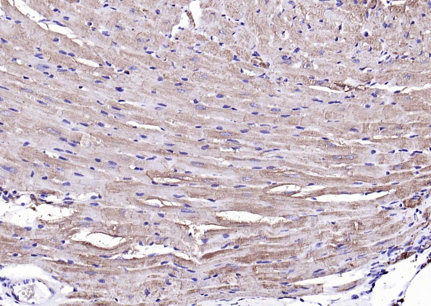 Paraformaldehyde-fixed, paraffin embedded (mouse heart); Antigen retrieval by boiling in sodium citrate buffer (pH6.0) for 15min; Block endogenous peroxidase by 3% hydrogen peroxide for 20 minutes; Blocking buffer (normal goat serum) at 37°C for 30min; Antibody incubation with (Desmin) Polyclonal Antibody, Unconjugated (SL1026R) at 1:200 overnight at 4°C, followed by operating according to SP Kit(Rabbit) (sp-0023) instructionsand DAB staining.
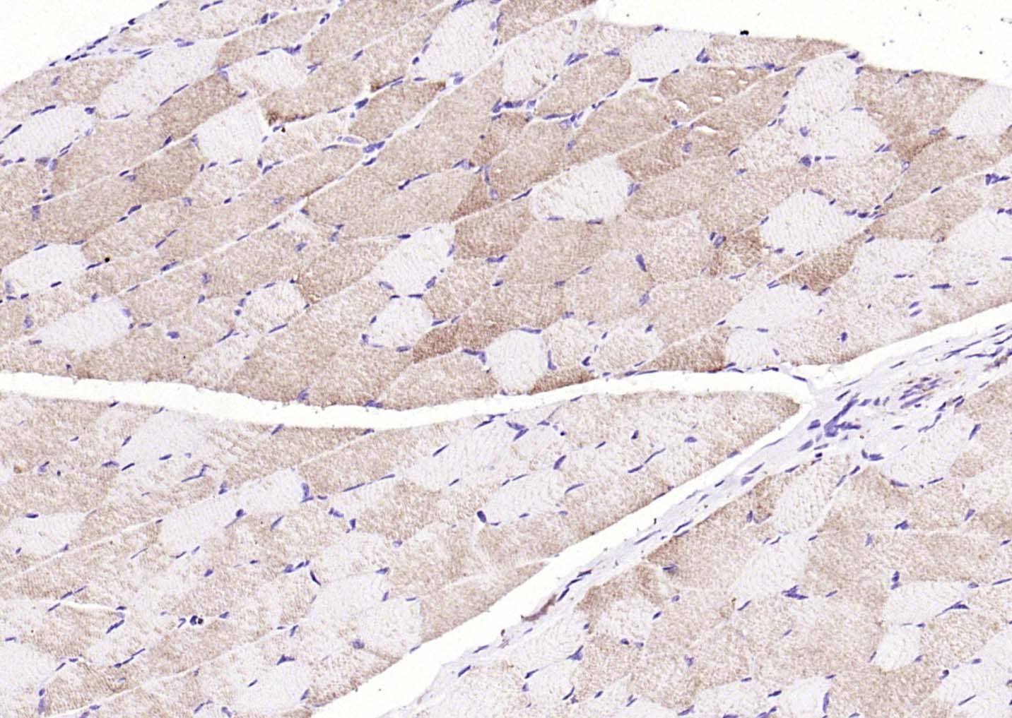 Paraformaldehyde-fixed, paraffin embedded (rat skeletal muscle); Antigen retrieval by boiling in sodium citrate buffer (pH6.0) for 15min; Block endogenous peroxidase by 3% hydrogen peroxide for 20 minutes; Blocking buffer (normal goat serum) at 37°C for 30min; Antibody incubation with (Desmin) Polyclonal Antibody, Unconjugated (SL1026R) at 1:200 overnight at 4°C, followed by operating according to SP Kit(Rabbit) (sp-0023) instructionsand DAB staining.
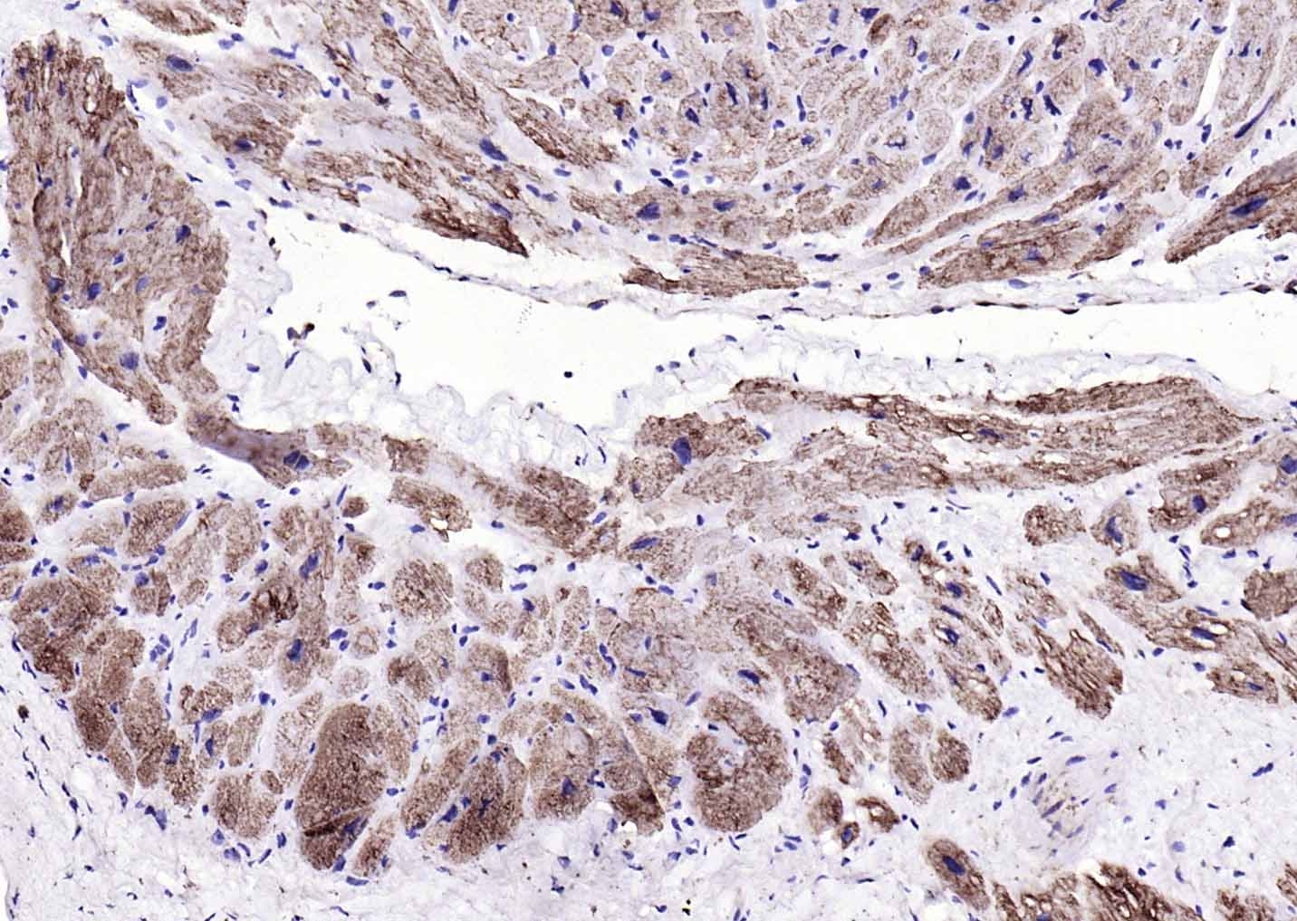 Paraformaldehyde-fixed, paraffin embedded (human myocardium); Antigen retrieval by boiling in sodium citrate buffer (pH6.0) for 15min; Block endogenous peroxidase by 3% hydrogen peroxide for 20 minutes; Blocking buffer (normal goat serum) at 37°C for 30min; Antibody incubation with (Desmin) Polyclonal Antibody, Unconjugated (SL1026R) at 1:200 overnight at 4°C, followed by operating according to SP Kit(Rabbit) (sp-0023) instructionsand DAB staining.
 Paraformaldehyde-fixed, paraffin embedded (mouse skeletal muscle); Antigen retrieval by boiling in sodium citrate buffer (pH6.0) for 15min; Block endogenous peroxidase by 3% hydrogen peroxide for 20 minutes; Blocking buffer (normal goat serum) at 37°C for 30min; Antibody incubation with (Desmin) Polyclonal Antibody, Unconjugated (SL1026R) at 1:200 overnight at 4°C, followed by operating according to SP Kit(Rabbit) (sp-0023) instructionsand DAB staining.
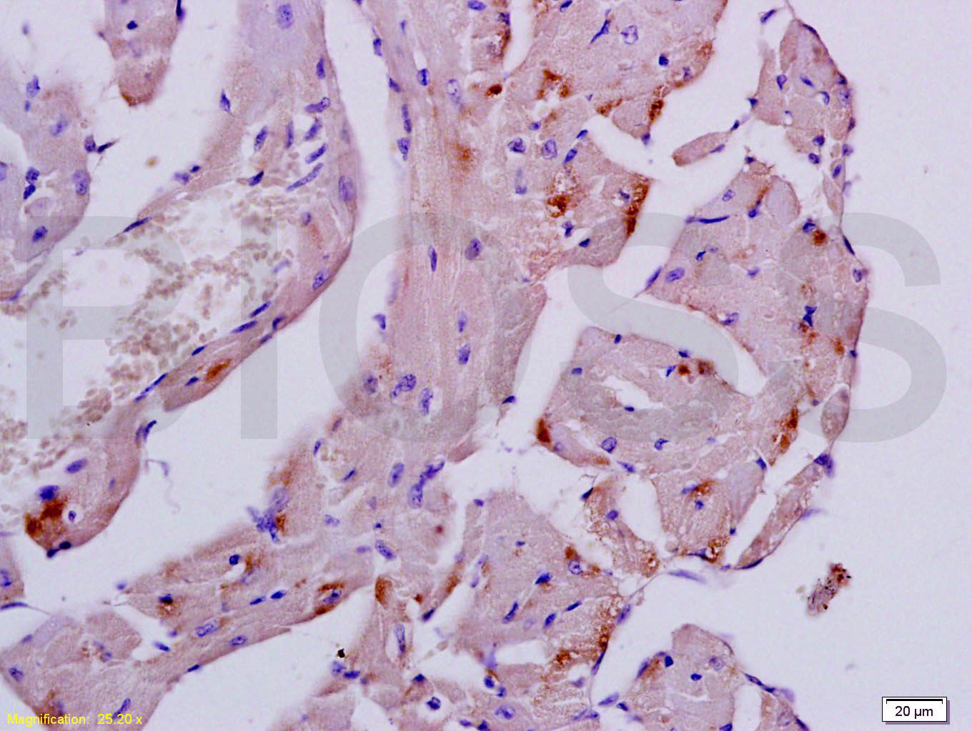 Tissue/cell: mouse heart tissue; 4% Paraformaldehyde-fixed and paraffin-embedded;
Antigen retrieval: citrate buffer ( 0.01M, pH 6.0 ), Boiling bathing for 15min; Block endogenous peroxidase by 3% Hydrogen peroxide for 30min; Blocking buffer (normal goat serum,C-0005) at 37℃ for 20 min;
Incubation: Anti-Desmin Polyclonal Antibody, Unconjugated(SL1026R) 1:200, overnight at 4°C, followed by conjugation to the secondary antibody(SP-0023) and DAB(C-0010) staining
 Tissue/cell: rat kidney tissue; 4% Paraformaldehyde-fixed and paraffin-embedded;
Antigen retrieval: citrate buffer ( 0.01M, pH 6.0 ), Boiling bathing for 15min; Block endogenous peroxidase by 3% Hydrogen peroxide for 30min; Blocking buffer (normal goat serum,C-0005) at 37℃ for 20 min;
Incubation: Anti-Desmin Polyclonal Antibody, Unconjugated(SL1026R) 1:200, overnight at 4°C, followed by conjugation to the secondary antibody(SP-0023) and DAB(C-0010) staining
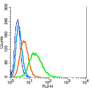 Blank control: Hela(blue).
Primary Antibody:Rabbit Anti- Desmin antibody(SL1026R), Dilution: 1μg in 100 μL 1X PBS containing 0.5% BSA;
Isotype Control Antibody: Rabbit IgG(orange) ,used under the same conditions );
Secondary Antibody: Goat anti-rabbit IgG-PE(white blue), Dilution: 1:200 in 1 X PBS containing 0.5% BSA.
Protocol
The cells were fixed with 2% paraformaldehyde (10 min) , then permeabilized with 90% ice-cold methanol for 30 min on ice. Antibody (SL1026R, 1μg /1x10^6 cells) were incubated for 30 min on the ice, followed by 1 X PBS containing 0.5% BSA + 1 0% goat serum (15 min) to block non-specific protein-protein interactions. Then the Goat Anti-rabbit IgG/PE antibody was added into the blocking buffer mentioned above to react with the primary antibody of SL1026R at 1/200 dilution for 30 min on ice. Acquisition of 20,000 events was performed.
Cartpieces
Totalgoods,subtotals:¥Checkout
Bought notes(bought amounts latest0)
No one bought this product
User Comment(Total0User Comment Num)
- No comment


 +86 571 56623320
 +86 18668110335

