
Rabbit Anti-TLR4 antibody
TLR-4; TLR 4; ARMD10; CD 284; CD284; CD284 antigen; Homolog of Drosophila toll; hTol; Toll (Drosophila) homolog; TOLL; Toll Endotoxin Hyporesponsiveness; Toll like receptor 4; Toll like receptor 4 precursor; TLR4_HUMAN.
View History [Clear]
Details
Product Name TLR4 Chinese Name Toll样受体4(CD284)抗体 Alias TLR-4; TLR 4; ARMD10; CD 284; CD284; CD284 antigen; Homolog of Drosophila toll; hTol; Toll (Drosophila) homolog; TOLL; Toll Endotoxin Hyporesponsiveness; Toll like receptor 4; Toll like receptor 4 precursor; TLR4_HUMAN. literatures Research Area Cardiovascular immunology Signal transduction The cell membrane受体 Immunogen Species Rabbit Clonality Polyclonal React Species Human, Rat, (predicted: Mouse, Dog, Pig, Cow, Sheep, ) Applications WB=1:500-2000 ELISA=1:5000-10000 IHC-P=1:100-500 IHC-F=1:100-500 Flow-Cyt=2ug/Test ICC=1:100 IF=1:100-500 (Paraffin sections need antigen repair)
not yet tested in other applications.
optimal dilutions/concentrations should be determined by the end user.Theoretical molecular weight 90kDa Detection molecular weight 110 kDa Cellular localization cytoplasmic The cell membrane Form Liquid Concentration 1mg/ml immunogen KLH conjugated synthetic peptide derived from rat TLR4: 751-835/835 <Cytoplasmic> Lsotype IgG Purification affinity purified by Protein A Buffer Solution 0.01M TBS(pH7.4) with 1% BSA, 0.03% Proclin300 and 50% Glycerol. Storage Shipped at 4℃. Store at -20 °C for one year. Avoid repeated freeze/thaw cycles. Attention This product as supplied is intended for research use only, not for use in human, therapeutic or diagnostic applications. PubMed PubMed Product Detail The protein encoded by this gene is a member of the Toll-like receptor (TLR) family which plays a fundamental role in pathogen recognition and activation of innate immunity. TLRs are highly conserved from Drosophila to humans and share structural and functional similarities. They recognize pathogen-associated molecular patterns that are expressed on infectious agents, and mediate the production of cytokines necessary for the development of effective immunity. The various TLRs exhibit different patterns of expression. In silico studies have found a particularly strong binding of surface TLR4 with the spike protein of severe acute respiratory syndrome coronavirus 2 (SARS-CoV-2), the causative agent of Coronavirus disease-2019 (COVID-19). This receptor has also been implicated in signal transduction events induced by lipopolysaccharide (LPS) found in most gram-negative bacteria. Mutations in this gene have been associated with differences in LPS responsiveness, and with susceptibility to age-related macular degeneration. Multiple transcript variants encoding different isoforms have been found for this gene. [provided by RefSeq, Aug 2020]
Function:
Cooperates with LY96 and CD14 to mediate the innate immune response to bacterial lipopolysaccharide (LPS). Acts via MYD88, TIRAP and TRAF6, leading to NF-kappa-B activation, cytokine secretion and the inflammatory response. Also involved in LPS-independent inflammatory responses triggered by Ni(2+). These responses require non-conserved histidines and are, therefore, species-specific.
Subcellular Location:
Membrane; Single-pass type I membrane protein.
Tissue Specificity:
Highly expressed in placenta, spleen and peripheral blood leukocytes. Detected in monocytes, macrophages, dendritic cells and several types of T-cells.
Post-translational modifications:
N-glycosylated. Glycosylation of Asn-526 and Asn-575 seems to be necessary for the expression of TLR4 on the cell surface and the LPS-response. Likewise, mutants lacking two or more of the other N-glycosylation sites were deficient in interaction with LPS.
DISEASE:
Genetic variation in TLR4 is associated with age-related macular degeneration type 10 (ARMD10) [MIM:611488]. ARMD is a multifactorial eye disease and the most common cause of irreversible vision loss in the developed world. In most patients, the disease is manifest as ophthalmoscopically visible yellowish accumulations of protein and lipid that lie beneath the retinal pigment epithelium and within an elastin-containing structure known as Bruch membrane.
Similarity:
Belongs to the Toll-like receptor family.
Contains 18 LRR (leucine-rich) repeats.
Contains 1 LRRCT domain.
Contains 1 TIR domain.
SWISS:
Q9QUK6
Gene ID:
7099
Database links:Entrez Gene: 7099 Human
Entrez Gene: 21898 Mouse
SwissProt: O00206 Human
SwissProt: Q9QUK6 Mouse
Unigene: 174312 Human
Unigene: 38049 Mouse
Toll样受体4(TLR4)通过激活天然免疫,参与特异性免疫应答的启动, Toll样受体4(TLR4)作为一种重要跨膜Signal transduction受体参与了内毒素诱发炎症反应的病理过程,对其调控机制的研究日益受到关注.Product Picture
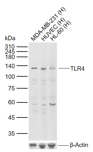
Lane 1: Human MDA-MB-231 cell lysates
Lane 2: Human HUVEC cell lysates
Lane 3: Human HL-60 cell lysates
Primary: Anti-TLR4 (SL1021R) at 1/1000 dilution
Secondary: IRDye800CW Goat Anti-Rabbit IgG at 1/20000 dilution
Predicted band size: 90 kDa
Observed band size: 120 kDa
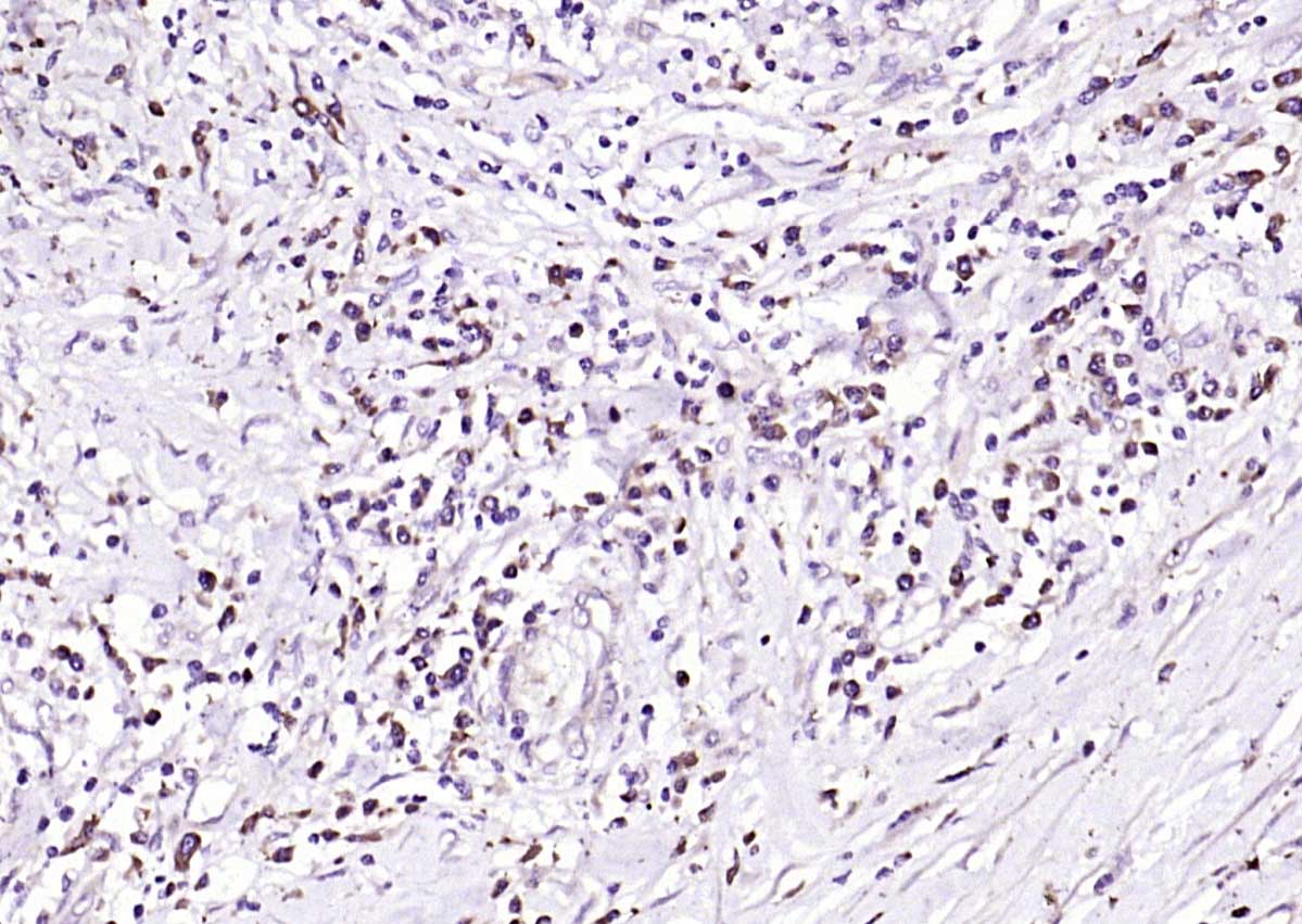
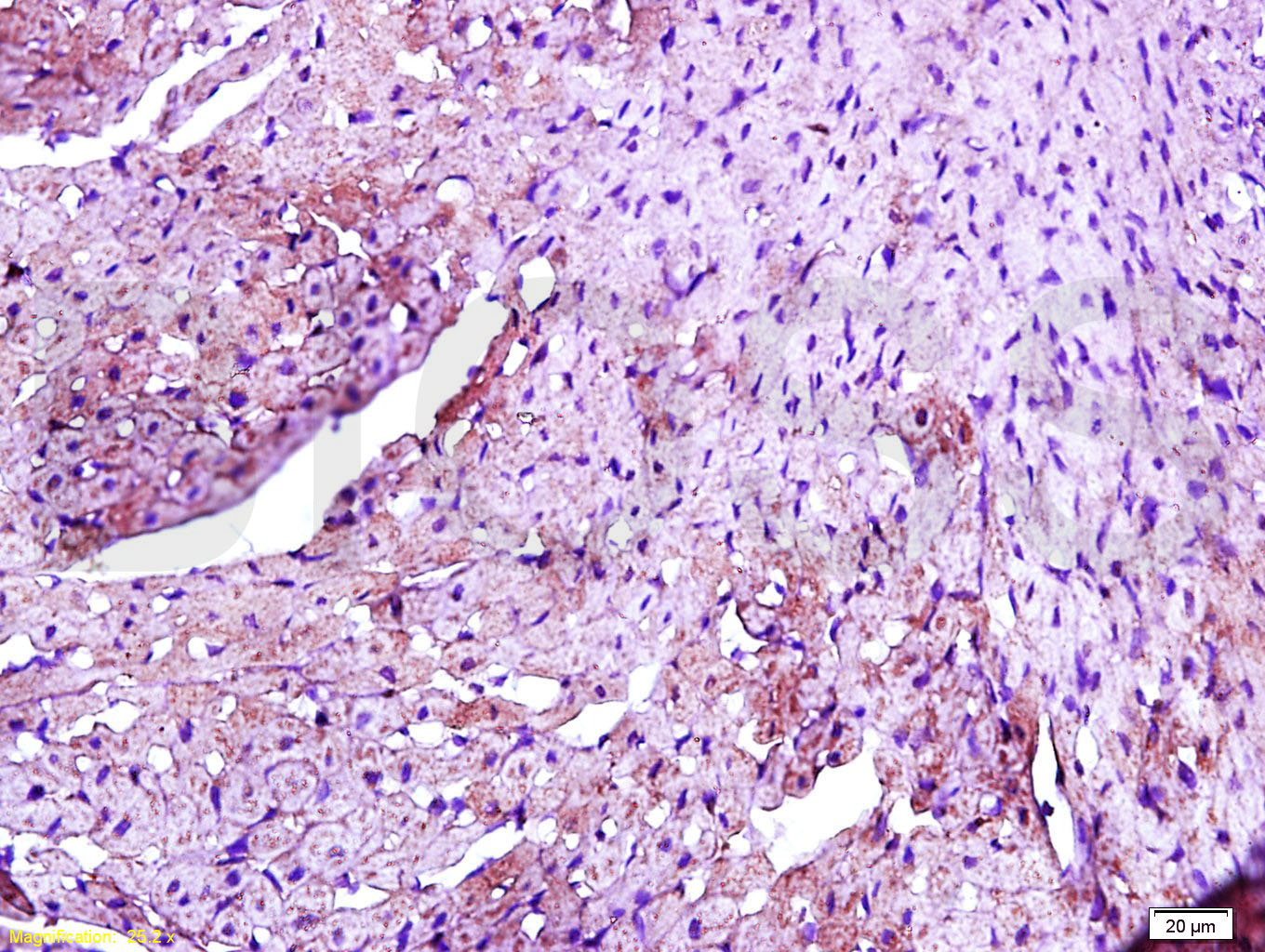
Paraformaldehyde-fixed, paraffin embedded (Human esophageal cancer); Antigen retrieval by boiling in sodium citrate buffer (pH6.0) for 15min; Block endogenous peroxidase by 3% hydrogen peroxide for 20 minutes; Blocking buffer (normal goat serum) at 37°C for 30min; Antibody incubation with (TLR4) Polyclonal Antibody, Unconjugated (SL1021R) at 1:200 overnight at 4°C, followed by operating according to SP Kit(Rabbit) (sp-0023) instructionsand DAB staining.Tissue/cell: rat heart tissue; 4% Paraformaldehyde-fixed and paraffin-embedded;
Antigen retrieval: citrate buffer ( 0.01M, pH 6.0 ), Boiling bathing for 15min; Block endogenous peroxidase by 3% Hydrogen peroxide for 30min; Blocking buffer (normal goat serum,C-0005) at 37℃ for 20 min;
Incubation: Anti-TLR4/CD284 Polyclonal Antibody, Unconjugated(SL1021R) 1:200, overnight at 4°C, followed by conjugation to the secondary antibody(SP-0023) and DAB(C-0010) staining
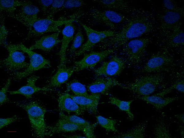
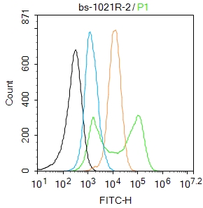
Image submitted by One World Lab validation program. HL60 and MCF-7 cells were stained with rabbit polyclonal antibody against TLR4 with two dilutions (1:100 and 1:250). 2nd antibody without primary antibody was used as control included here.Blank control:K562.
Primary Antibody (green line): Rabbit Anti-TLR4 antibody (SL1021R)
Dilution: 0.5ug/Test;
Secondary Antibody (white blue line) : Goat anti-rabbit IgG-AF488
Dilution: 0.5ug/Test.
Isotype control(orange line):Normal Rabbit IgG
Protocol
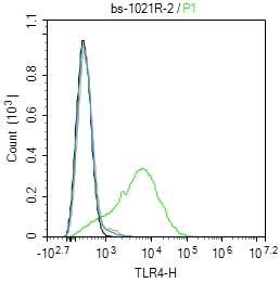
The cells were incubated in 5%BSA to block non-specific protein-protein interactions for 30 min at room temperature .Cells stained with Primary Antibody for 30 min at room temperature. The secondary antibody used for 40 min at room temperature. Acquisition of 20,000 events was performed.Blank control: Raw264.7.
Primary Antibody (green line): Rabbit Anti-TLR4 antibody (SL1021R)
Dilution: 2μg /10^6 cells;
Isotype Control Antibody (orange line): Rabbit IgG .
Secondary Antibody : Goat anti-rabbit IgG-AF488
Dilution: 1μg /test.
Protocol
The cells were fixed with 4% PFA (10min at room temperature)and then permeabilized with 0.1% PBST for 20 min at at room temperature. The cells were then incubated in 5%BSA to block non-specific protein-protein interactions for 30 min at room temperature .Cells stained with Primary Antibody for 30 min at room temperature.The secondary antibody used for 40 min at room temperature.Acquisition of 20,000 events was performed.
Partial purchase records(bought amounts latest0)
No one bought this product
User Comment(Total0User Comment Num)
- No comment


 +86 571 56623320


