Rabbit Anti-EphA2 antibody
ARCC2; Eck; Eph receptor A2 antibody EphA2; Ephrin receptor; Ephrin receptor EphA2; Ephrin type A receptor 2; Epithelial cell kinase; Epithelial cell receptor protein tyrosine kinase antibody Myk 2; Myk2; Sek 2; Sek2; Tyrosine protein kinase receptor ECK;
View History [Clear]
Details
Product Name EphA2 Chinese Name endothelial cells受体蛋白酪氨酸激酶抗体 Alias ARCC2; Eck; Eph receptor A2 antibody EphA2; Ephrin receptor; Ephrin receptor EphA2; Ephrin type A receptor 2; Epithelial cell kinase; Epithelial cell receptor protein tyrosine kinase antibody Myk 2; Myk2; Sek 2; Sek2; Tyrosine protein kinase receptor ECK; Tyrosine-protein kinase receptor MPK-5; Tyrosine-protein kinase receptor SEK-2; EPHA2_HUMAN. Research Area Tumour Cell biology Signal transduction Growth factors and hormones Kinases and Phosphatases Immunogen Species Rabbit Clonality Polyclonal React Species Human, Rat, (predicted: Mouse, Dog, Pig, Cow, Horse, Rabbit, Sheep, ) Applications WB=1:500-2000 ELISA=1:5000-10000 IHC-P=1:100-500 IHC-F=1:100-500 ICC=1:100 IF=1:100-500 (Paraffin sections need antigen repair)
not yet tested in other applications.
optimal dilutions/concentrations should be determined by the end user.Theoretical molecular weight 105kDa Cellular localization The cell membrane Form Liquid Concentration 1mg/ml immunogen KLH conjugated synthetic peptide derived from human EphA2: 401-500/976 <Extracellular> Lsotype IgG Purification affinity purified by Protein A Buffer Solution 0.01M TBS(pH7.4) with 1% BSA, 0.03% Proclin300 and 50% Glycerol. Storage Shipped at 4℃. Store at -20 °C for one year. Avoid repeated freeze/thaw cycles. Attention This product as supplied is intended for research use only, not for use in human, therapeutic or diagnostic applications. PubMed PubMed Product Detail This gene belongs to the ephrin receptor subfamily of the protein-tyrosine kinase family. EPH and EPH-related receptors have been implicated in mediating developmental events, particularly in the nervous system. Receptors in the EPH subfamily typically have a single kinase domain and an extracellular region containing a Cys-rich domain and 2 fibronectin type III repeats. The ephrin receptors are divided into 2 groups based on the similarity of their extracellular domain sequences and their affinities for binding ephrin-A and ephrin-B ligands. This gene encodes a protein that binds ephrin-A ligands. Mutations in this gene are the cause of certain genetically-related cataract disorders.[provided by RefSeq, May 2010].
Function:
Receptor tyrosine kinase which binds promiscuously membrane-bound ephrin-A family ligands residing on adjacent cells, leading to contact-dependent bidirectional signaling into neighboring cells. The signaling pathway downstream of the receptor is referred to as forward signaling while the signaling pathway downstream of the ephrin ligand is referred to as reverse signaling. Activated by the ligand ephrin-A1/EFNA1 regulates migration, integrin-mediated adhesion, proliferation and differentiation of cells. Regulates cell adhesion and differentiation through DSG1/desmoglein-1 and inhibition of the ERK1/ERK2 (MAPK3/MAPK1, respectively) signaling pathway. May also participate in UV radiation-induced apoptosis and have a ligand-independent stimulatory effect on chemotactic cell migration. During development, may function in distinctive aspects of pattern formation and subsequently in development of several fetal tissues. Involved for instance in angiogenesis, in early hindbrain development and epithelial proliferation and branching morphogenesis during mammary gland development. Engaged by the ligand ephrin-A5/EFNA5 may regulate lens fiber cells shape and interactions and be important for lens transparency development and maintenance. With ephrin-A2/EFNA2 may play a role in bone remodeling through regulation of osteoclastogenesis and osteoblastogenesis.
Subunit:
Homodimer. Interacts with SLA. Interacts (phosphorylated form) with VAV2, VAV3 and PI3-kinase p85 subunit (PIK3R1, PIK3R2 or PIK3R3); critical for the EFNA1-induced activation of RAC1 which stimulates cell migration. Interacts with ANKS1A. Interacts with INPPL1; regulates activated EPHA2 endocytosis and degradation. Interacts (inactivated form) with PTK2/FAK1 and interacts (EFNA1 ligand-activated form) with PTPN11; regulates integrin-mediated adhesion. Interacts with ARHGEF16, DOCK4 and ELMO2; mediates ligand-independent activation of RAC1 which stimulates cell migration. Interacts with CLDN4; phosphorylates CLDN4 and may regulate tight junctions. Interacts with ACP1.
Subcellular Location:
Cell membrane; Single-pass type I membrane protein. Cell projection, ruffle membrane; Single-pass type I membrane protein. Cell projection, lamellipodium membrane; Single-pass type I membrane protein. Cell junction, focal adhesion. Note=Present at regions of cell-cell contacts but also at the leading edge of migrating cells.
Tissue Specificity:
Expressed in brain and glioma tissue and glioma cell lines (at protein level). Expressed most highly in tissues that contain a high proportion of epithelial cells, e.g., skin, intestine, lung, and ovary.
Post-translational modifications:
Autophosphorylates. Phosphorylated on tyrosine upon binding and activation by EFNA1. Phosphorylated residues Tyr-588 and Tyr-594 are required for binding VAV2 and VAV3 while phosphorylated residues Tyr-735 and Tyr-930 are required for binding PI3-kinase p85 subunit (PIK3R1, PIK3R2 or PIK3R3). These phosphorylated residues are critical for recruitment of VAV2 and VAV3 and PI3-kinase p85 subunit which transduce downstream signaling to activate RAC1 GTPase and cell migration. Phosphorylated at Ser-897 by PKB; serum-induced phosphorylation which targets EPHA2 to the cell leading edge and stimulates cell migration. Phosphorylation by PKB is inhibited by EFNA1-activated EPHA2 which regulates PKB activity via a reciprocal regulatory loop. Dephosphorylated by ACP1.
Ubiquitinated by CHIP/STUB1. Ubiquitination is regulated by the HSP90 chaperone and regulates the receptor stability and activity through proteasomal degradation. ANKS1A prevents ubiquitination and degradation.
DISEASE:
Genetic variations in EPHA2 are the cause of susceptibility to cataract cortical age-related type 2 (ARCC2) [MIM:613020]. A developmental punctate opacity common in the cortex and present in most lenses. The cataract is white or cerulean, increases in number with age, but rarely affects vision.
Defects in EPHA2 are the cause of cataract posterior polar type 1 (CTPP1) [MIM:116600]. A subcapsular opacity, usually disk-shaped, located at the back of the lens. It can have a marked effect on visual acuity.
Note=Overexpressed in several cancer types and promotes malignancy.
Similarity:
Belongs to the protein kinase superfamily. Tyr protein kinase family. Ephrin receptor subfamily.
Contains 1 Eph LBD (Eph ligand-binding) domain.
Contains 2 fibronectin type-III domains.
Contains 1 protein kinase domain.
Contains 1 SAM (sterile alpha motif) domain.
SWISS:
P29317
Gene ID:
1969
Database links:Entrez Gene: 1969 Human
Entrez Gene: 13836 Mouse
Omim: 176946 Human
SwissProt: P29317 Human
SwissProt: Q03145 Mouse
Unigene: 171596 Human
Unigene: 2581 Mouse
Unigene: 23415 Rat
Product Picture
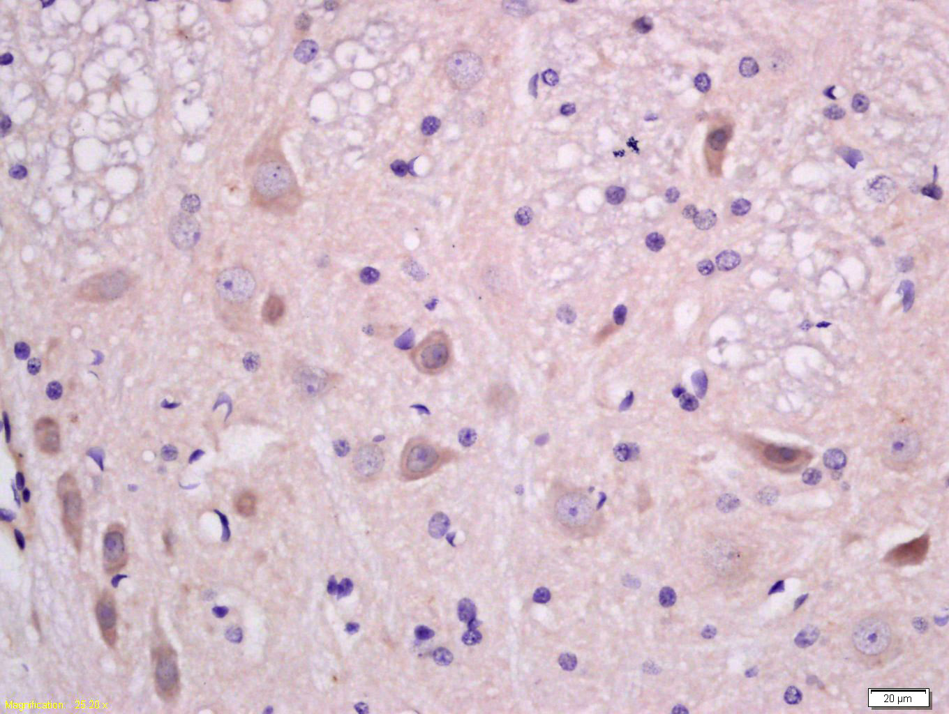
Antigen retrieval: citrate buffer ( 0.01M, pH 6.0 ), Boiling bathing for 15min; Block endogenous peroxidase by 3% Hydrogen peroxide for 30min; Blocking buffer (normal goat serum,C-0005) at 37℃ for 20 min;
Incubation: Anti-EphA2 Polyclonal Antibody, Unconjugated(SL10209R) 1:200, overnight at 4°C, followed by conjugation to the secondary antibody(SP-0023) and DAB(C-0010) staining
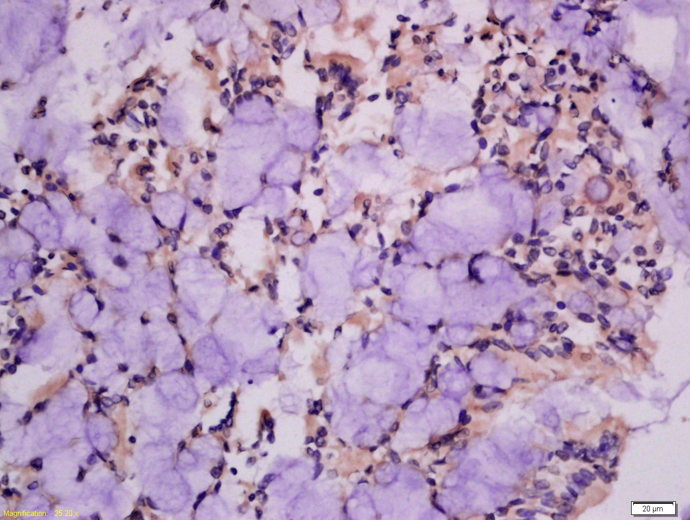
Tissue/cell: rat intestine tissue; 4% Paraformaldehyde-fixed and paraffin-embedded;
Antigen retrieval: citrate buffer ( 0.01M, pH 6.0 ), Boiling bathing for 15min; Block endogenous peroxidase by 3% Hydrogen peroxide for 30min; Blocking buffer (normal goat serum,C-0005) at 37℃ for 20 min;
Incubation: Anti-EphA2 Polyclonal Antibody, Unconjugated(SL10209R) 1:200, overnight at 4°C, followed by conjugation to the secondary antibody(SP-0023) and DAB(C-0010) staining
Partial purchase records(bought amounts latest0)
No one bought this product
User Comment(Total0User Comment Num)
- No comment


 +86 571 56623320


