
Rabbit Anti-c-fos antibody
Cellular oncogene fos; FBJ murine osteosarcoma viral v fos oncogene homolog antibody FBJ Osteosarcoma Virus; FOS; FOS protein; G0 G1 switch regulatory protein 7; G0S7; Oncogene FOS; Proto oncogene protein c fos; v fos FBJ murine osteosarcoma viral oncogen
View History [Clear]
Details
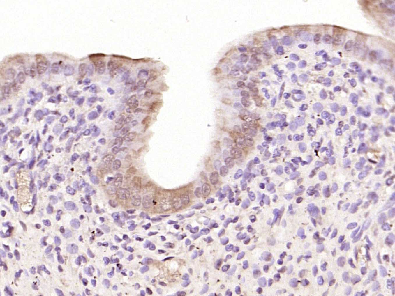
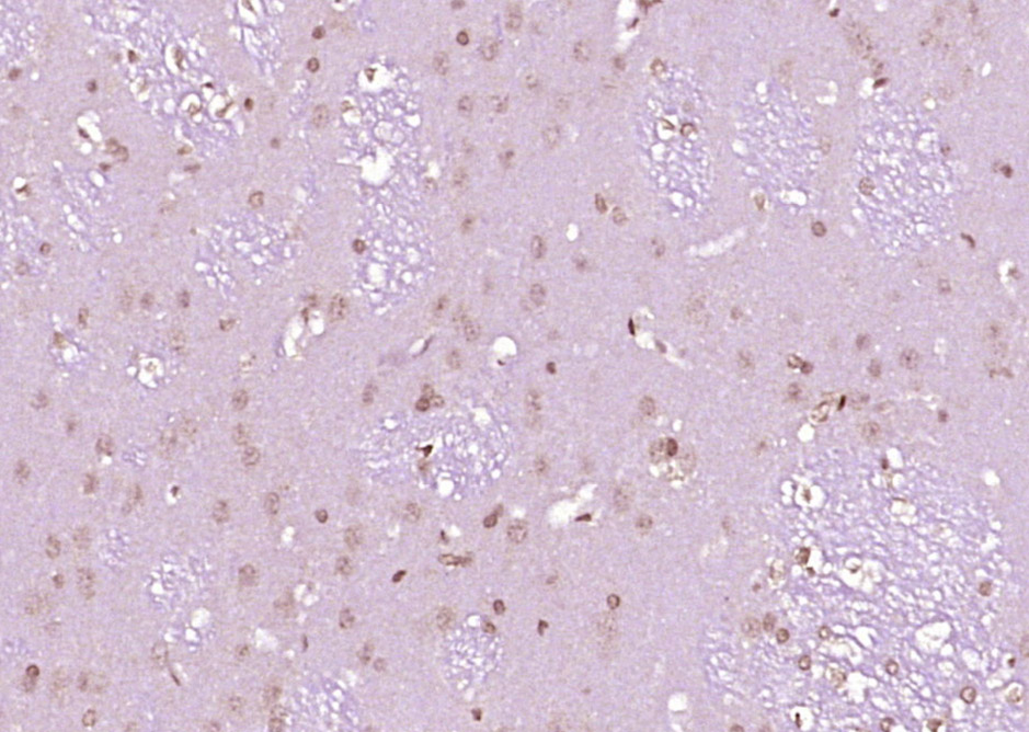
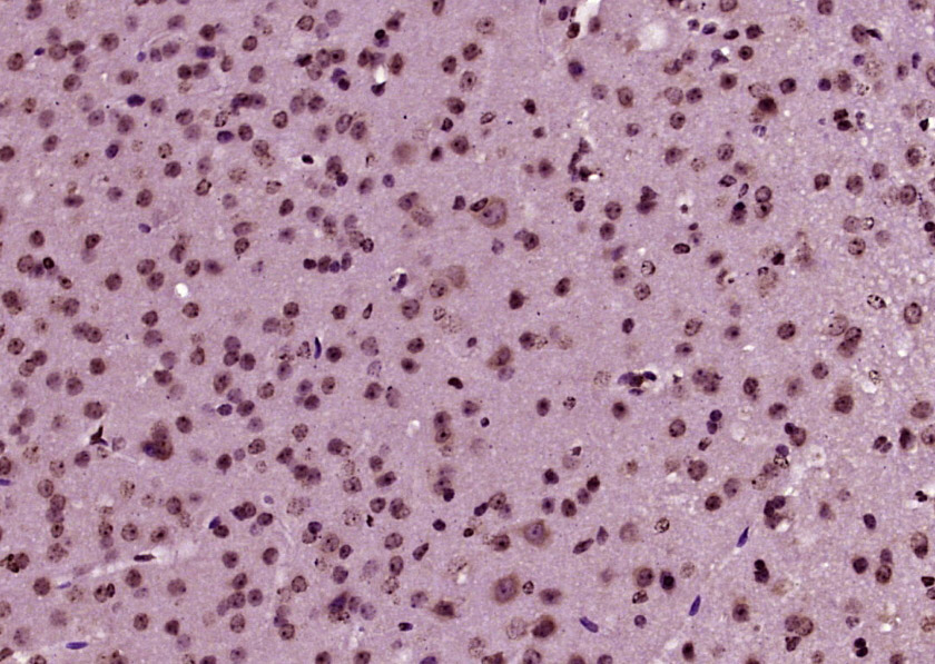
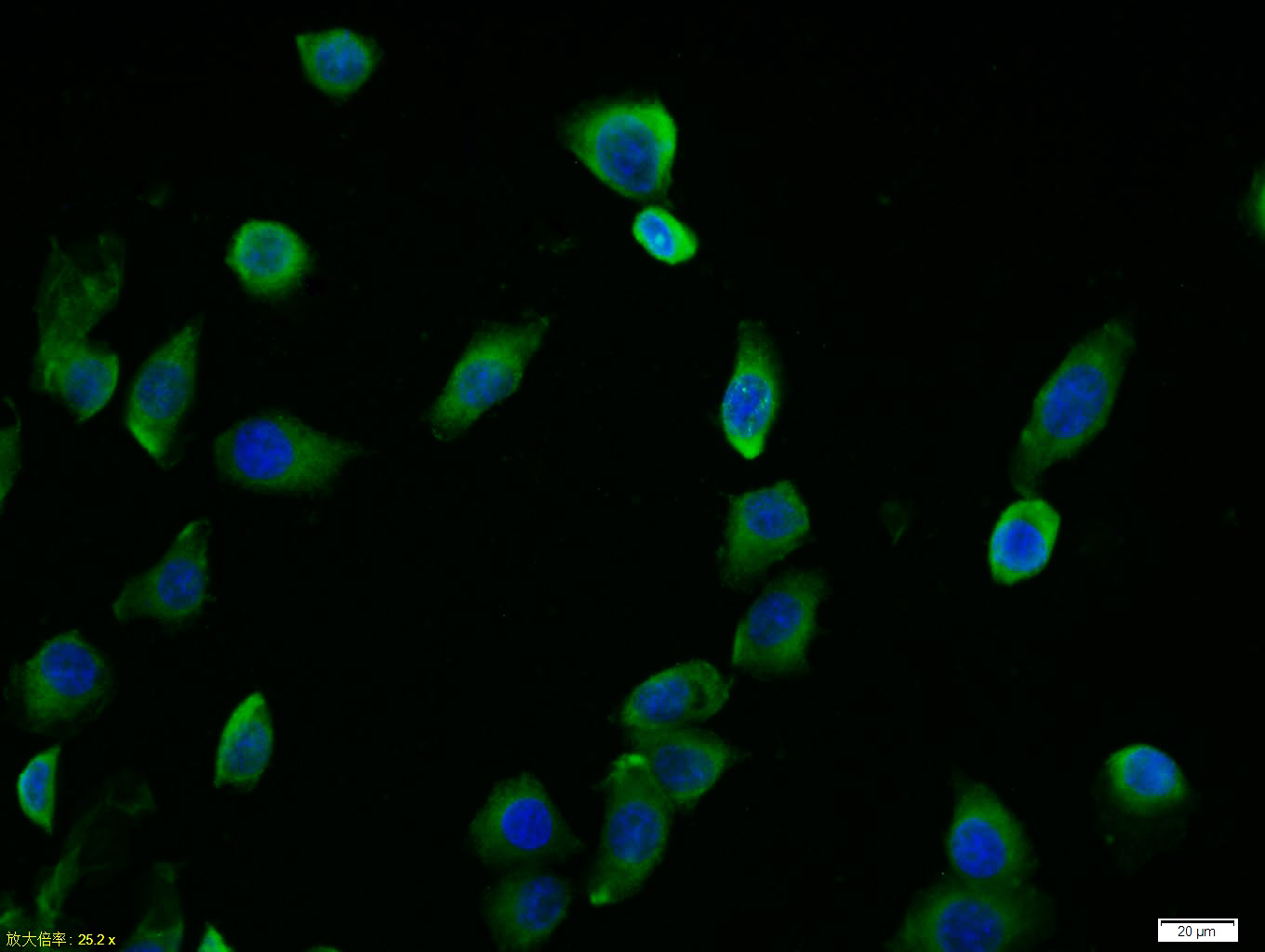
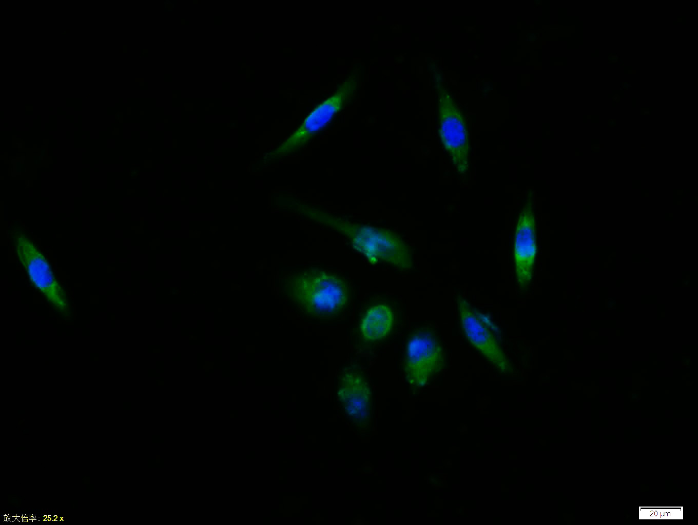
Product Name c-fos Chinese Name c-fos抗体 Alias Cellular oncogene fos; FBJ murine osteosarcoma viral v fos oncogene homolog antibody FBJ Osteosarcoma Virus; FOS; FOS protein; G0 G1 switch regulatory protein 7; G0S7; Oncogene FOS; Proto oncogene protein c fos; v fos FBJ murine osteosarcoma viral oncogene homolog; AP-1; p55; FOS_HUMAN; Proto-oncogene c-Fos; G0/G1 switch regulatory protein 7. literatures Research Area Tumour Cell biology immunology Neurobiology Signal transduction transcriptional regulatory factor TumourCell biologyMaker Immunogen Species Rabbit Clonality Polyclonal React Species Human, Mouse, Rat, (predicted: Dog, Pig, Cow, Horse, Rabbit, ) Applications ELISA=1:5000-10000 IHC-P=1:100-500 IHC-F=1:100-500 ICC=1:100 IF=1:100-500 (Paraffin sections need antigen repair)
not yet tested in other applications.
optimal dilutions/concentrations should be determined by the end user.Theoretical molecular weight 41kDa Cellular localization The nucleus Form Liquid Concentration 1mg/ml immunogen KLH conjugated synthetic peptide derived from human c-fos: 145-260/380 Lsotype IgG Purification affinity purified by Protein A Buffer Solution 0.01M TBS(pH7.4) with 1% BSA, 0.03% Proclin300 and 50% Glycerol. Storage Shipped at 4℃. Store at -20 °C for one year. Avoid repeated freeze/thaw cycles. Attention This product as supplied is intended for research use only, not for use in human, therapeutic or diagnostic applications. PubMed PubMed Product Detail The Fos gene family consists of 4 members: FOS, FOSB, FOSL1, and FOSL2. These genes encode leucine zipper proteins that can dimerize with proteins of the JUN family, thereby forming the transcription factor complex AP-1. As such, the FOS proteins have been implicated as regulators of cell proliferation, differentiation, and transformation. In some cases, expression of the FOS gene has also been associated with apoptotic cell death. [provided by RefSeq, Jul 2008].
Function:
Nuclear phosphoprotein which forms a tight butnon-covalently linked complex with the JUN/AP-1 transcriptionfactor. In the heterodimer, FOS and JUN/AP-1 basic regions eachseems to interact with symmetrical DNA half sites. On TGF-betaactivation, forms a multimeric SMAD3/SMAD4/JUN/FOS complex at theAP1/SMAD-binding site to regulate TGF-beta-mediated signaling. Hasa critical function in regulating the Has a critical function inregulating the development of cells destined to form and maintainthe skeleton. It is thought to have an important role in signaltransduction, cell proliferation and differentiation.
Subunit:
Heterodimer; with JUN (By similarity). Interacts withMAFB. Component of the SMAD3/SMAD4/JUN/FOS complexrequired for syngernistic TGF-beta-mediated transcription at theAP1 promoter site. Interacts with SMAD3; the interaction is weakeven on TGF-beta activation. Interacts with MAFB. Interacts withDSIPI; this interaction inhibits the binding of active AP1 to itstarget DNA.
Subcellular Location:
Nucleus.
Post-translational modifications:
Phosphorylated in the C-terminal upon stimulation by nervegrowth factor (NGF) and epidermal growth factor (EGF).Phosphorylated, in vitro, by MAPK and RSK1. Phosphorylation on bothSer-362 and Ser-374 by MAPK1/2 and RSK1/2 leads to proteinstabilization with phosphorylation on Ser-374 being the major sitefor protein stabilization on NGF stimulation. Phosphorylation onSer-362 and Ser-374 primes further phosphorylations on Thr-325 andThr-331 through promoting docking of MAPK to the DEF domain.Phosphorylation on Thr-232, induced by HA-RAS, activates thetranscriptional activity and antagonizes sumoylation.Phosphorylation on Ser-362 by RSK2 in osteoblasts contributes toosteoblast transformation (By similarity). [PTM] Constitutively sumoylated by SUMO1, SUMO2 and SUMO3.Desumoylated by SENP2. Sumoylation requires heterodimerization withJUN and is enhanced by mitogen stimulation. Sumoylation inhibitsthe AP-1 transcriptional activity and is, itself, inhibited byRas-activated phosphorylation on Thr-232.
Similarity:
Belongs to the bZIP family. Fos subfamily.
Contains 1 bZIP domain.
SWISS:
P01100
Gene ID:
2353
Database links:Entrez Gene: 2353 Human
Entrez Gene: 14281 Mouse
Omim: 164810 Human
SwissProt: P01100 Human
SwissProt: P01101 Mouse
Unigene: 246513 Mouse
Unigene: 103750 Rat
Product Picture
Partial purchase records(bought amounts latest0)
No one bought this product
User Comment(Total0User Comment Num)
- No comment


 +86 571 56623320


