
Rabbit Anti-DC-SIGN antibody
CLEC4L; Dendritic cell-specific ICAM-3-grabbing non-integrin 1; C type lectin domain family 4 member L; CD 209; CD209; CD209 antigen; CD209 antigen-like protein A; CD209 molecule; Cd209a; CDSIGN; CIRE; DC SIGN1; DCSIGN; Dendritic cell specific ICAM 3 grab
View History [Clear]
Details
Product Name DC-SIGN Chinese Name 细胞间粘附分子非整合素蛋白3抗体 Alias CLEC4L; Dendritic cell-specific ICAM-3-grabbing non-integrin 1; C type lectin domain family 4 member L; CD 209; CD209; CD209 antigen; CD209 antigen-like protein A; CD209 molecule; Cd209a; CDSIGN; CIRE; DC SIGN1; DCSIGN; Dendritic cell specific ICAM 3 grabbing nonintegrin 1; Dendritic cell specific ICAM3 grabbing nonintegrin 1; Dendritic cell-specific intracellular adhesion molecules (ICAM)-3 grabbing non-integrin; Dengue fever, protection against, included; Dentritic Cell Specific ICAM3 Grabbing Nonintegrin; HIV GP120 Binding Protein; MGC129965; MGC130443; SIGN-R1; SIGNR5; CD209_HUMAN. Immunogen Species Rabbit Clonality Polyclonal React Species Human, Applications WB=1:500-2000 ELISA=1:5000-10000 IHC-P=1:100-500 IHC-F=1:100-500 Flow-Cyt=1μg/Test ICC=1:100-500 IF=1:100-500 (Paraffin sections need antigen repair)
not yet tested in other applications.
optimal dilutions/concentrations should be determined by the end user.Theoretical molecular weight 45kDa Cellular localization The cell membrane Secretory protein Form Liquid Concentration 1mg/ml immunogen KLH conjugated synthetic peptide derived from human DC-SIGN/CD209: 51-150/1404 <Extracellular> Lsotype IgG Purification affinity purified by Protein A Buffer Solution 0.01M TBS(pH7.4) with 1% BSA, 0.03% Proclin300 and 50% Glycerol. Storage Shipped at 4℃. Store at -20 °C for one year. Avoid repeated freeze/thaw cycles. Attention This product as supplied is intended for research use only, not for use in human, therapeutic or diagnostic applications. PubMed PubMed Product Detail Dendritic cells (DCs) that control immune responses were recently found to capture and transport HIV from the mucosal area to remote lymph nodes, where DCs hand over HIV to CD4+ T lymphocytes. DCs also amplify the amount of virus and extend the duration of viral infectivity. Multiple strains of HIV1, HIV2 and SIV bind to DCs via DCSIGN. ICAM3 is the natural ligand for DC-SIGN. A DC-SIGN homologue (termed CD299, DC-SIGNR, L-SIGN and DCSIGN2) was identified recently. DC-SIGN forms a novel gene family with DC-SIGNR and many alternatively spliced isoforms of DC-SIGN and DC-SIGNR. The expression of DC-SIGN was found in mucosal tissues including placenta, small intestine, and rectum.
Function:
Pathogen-recognition receptor expressed on the surface of immature dendritic cells (DCs) and involved in initiation of primary immune response. Thought to mediate the endocytosis of pathogens which are subsequently degraded in lysosomal compartments. The receptor returns to the cell membrane surface and the pathogen-derived antigens are presented to resting T-cells via MHC class II proteins to initiate the adaptive immune response. Probably recognizes in a calcium-dependent manner high mannose N-linked oligosaccharides in a variety of pathogen antigens, including HIV-1 gp120, HIV-2 gp120, SIV gp120, ebolavirus glycoproteins, cytomegalovirus gB, HCV E2, dengue virus gE, Leishmania pifanoi LPG, Lewis-x antigen in Helicobacter pylori LPS, mannose in Klebsiella pneumonae LPS, di-mannose and tri-mannose in Mycobacterium tuberculosis ManLAM and Lewis-x antigen in Schistosoma mansoni SEA.
On DCs it is a high affinity receptor for ICAM2 and ICAM3 by binding to mannose-like carbohydrates. May act as a DC rolling receptor that mediates transendothelial migration of DC presursors from blood to tissues by binding endothelial ICAM2. Seems to regulate DC-induced T-cell proliferation by binding to ICAM3 on T-cells in the immunological synapse formed between DC and T-cells.
Subunit:
Homotetramer. Binds to many viral surface glycoproteins such as HIV-1 gp120, HIV-2 gp120, SIV gp120, ebolavirus envelope glycoproteins, cytomegalovirus gB, HCV E2 and dengue virus major envelope protein E.
Subcellular Location:
Isoform 1, 2, 3, 4, 5, : Cell membrane; Single-pass type II membrane protein (Probable). Isoform 6, 7, 8, 9, 10, 11, 12: Secreted (Probable).
Tissue Specificity:
Predominantly expressed in dendritic cells and in DC-residing tissues. Also found in placental macrophages, endothelial cells of placental vascular channels, peripheral blood mononuclear cells, and THP-1 monocytes.
Similarity:
Contains 1 C-type lectin domain.
SWISS:
Q9NNX6
Gene ID:
30835
Database links:Entrez Gene: 30835 Human
Omim: 604672 Human
SwissProt: Q9NNX6 Human
Product Picture 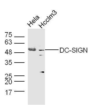 Sample:
Hela Cell Lysate at 40 ug
Hcclm3 Cell Lysate at 40 ug
Primary: Anti-DC-SIGN (SL10053R) at 1/300 dilution
Secondary: IRDye800CW Goat Anti-Rabbit IgG at 1/20000 dilution
Predicted band size: 45 kD
Observed band size: 50 kD
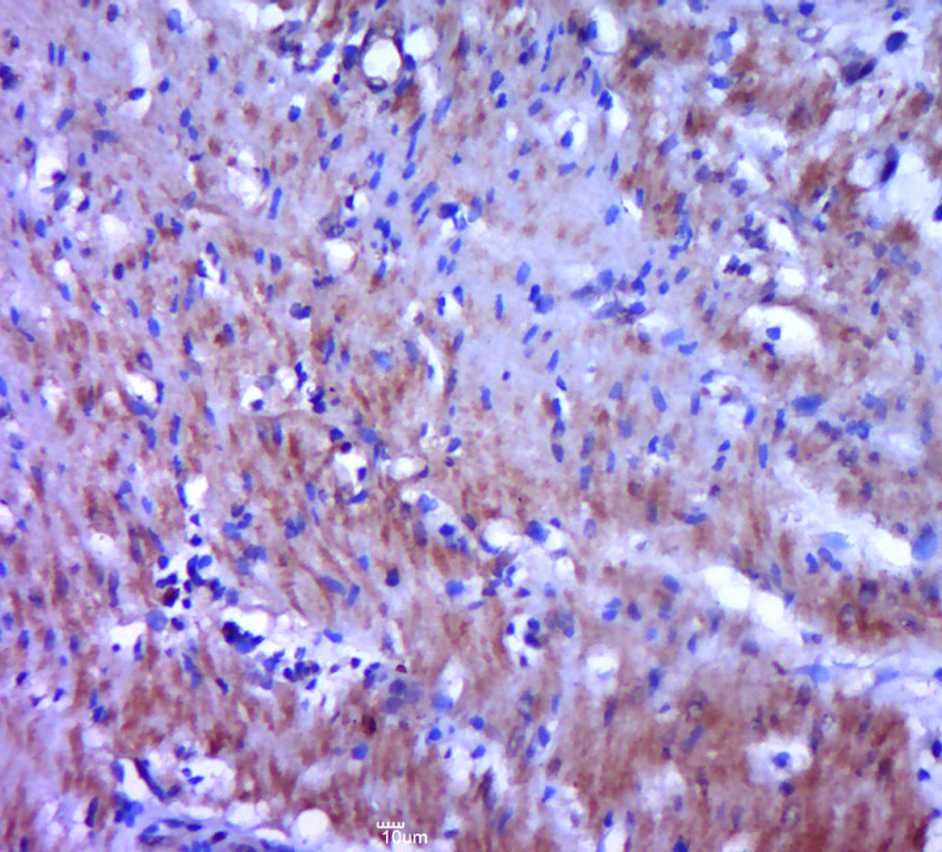 Paraformaldehyde-fixed, paraffin embedded (human cervical cancer); Antigen retrieval by boiling in sodium citrate buffer (pH6.0) for 15min; Block endogenous peroxidase by 3% hydrogen peroxide for 20 minutes; Blocking buffer (normal goat serum) at 37°C for 30min; Antibody incubation with (DC-SIGN) Polyclonal Antibody, Unconjugated (SL10053R) at 1:400 overnight at 4°C, followed by a conjugated secondary (sp-0023) for 20 minutes and DAB staining.
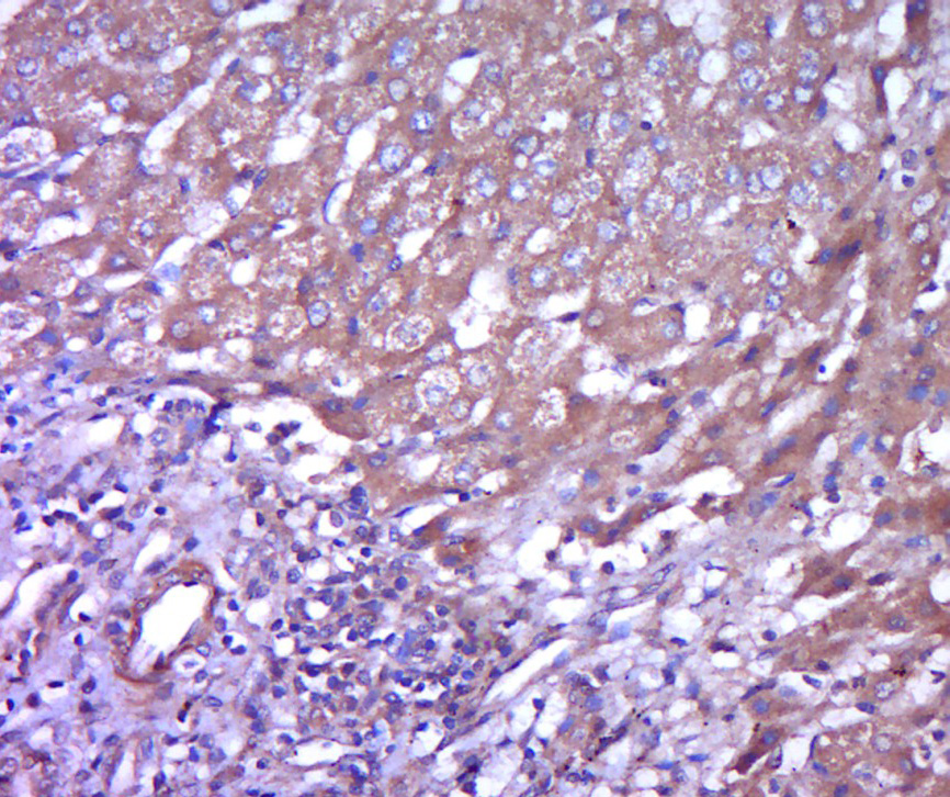 Paraformaldehyde-fixed, paraffin embedded (human liver carcinoma); Antigen retrieval by boiling in sodium citrate buffer (pH6.0) for 15min; Block endogenous peroxidase by 3% hydrogen peroxide for 20 minutes; Blocking buffer (normal goat serum) at 37°C for 30min; Antibody incubation with (DC-SIGN) Polyclonal Antibody, Unconjugated (SL10053R) at 1:400 overnight at 4°C, followed by a conjugated secondary (sp-0023) for 20 minutes and DAB staining.
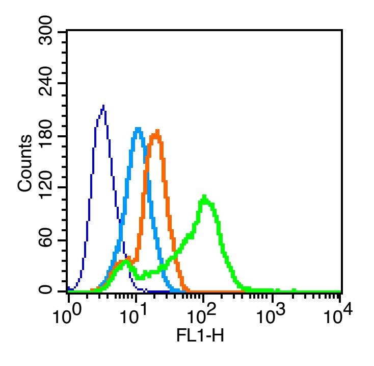 Blank control (blue line): MCF7 (blue).
Primary Antibody (green line): Rabbit Anti-DC-SIGN antibody(SL10053)
Dilution: 1μg /10^6 cells;
Isotype Control Antibody (orange line): Rabbit IgG .
Secondary Antibody (white blue line): F(ab’)2 fragment goat anti-rabbit IgG-FITC.
Dilution: 1μg /test.
Protocol
The cells were fixed with 2% paraformaldehyde for 10 min at room temperature.Cells stained with Primary Antibody for 30 min at room temperature. The cells were then incubated in 1 X PBS/2%BSA/10% goat serum to block non-specific protein-protein interactions followed by the antibody for 15 min at room temperature. The secondary antibody used for 40 min at room temperature. Acquisition of 20,000 events was performed.
Cartpieces
Totalgoods,subtotals:¥Checkout
Bought notes(bought amounts latest0)
No one bought this product
User Comment(Total0User Comment Num)
- No comment


 +86 571 56623320
 +86 18668110335

