
Rabbit Anti-Von Willebrand Factor antibody
Von Willebrand Factor; Coagulation factor VIII; F8VWF; Factor VIII related antigen; von Willebrand antigen 2; Von Willebrand antigen II; Von Willebrand disease; VWD; Coagulation factor VIII; Coagulation factor VIII VWF; F8VWF; VWF_HUMAN.
View History [Clear]
Details
Product Name Von Willebrand Factor Chinese Name 血管假性血友病因子/血管性血友病因子抗体 Alias Von Willebrand Factor; Coagulation factor VIII; F8VWF; Factor VIII related antigen; von Willebrand antigen 2; Von Willebrand antigen II; Von Willebrand disease; VWD; Coagulation factor VIII; Coagulation factor VIII VWF; F8VWF; VWF_HUMAN. literatures Research Area Tumour Cardiovascular Cell biology Developmental biology Growth factors and hormones Immunogen Species Rabbit Clonality Polyclonal React Species Human, Mouse, Rat, Applications IHC-P=1:100-500 IHC-F=1:100-500 IF=1:100-500 (Paraffin sections need antigen repair)
not yet tested in other applications.
optimal dilutions/concentrations should be determined by the end user.Theoretical molecular weight 226/309kDa Cellular localization Extracellular matrix Secretory protein Form Liquid Concentration 1mg/ml immunogen KLH conjugated synthetic peptide derived from human VWF/Von Willebrand Factor: 1651-1800/2813 Lsotype IgG Purification affinity purified by Protein A Buffer Solution 0.01M TBS(pH7.4) with 1% BSA, 0.03% Proclin300 and 50% Glycerol. Storage Shipped at 4℃. Store at -20 °C for one year. Avoid repeated freeze/thaw cycles. Attention This product as supplied is intended for research use only, not for use in human, therapeutic or diagnostic applications. PubMed PubMed Product Detail Von Willebrand Factor (VWF) was previously known as Factor VIII related antigen. VWF is synthesized exclusively by endothelial cells and megakaryocytes, and stored in the intracellular granules or constitutively secreted into plasma. This glycoprotein functions as both an antihemophilic factor carrier and a platelet vessel wall mediator in the blood coagulation system. Important in the maintenance of homeostasis, it participates in platelet vessel wall interactions by forming a noncovalent complex with coagulation factor VIII at the site of vascular injury. The Von Willebrand factor has functional binding domains to platelet glycoprotein Ib, glycoprotein IIb/IIIa, collagen and heparin. Mutations in this gene or deficiencies in this protein result in Von Willebrand's disease. VWD is characterized by frequent bleeding (gingival, minor skin quantitative lacerations, menorrhagia, etc.).
Function:
Important in the maintenance of hemostasis, it promotes adhesion of platelets to the sites of vascular injury by forming a molecular bridge between sub-endothelial collagen matrix and platelet-surface receptor complex GPIb-IX-V. Also acts as a chaperone for coagulation factor VIII, delivering it to the site of injury, stabilizing its heterodimeric structure and protecting it from premature clearance from plasma.
Subunit:
Multimeric. Interacts with F8.
Subcellular Location:
Secreted. Secreted, extracellular space, extracellular matrix. Note=Localized to storage granules.
Tissue Specificity:
Plasma.
Post-translational modifications:
All cysteine residues are involved in intrachain or interchain disulfide bonds.
N- and O-glycosylated.
DISEASE:
Defects in VWF are the cause of von Willebrand disease type 1 (VWD1) [MIM:193400]. A common hemorrhagic disorder due to defects in von Willebrand factor protein and resulting in impaired platelet aggregation. Von Willebrand disease type 1 is characterized by partial quantitative deficiency of circulating von Willebrand factor, that is otherwise structurally and functionally normal. Clinical manifestations are mucocutaneous bleeding, such as epistaxis and menorrhagia, and prolonged bleeding after surgery or trauma.
Defects in VWF are the cause of von Willebrand disease type 2 (VWD2) [MIM:613554]. A hemorrhagic disorder due to defects in von Willebrand factor protein and resulting in impaired platelet aggregation. Von Willebrand disease type 2 is characterized by qualitative deficiency and functional anomalies of von Willebrand factor. It is divided in different subtypes including 2A, 2B, 2M and 2N (Normandy variant). The mutant VWF protein in types 2A, 2B and 2M are defective in their platelet-dependent function, whereas the mutant protein in type 2N is defective in its ability to bind factor VIII. Clinical manifestations are mucocutaneous bleeding, such as epistaxis and menorrhagia, and prolonged bleeding after surgery or trauma.
Defects in VWF are the cause of von Willebrand disease type 3 (VWD3) [MIM:277480]. A severe hemorrhagic disorder due to a total or near total absence of von Willebrand factor in the plasma and cellular compartments, also leading to a profound deficiency of plasmatic factor VIII. Bleeding usually starts in infancy and can include epistaxis, recurrent mucocutaneous bleeding, excessive bleeding after minor trauma, and hemarthroses.
Similarity:
Contains 1 CTCK (C-terminal cystine knot-like) domain. Contains 4 TIL (trypsin inhibitory-like) domains.
Contains 3 VWFA domains.
Contains 3 VWFC domains.
Contains 4 VWFD domains.
SWISS:
P04275
Gene ID:
7450
Database links:Entrez Gene: 7450 Human
Omim: 193400 Human
SwissProt: P04275 Human
Unigene: 440848 Human
血管性血友病因子(vWF)是vascular endothelial cell和骨髓巨核细胞合成的一种glycoprotein,在1期和2期止血中都起着重要作用,如缺乏将导致患者出现血管性血友病(vWD)。vWF可被ADAMTS13裂解以失去活性,血小板反应蛋白/凝血酶敏感蛋白-1(Thrombospondin,TSP-1))可参与了这个调节过程。vWF水平受多种遗传和环境因素影响,其中ABO血型影响较大。vWF主要通过A1和A3区与血小板GP 1b和胶原结合,在止血和血栓形成过程中起重要作用,并与心、脑血管疾病及血管新生密切相关,因此研究vWF的生物学特性和功能具有重要的意Product Picture 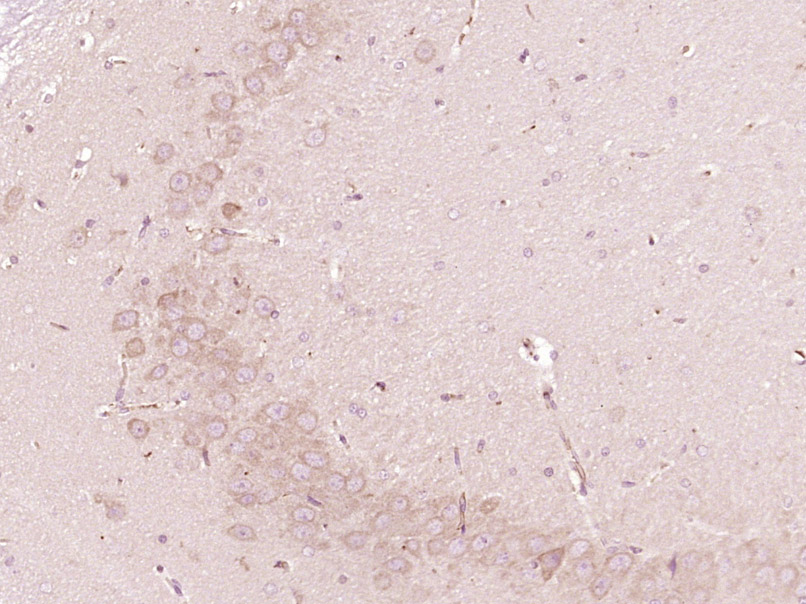 Paraformaldehyde-fixed, paraffin embedded (Rat brain); Antigen retrieval by boiling in sodium citrate buffer (pH6.0) for 15min; Block endogenous peroxidase by 3% hydrogen peroxide for 20 minutes; Blocking buffer (normal goat serum) at 37°C for 30min; Antibody incubation with (Von Willebrand Factor) Polyclonal Antibody, Unconjugated (SL10048R) at 1:400 overnight at 4°C, followed by operating according to SP Kit(Rabbit) (sp-0023) instructionsand DAB staining.
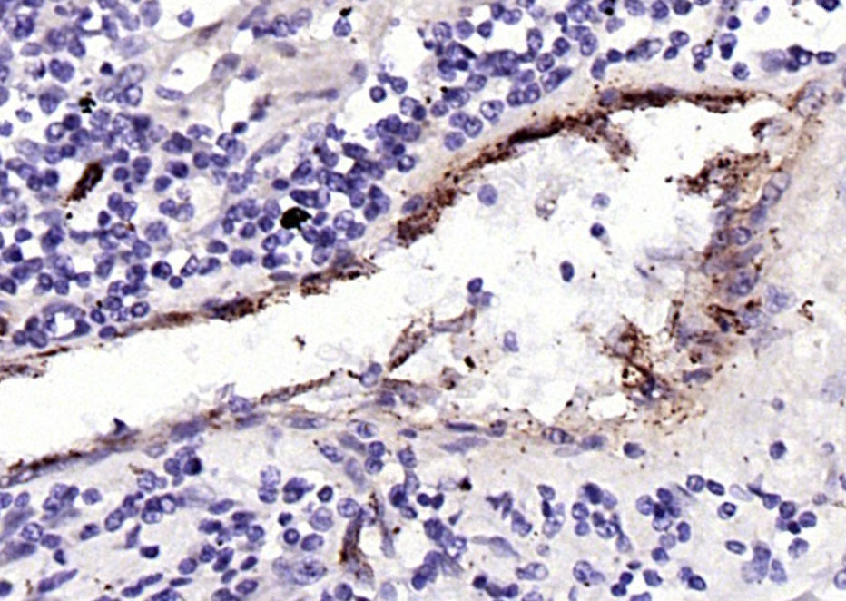 Paraformaldehyde-fixed, paraffin embedded (human lung carcinoma); Antigen retrieval by boiling in sodium citrate buffer (pH6.0) for 15min; Block endogenous peroxidase by 3% hydrogen peroxide for 20 minutes; Blocking buffer (normal goat serum) at 37°C for 30min; Antibody incubation with (Von Willebrand Factor) Polyclonal Antibody, Unconjugated (SL10048R) at 1:400 overnight at 4°C, followed by operating according to SP Kit(Rabbit) (sp-0023) instructionsand DAB staining.
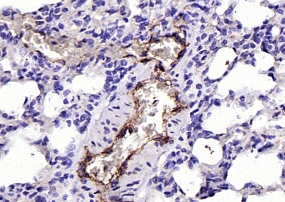 Paraformaldehyde-fixed, paraffin embedded (rat lung); Antigen retrieval by boiling in sodium citrate buffer (pH6.0) for 15min; Block endogenous peroxidase by 3% hydrogen peroxide for 20 minutes; Blocking buffer (normal goat serum) at 37°C for 30min; Antibody incubation with (Von Willebrand Factor) Polyclonal Antibody, Unconjugated (SL10048R) at 1:400 overnight at 4°C, followed by operating according to SP Kit(Rabbit) (sp-0023) instructionsand DAB staining.
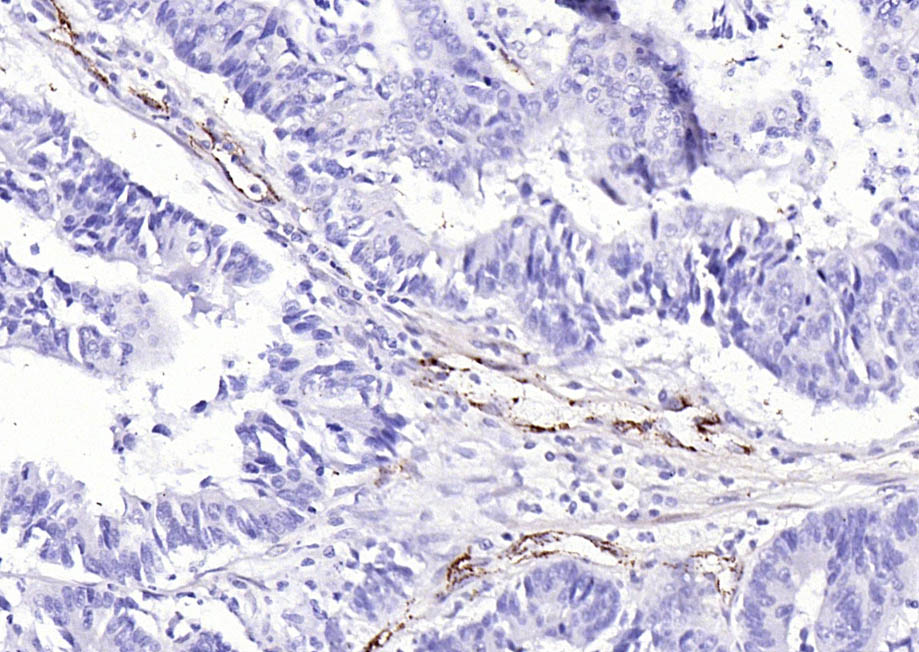 Paraformaldehyde-fixed, paraffin embedded (human colon carcinoma); Antigen retrieval by boiling in sodium citrate buffer (pH6.0) for 15min; Block endogenous peroxidase by 3% hydrogen peroxide for 20 minutes; Blocking buffer (normal goat serum) at 37°C for 30min; Antibody incubation with (Von Willebrand Factor) Polyclonal Antibody, Unconjugated (SL10048R) at 1:400 overnight at 4°C, followed by operating according to SP Kit(Rabbit) (sp-0023) instructionsand DAB staining.
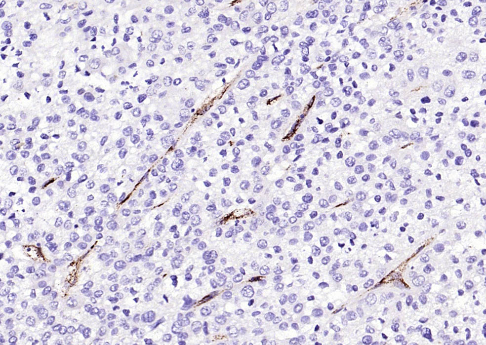 Paraformaldehyde-fixed, paraffin embedded (human liver carcinoma); Antigen retrieval by boiling in sodium citrate buffer (pH6.0) for 15min; Block endogenous peroxidase by 3% hydrogen peroxide for 20 minutes; Blocking buffer (normal goat serum) at 37°C for 30min; Antibody incubation with (Von Willebrand Factor) Polyclonal Antibody, Unconjugated (SL10048R) at 1:400 overnight at 4°C, followed by operating according to SP Kit(Rabbit) (sp-0023) instructionsand DAB staining.
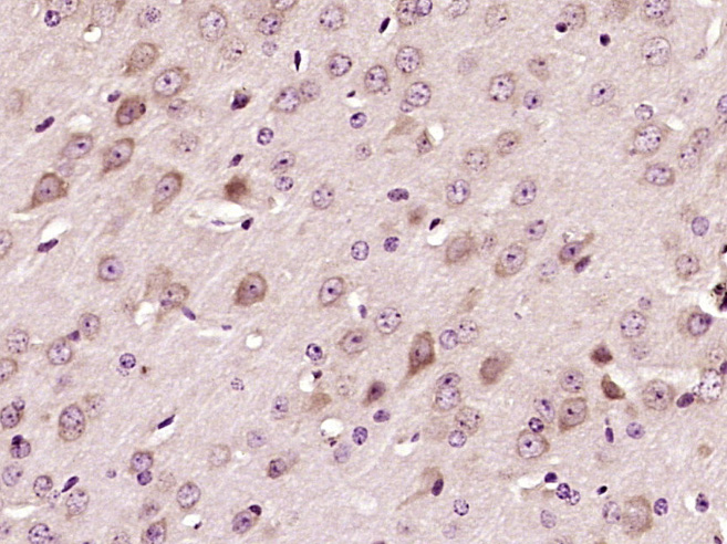 Paraformaldehyde-fixed, paraffin embedded (Mouse brain); Antigen retrieval by boiling in sodium citrate buffer (pH6.0) for 15min; Block endogenous peroxidase by 3% hydrogen peroxide for 20 minutes; Blocking buffer (normal goat serum) at 37°C for 30min; Antibody incubation with (Von Willebrand Factor) Polyclonal Antibody, Unconjugated (SL10048R) at 1:400 overnight at 4°C, followed by operating according to SP Kit(Rabbit) (sp-0023) instructionsand DAB staining.
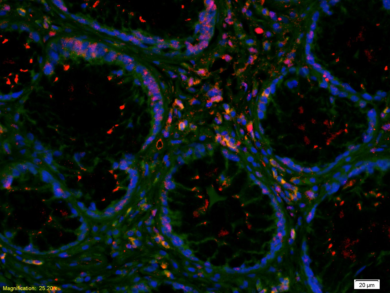 Tissue/cell: human colon carcinoma;4% Paraformaldehyde-fixed and paraffin-embedded;
Antigen retrieval: citrate buffer ( 0.01M, pH 6.0 ), Boiling bathing for 15min; Blocking buffer (normal goat serum,C-0005) at 37℃ for 20 min;
Incubation: Anti-VWF Polyclonal Antibody, Unconjugated(SL10048R) 1:200, overnight at 4°C; The secondary antibody was Goat Anti-Rabbit IgG, Cy3 conjugated(SL0295G-Cy3)used at 1:200 dilution for 40 minutes at 37°C. DAPI(5ug/ml,blue,C-0033) was used to stain the cell nuclei
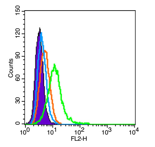 Blank control (Black line):HUVEC(Black).
Primary Antibody (green line): Rabbit Anti-VWF antibody (SL10048R)
Dilution: 3μg /10^6 cells;
Isotype Control Antibody (orange line): Rabbit IgG .
Secondary Antibody (white blue line): Goat anti-rabbit IgG-PE
Dilution: 1μg /test.
Protocol
The cells were fixed with 4% PFA (10min at room temperature)and then permeabilized with PBST for 20 min at room temperature. The cells were then incubated in 5%BSA to block non-specific protein-protein interactions for 30 min at room temperature .Cells stained with Primary Antibody for 30 min at room temperature. The secondary antibody used for 40 min at room temperature. Acquisition of 20,000 events was performed.
Cartpieces
Totalgoods,subtotals:¥Checkout
Bought notes(bought amounts latest0)
No one bought this product
User Comment(Total0User Comment Num)
- No comment


 +86 571 56623320
 +86 18668110335

