
Rabbit Anti-HDAC3 antibody
HDAC3_HUMAN; Histone deacetylase 3; EC:3.5.1.98; HD3; Protein deacetylase HDAC3; EC:3.5.1.; Protein deacylase HDAC3; RPD3-2; SMAP45; SMAP 45; SMAP-45; RPD3; KDAC3;
View History [Clear]
Details
Product Name HDAC3 Chinese Name 组蛋白去乙酰化酶3抗体 Alias HDAC3_HUMAN; Histone deacetylase 3; EC:3.5.1.98; HD3; Protein deacetylase HDAC3; EC:3.5.1.; Protein deacylase HDAC3; RPD3-2; SMAP45; SMAP 45; SMAP-45; RPD3; KDAC3; Research Area Tumour Cell biology Developmental biology Signal transduction Stem cells Epigenetics Immunogen Species Rabbit Clonality Polyclonal React Species Human, Mouse, Rat, (predicted: Chicken, Pig, Cow, Horse, ) Applications WB=1:500-2000 ELISA=1:5000-10000 IHC-P=1:100-500 IHC-F=1:100-500 ICC=1:100 IF=1:100-500 (Paraffin sections need antigen repair)
not yet tested in other applications.
optimal dilutions/concentrations should be determined by the end user.Theoretical molecular weight 47kDa Cellular localization The nucleus Form Liquid Concentration 1mg/ml immunogen KLH conjugated synthetic peptide derived from human HDAC3/HD3: 31-130/428 Lsotype IgG Purification affinity purified by Protein A Buffer Solution 0.01M TBS(pH7.4) with 1% BSA, 0.03% Proclin300 and 50% Glycerol. Storage Shipped at 4℃. Store at -20 °C for one year. Avoid repeated freeze/thaw cycles. Attention This product as supplied is intended for research use only, not for use in human, therapeutic or diagnostic applications. PubMed PubMed Product Detail Histones play a critical role in transcriptional regulation, cell cycle progression, and developmental events. Histone acetylation/deacetylation alters chromosome structure and affects transcription factor access to DNA. The protein encoded by this gene belongs to the histone deacetylase/acuc/apha family. It has histone deacetylase activity and represses transcription when tethered to a promoter. It may participate in the regulation of transcription through its binding with the zinc-finger transcription factor YY1. This protein can also down-regulate p53 function and thus modulate cell growth and apoptosis. This gene is regarded as a potential tumor suppressor gene. [provided by RefSeq, Jul 2008]
Function:
Responsible for the deacetylation of lysine residues on the N-terminal part of the core histones (H2A, H2B, H3 and H4), and some other non-histone substrates. Histone deacetylation gives a tag for epigenetic repression and plays an important role in transcriptional regulation, cell cycle progression and developmental events. Histone deacetylases act via the formation of large multiprotein complexes. Probably participates in the regulation of transcription through its binding to the zinc-finger transcription factor YY1; increases YY1 repression activity. Required to repress transcription of the POU1F1 transcription factor. Acts as a molecular chaperone for shuttling phosphorylated NR2C1 to PML bodies for sumoylation.
Subunit:
Interacts with HDAC7 and HDAC9. Forms a heterologous complex at least with YY1. Interacts with DAXX, HDAC10 and DACH1. Found in a complex with NCOR1 and NCOR2. Component of the N-Cor repressor complex, at least composed of NCOR1, NCOR2, HDAC3, TBL1X, TBL1R, CORO2A and GPS2. Interacts with BCOR, MJD2A/JHDM3A, NRIP1, PRDM6 and SRY. Interacts with BTBD14B. Interacts with GLIS2. Interacts (via the DNA-binding domain) with NR2C1; the interaction recruits phosphorylated NR2C1 to PML bodies for sumoylation. Component of the Notch corepressor complex. Interacts with CBFA2T3 and NKAP. Interacts with APEX1; the interaction is not dependent on the acetylated status of APEX1. Interacts with and deacetylates MAPK14. Interacts with ZMYND15.
Subcellular Location:
Nucleus.
Tissue Specificity:
Widely expressed.
Post-translational modifications:
Sumoylated in vitro.
Similarity:
Belongs to the histone deacetylase family. HD type 1 subfamily.
SWISS:
O15379
Gene ID:
8841
Database links:Entrez Gene: 8841 Human
Entrez Gene: 15183 Mouse
Omim: 605166 Human
SwissProt: O15379 Human
SwissProt: O88895 Mouse
组蛋白去乙酰化酶(HDACs)是一组在细胞染色质水平、通过诱导组蛋白去乙酰化来调控包括染色质重组、转录活化或抑制、细胞周期、Cell differentiation及Apoptosis等一系列生物学效应的酶,特别是与细胞活化后的基因转录表达调控有关。Product Picture 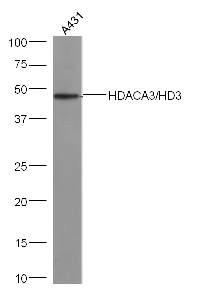 Sample: A431 (human)Cell Lysate at 40 ug
Primary: Anti-HDAC3/HD3(SL10024R) at 1/300 dilution
Secondary: IRDye800CW Goat Anti-Rabbit IgG at 1/20000 dilution
Predicted band size: 47 kD
Observed band size: 47 kD
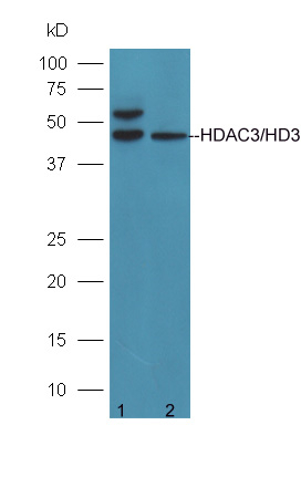 Protein:
293T(human)cell lysates;
A431(human)cell lysates;
Primary: rabbit Anti-HDAC3/HD3 (SL10024R) at 1:300;
Secondary: HRP conjugated Goat-Anti-rabbit IgG(SL0295G-HRP) at 1: 5000;
Predicted band size:47 kD
Observed band size:47 kD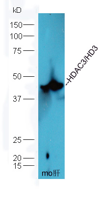 Protein: liver(mouse) lysates at 40ug;
Primary: rabbit Anti-HDAC3/HD3 (SL10024R) at 1:300;
Secondary: HRP conjugated Goat-Anti-rabbit IgG(SL0295G-HRP) at 1: 5000;
Predicted band size:47 kD
Observed band size: 47 kD Sample:
Lane 1: Mouse Cerebrum tissue lysates
Lane 2: Rat Cerebrum tissue lysates
Primary: Anti-HDAC3/HD3 (SL10024R) at 1/500 dilution
Secondary: IRDye800CW Goat Anti-Rabbit IgG at 1/20000 dilution
Predicted band size: 47 kDa
Observed band size: 47 kDa
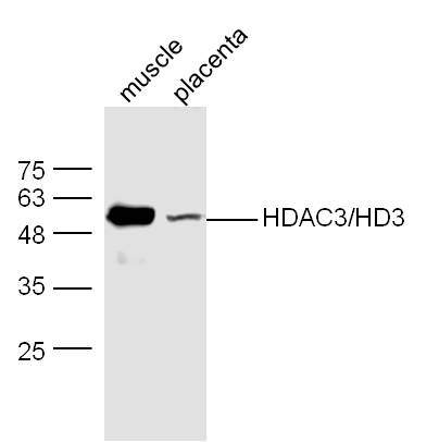 Sample:
muscle (Mouse) Lysate at 40 ug
placenta (Mouse) Lysate at 40 ug
Primary: Anti-HDAC3/HD3(SL10024R) at 1/300 dilution
Secondary: IRDye800CW Goat Anti-Rabbit IgG at 1/20000 dilution
Predicted band size: 47 kD
Observed band size: 52 kD
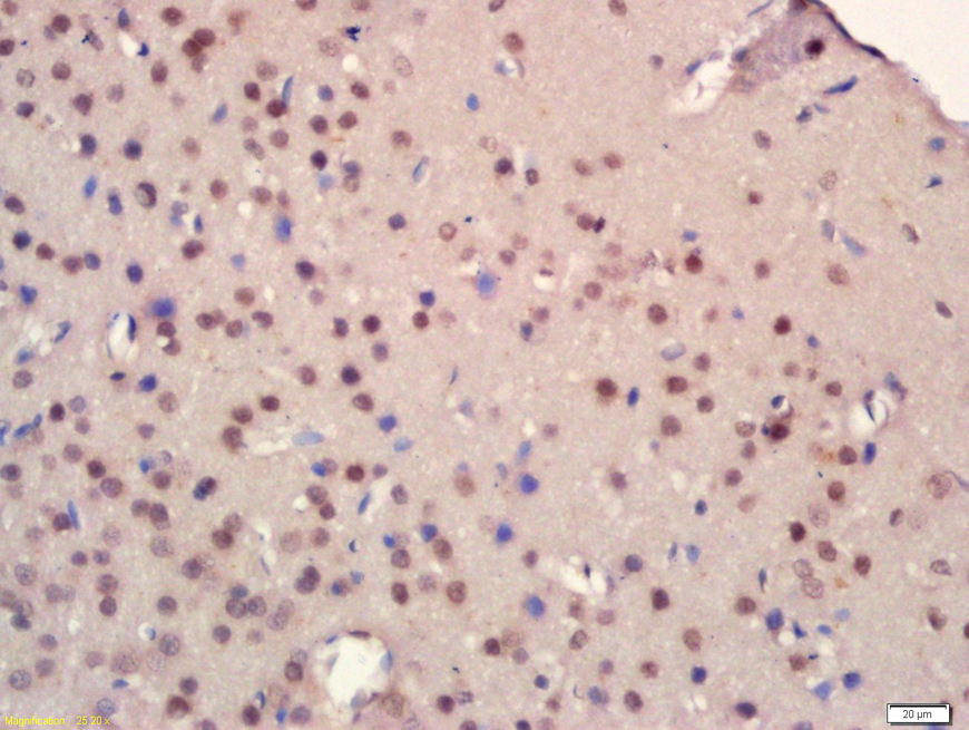 Tissue/cell: rat brain tissue; 4% Paraformaldehyde-fixed and paraffin-embedded;
Antigen retrieval: citrate buffer ( 0.01M, pH 6.0 ), Boiling bathing for 15min; Block endogenous peroxidase by 3% Hydrogen peroxide for 30min; Blocking buffer (normal goat serum,C-0005) at 37℃ for 20 min;
Incubation: Anti-HDAC3 Polyclonal Antibody, Unconjugated(SL10024R) 1:200, overnight at 4°C, followed by conjugation to the secondary antibody(SP-0023) and DAB(C-0010) staining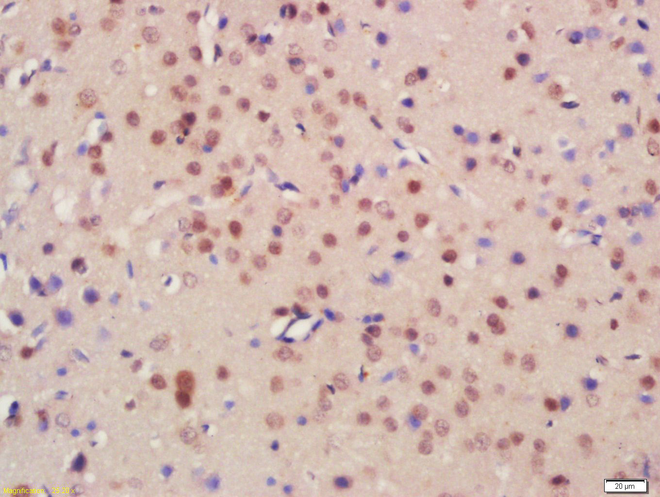
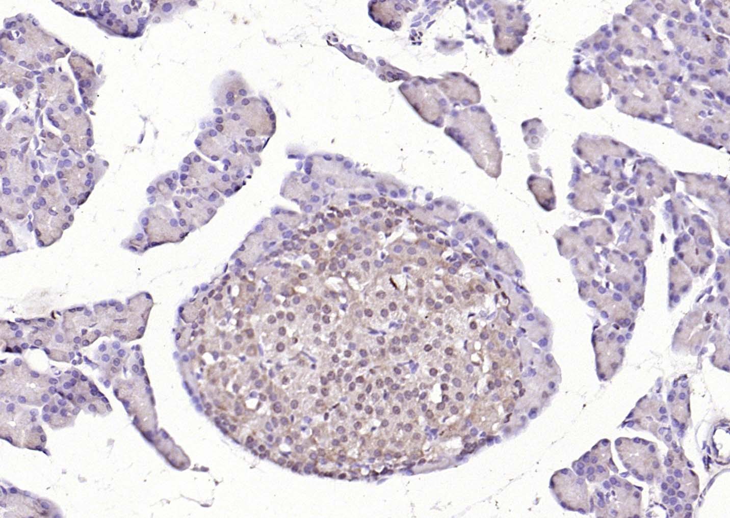 Paraformaldehyde-fixed, paraffin embedded (rat pancreas); Antigen retrieval by boiling in sodium citrate buffer (pH6.0) for 15min; Block endogenous peroxidase by 3% hydrogen peroxide for 20 minutes; Blocking buffer (normal goat serum) at 37°C for 30min; Antibody incubation with (HDAC3/HD3 ) Polyclonal Antibody, Unconjugated (SL10024R) at 1:200 overnight at 4°C, followed by operating according to SP Kit(Rabbit) (sp-0023) instructionsand DAB staining.
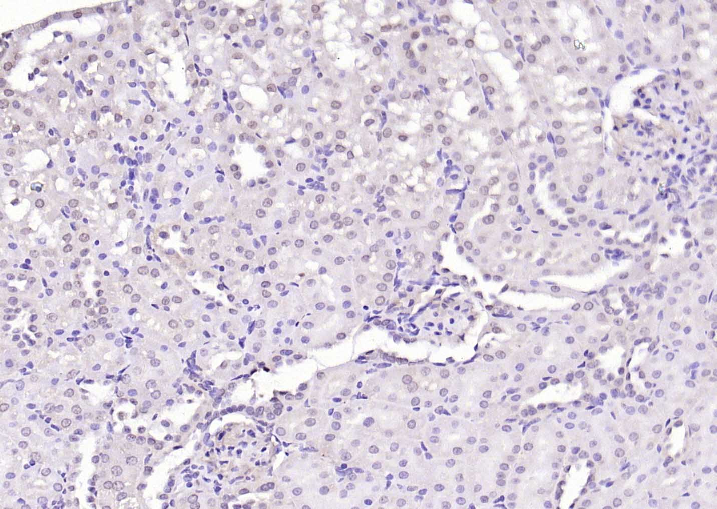 Paraformaldehyde-fixed, paraffin embedded (rat kidney); Antigen retrieval by boiling in sodium citrate buffer (pH6.0) for 15min; Block endogenous peroxidase by 3% hydrogen peroxide for 20 minutes; Blocking buffer (normal goat serum) at 37°C for 30min; Antibody incubation with (HDAC3/HD3 ) Polyclonal Antibody, Unconjugated (SL10024R) at 1:200 overnight at 4°C, followed by operating according to SP Kit(Rabbit) (sp-0023) instructionsand DAB staining.
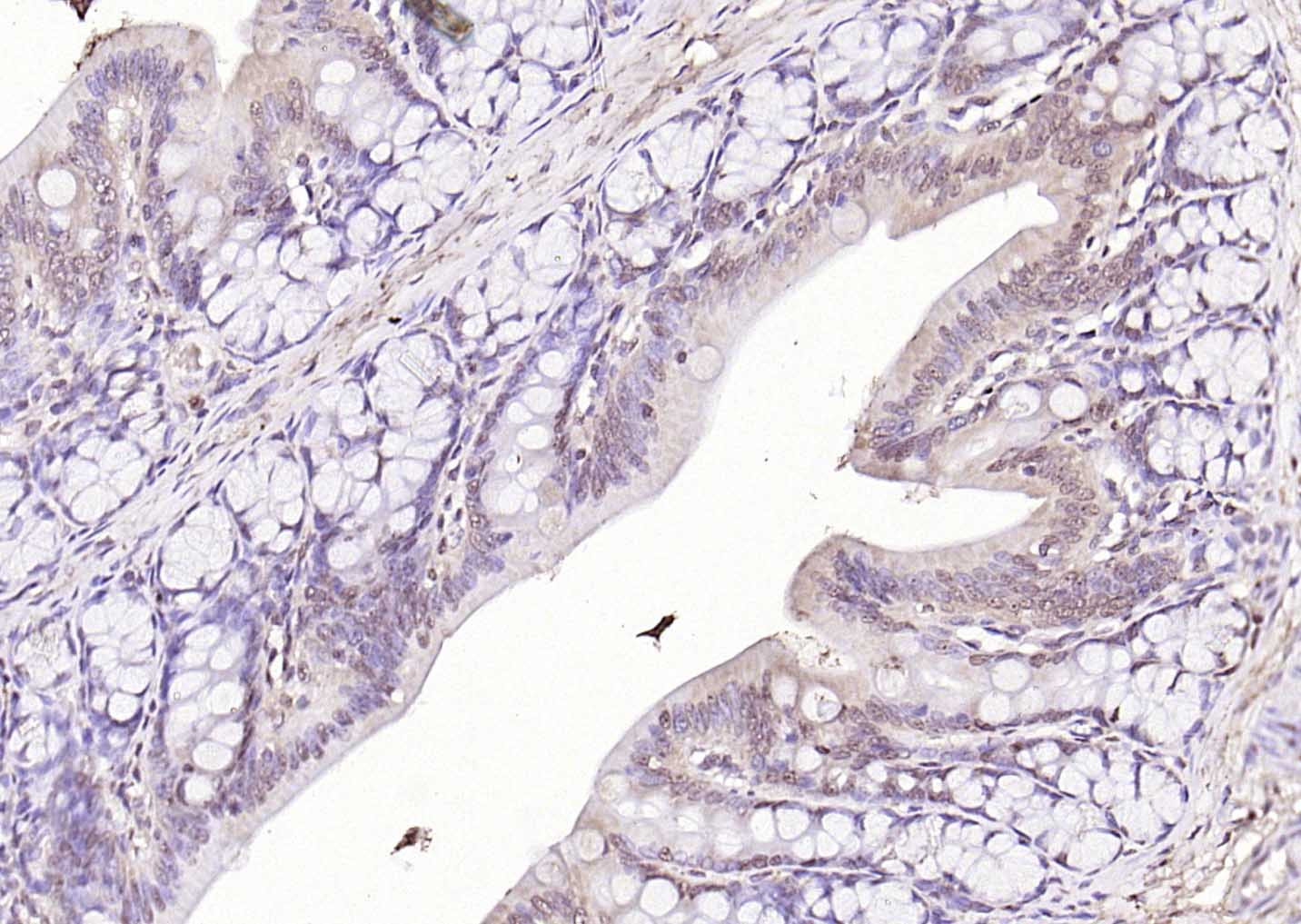 Paraformaldehyde-fixed, paraffin embedded (rat colon); Antigen retrieval by boiling in sodium citrate buffer (pH6.0) for 15min; Block endogenous peroxidase by 3% hydrogen peroxide for 20 minutes; Blocking buffer (normal goat serum) at 37°C for 30min; Antibody incubation with (HDAC3/HD3 ) Polyclonal Antibody, Unconjugated (SL10024R) at 1:200 overnight at 4°C, followed by operating according to SP Kit(Rabbit) (sp-0023) instructionsand DAB staining.
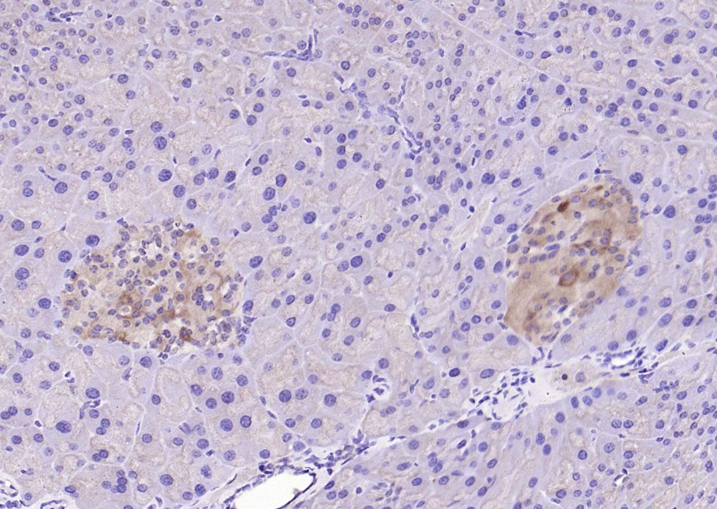 Paraformaldehyde-fixed, paraffin embedded (mouse pancreas); Antigen retrieval by boiling in sodium citrate buffer (pH6.0) for 15min; Block endogenous peroxidase by 3% hydrogen peroxide for 20 minutes; Blocking buffer (normal goat serum) at 37°C for 30min; Antibody incubation with (HDAC3/HD3 ) Polyclonal Antibody, Unconjugated (SL10024R) at 1:200 overnight at 4°C, followed by operating according to SP Kit(Rabbit) (sp-0023) instructionsand DAB staining.
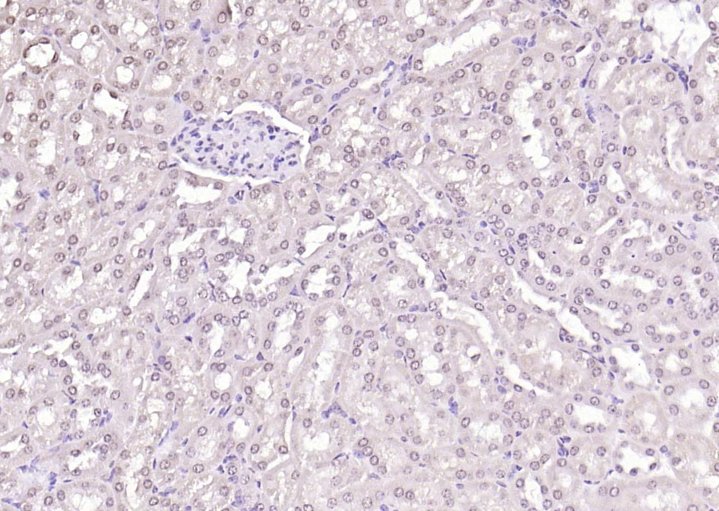 Paraformaldehyde-fixed, paraffin embedded (mouse kidney); Antigen retrieval by boiling in sodium citrate buffer (pH6.0) for 15min; Block endogenous peroxidase by 3% hydrogen peroxide for 20 minutes; Blocking buffer (normal goat serum) at 37°C for 30min; Antibody incubation with (HDAC3/HD3 ) Polyclonal Antibody, Unconjugated (SL10024R) at 1:200 overnight at 4°C, followed by operating according to SP Kit(Rabbit) (sp-0023) instructionsand DAB staining.
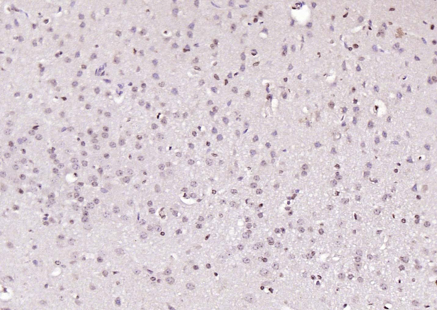 Paraformaldehyde-fixed, paraffin embedded (mouse brain); Antigen retrieval by boiling in sodium citrate buffer (pH6.0) for 15min; Block endogenous peroxidase by 3% hydrogen peroxide for 20 minutes; Blocking buffer (normal goat serum) at 37°C for 30min; Antibody incubation with (HDAC3/HD3 ) Polyclonal Antibody, Unconjugated (SL10024R) at 1:200 overnight at 4°C, followed by operating according to SP Kit(Rabbit) (sp-0023) instructionsand DAB staining.
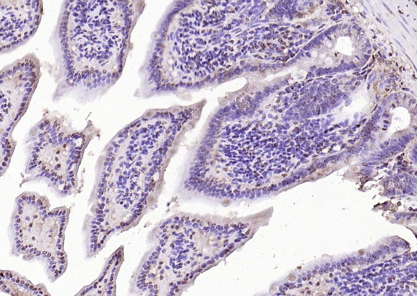 Paraformaldehyde-fixed, paraffin embedded (mouse colon); Antigen retrieval by boiling in sodium citrate buffer (pH6.0) for 15min; Block endogenous peroxidase by 3% hydrogen peroxide for 20 minutes; Blocking buffer (normal goat serum) at 37°C for 30min; Antibody incubation with (HDAC3/HD3 ) Polyclonal Antibody, Unconjugated (SL10024R) at 1:200 overnight at 4°C, followed by operating according to SP Kit(Rabbit) (sp-0023) instructionsand DAB staining.
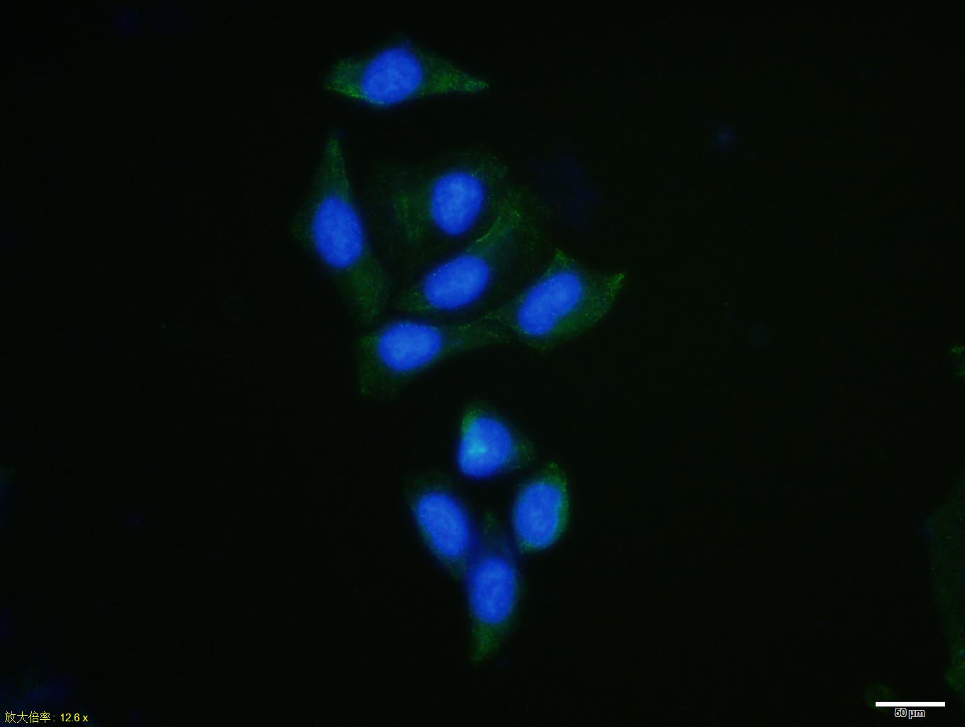 Hela cell; 4% Paraformaldehyde-fixed; Triton X-100 at room temperature for 20 min; Blocking buffer (normal goat serum, C-0005) at 37°C for 20 min; Antibody incubation with (HDAC3) polyclonal Antibody, Unconjugated (SL10024R) 1:100, 90 minutes at 37°C; followed by a conjugated Goat Anti-Rabbit IgG antibody at 37°C for 90 minutes, DAPI (blue, C02-04002) was used to stain the cell nuclei.
Cartpieces
Totalgoods,subtotals:¥Checkout
Bought notes(bought amounts latest0)
No one bought this product
User Comment(Total0User Comment Num)
- No comment


 +86 571 56623320
 +86 18668110335

