
Rabbit Anti-NALP3/CIAS1 antibody
LRR and PYD domains-containing protein 3; AGTAVPRL; AII/AVP antibody Angiotensin/vasopressin receptor AII/AVP like; Angiotensin/vasopressin receptor AII/AVP-like; C1orf7; Caterpiller protein 1.1; CIAS 1; CIAS1; CLR1.1; Cold autoinflammatory syndrome 1; Co
View History [Clear]
Details
Product Name NALP3/CIAS1 Chinese Name Apoptosis诱导蛋白NALP3抗体 Alias LRR and PYD domains-containing protein 3; AGTAVPRL; AII/AVP antibody Angiotensin/vasopressin receptor AII/AVP like; Angiotensin/vasopressin receptor AII/AVP-like; C1orf7; Caterpiller protein 1.1; CIAS 1; CIAS1; CLR1.1; Cold autoinflammatory syndrome 1; Cold autoinflammatory syndrome 1 protein; Cryopyrin; Familial cold autoinflammatory syndrome; FCAS; FCU; Muckle-Wells syndrome; MWS; NACHT; NACHT LRR and PYD containing protein 3; NALP 3; NALP3; NALP3_HUMAN; NLRP3; PYPAF 1; PYPAF1 antibody PYRIN containing APAF1 like protein 1; PYRIN-containing APAF1-like protein 1. literatures Research Area Tumour Cardiovascular Cell biology immunology Signal transduction Apoptosis transcriptional regulatory factor Immunogen Species Rabbit Clonality Polyclonal React Species Human, Mouse, Rat, Dog, Applications WB=1:500-2000 ELISA=1:5000-10000 IHC-P=1:100-500 IHC-F=1:100-500 Flow-Cyt=2ug/Test IF=1:200-800 (Paraffin sections need antigen repair)
not yet tested in other applications.
optimal dilutions/concentrations should be determined by the end user.Theoretical molecular weight 114kDa Cellular localization The nucleus cytoplasmic Secretory protein Form Liquid Concentration 1mg/ml immunogen KLH conjugated synthetic peptide derived from human Cryopyrin: 15-120/1036 Lsotype IgG Purification affinity purified by Protein A Buffer Solution 0.01M TBS(pH7.4) with 1% BSA, 0.03% Proclin300 and 50% Glycerol. Storage Shipped at 4℃. Store at -20 °C for one year. Avoid repeated freeze/thaw cycles. Attention This product as supplied is intended for research use only, not for use in human, therapeutic or diagnostic applications. PubMed PubMed Product Detail May function as an inducer of apoptosis. Interacts selectively with ASC and this complex may function as an upstream activator of NF-kappa-B signaling. Inhibits TNF-alpha induced activation and nuclear translocation of RELA/NF-KB p65. Also inhibits transcriptional activity of RELA. Activates caspase-1 in response to a number of triggers including bacterial or viral infection which leads to processing and release of IL1B and IL18.
Function:
May function as an inducer of apoptosis. Interacts selectively with ASC and this complex may function as an upstream activator of NF-kappa-B signaling. Inhibits TNF-alpha induced activation and nuclear translocation of RELA/NF-KB p65. Also inhibits transcriptional activity of RELA. Activates caspase-1 in response to a number of triggers including bacterial or viral infection which leads to processing and release of IL1B and IL18.
Subcellular Location:
Cytoplasm.
Tissue Specificity:
Expressed in blood leukocytes. Strongly expressed in polymorphonuclear cells and osteoblasts. Undetectable or expressed at a lower magnitude in B- and T-lymphoblasts, respectively. High level of expression detected in chondrocytes. Detected in non-keratinizing epithelia of oropharynx, esophagus and ectocervix and in the urothelial layer of the bladder.
DISEASE:
Defects in NLRP3 are the cause of familial cold autoinflammatory syndrome type 1 (FCAS1) [MIM:120100]; also known as familial cold urticaria. FCAS are rare autosomal dominant systemic inflammatory diseases characterized by episodes of rash, arthralgia, fever and conjunctivitis after generalized exposure to cold.
Defects in NLRP3 are a cause of Muckle-Wells syndrome (MWS) [MIM:191900]; also known as urticaria-deafness-amyloidosis syndrome. MWS is a hereditary periodic fever syndrome characterized by fever, chronic recurrent urticaria, arthralgias, progressive sensorineural deafness, and reactive renal amyloidosis. The disease may be severe if generalized amyloidosis occurs.
Defects in NLRP3 are the cause of chronic infantile neurologic cutaneous and articular syndrome (CINCA) [MIM:607115]; also known as neonatal onset multisystem inflammatory disease (NOMID). CINCA is a rare congenital inflammatory disorder characterized by a triad of neonatal onset of cutaneous symptoms, chronic meningitis and joint manifestations with recurrent fever and inflammation.
Similarity:
Belongs to the NLRP family.
Contains 1 DAPIN domain.
Contains 9 LRR (leucine-rich) repeats.
Contains 1 NACHT domain.
SWISS:
Q96P20
Gene ID:
114548
Database links:
Entrez Gene: 114548 Human
Entrez Gene: 216799 Mouse
Omim: 606416 Human
SwissProt: Q96P20 Human
SwissProt: Q8R4B8 Mouse
Unigene: 159483 Human
Unigene: 54174 Mouse
Product Picture 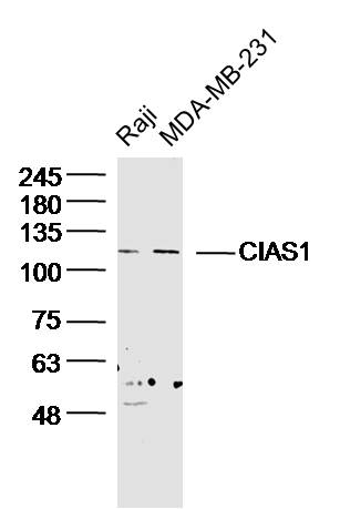 Sample:
Raji Cell (Human) Lysate at 30 ug
MDA-MB-231 Cell (Human) Lysate at 30 ug
Primary: Anti- CIAS1 (SL10021R) at 1/300 dilution
Secondary: IRDye800CW Goat Anti-Rabbit IgG at 1/20000 dilution
Predicted band size: 114kD
Observed band size: 114kD
 Sample:
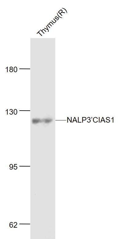
Thymus (Rat) Lysate at 40 ug
Primary: Anti-NALP3’CIAS1 (SL10021R) at 1/1000 dilution
Secondary: IRDye800CW Goat Anti-Rabbit IgG at 1/20000 dilution
Predicted band size: 118 kD
Observed band size: 120 kD
 Sample:
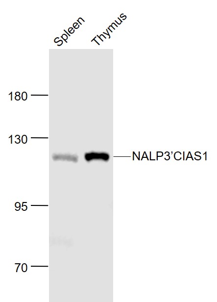
Spleen (Mouse) Lysate at 40 ug
Thymus (Mouse) Lysate at 40 ug
Primary: Anti- NALP3’CIAS1 (SL10021R) at 1/1000 dilution
Secondary: IRDye800CW Goat Anti-Rabbit IgG at 1/20000 dilution
Predicted band size: 118 kD
Observed band size: 118 kD
 Sample:
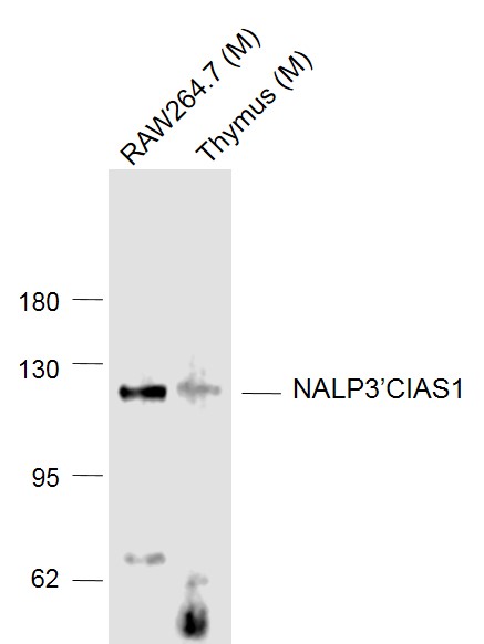
Lane 1: RAW264.7 (Mouse) Cell Lysate at 30 ug
Lane 2: Thymus (Mouse) Lysate at 40 ug
Primary: Anti- NALP3’CIAS1 (SL10021R) at 1/1000 dilution
Secondary: IRDye800CW Goat Anti-Rabbit IgG at 1/20000 dilution
Predicted band size: 118 kD
Observed band size: 118 kD
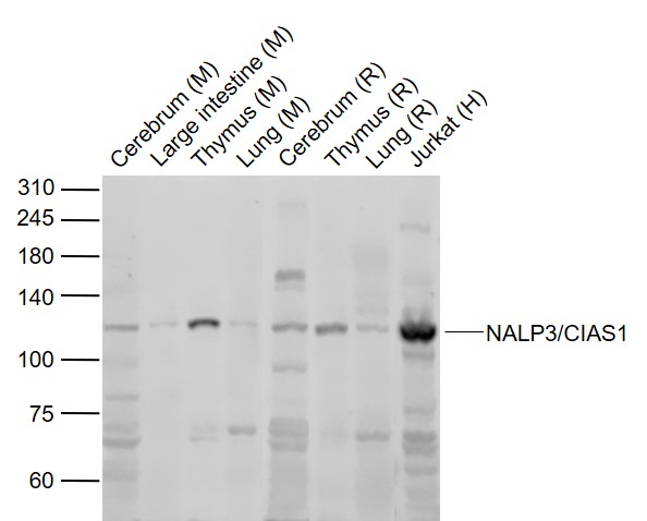 Sample:
Lane 1: Cerebrum (Mouse) Lysate at 40 ug
Lane 2: Large intestine (Mouse) Lysate at 40 ug
Lane 3: Thymus (Mouse) Lysate at 40 ug
Lane 4: Lung (Mouse) Lysate at 40 ug
Lane 5: Cerebrum (Rat) Lysate at 40 ug
Lane 6: Thymus (Rat) Lysate at 40 ug
Lane 7: Lung (Rat) Lysate at 40 ug
Lane 8: Jurkat (Human) cell Lysate at 30 ug
Primary: Anti-NALP3/CIAS1 (SL10021R) at 1/1000 dilution
Secondary: IRDye800CW Goat Anti-Rabbit IgG at 1/20000 dilution
Predicted band size: 118 kD
Observed band size: 118 kD
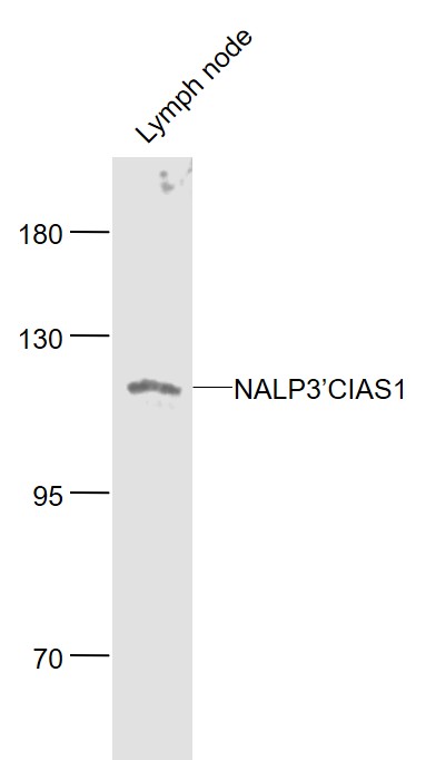 Sample:
Lymph node (Mouse) Lysate at 40 ug
Primary: Anti- NALP3’CIAS1 (SL10021R) at 1/1000 dilution
Secondary: IRDye800CW Goat Anti-Rabbit IgG at 1/20000 dilution
Predicted band size: 114 kD
Observed band size: 114 kD
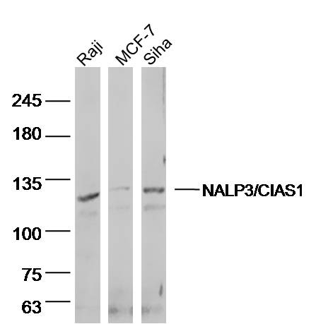 Sample:
Raji (human)cell Lysate at 40 ug
MCF-7 (human)cell Lysate at 40 ug
siha (human)cell Lysate at 40 ug
Primary: Anti- NALP3'CIAS1 (SL10021R)at 1/300 dilution
Secondary: IRDye800CW Goat Anti-Rabbit IgG at 1/20000 dilution
Predicted band size: 114kD
Observed band size: 114 kD
 Tissue/cell: mouse embryo tissue; 4% Paraformaldehyde-fixed and paraffin-embedded;
Antigen retrieval: citrate buffer ( 0.01M, pH 6.0 ), Boiling bathing for 15min; Block endogenous peroxidase by 3% Hydrogen peroxide for 30min; Blocking buffer (normal goat serum,C-0005) at 37℃ for 20 min;
Incubation: Anti-Cryopyrin/CIAS1/NALP3 Polyclonal Antibody, Unconjugated(SL10021R) 1:200, overnight at 4°C, followed by conjugation to the secondary antibody(SP-0023) and DAB(C-0010) staining
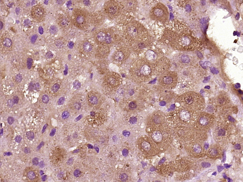 Paraformaldehyde-fixed, paraffin embedded (Human liver cancer); Antigen retrieval by boiling in sodium citrate buffer (pH6.0) for 15min; Block endogenous peroxidase by 3% hydrogen peroxide for 20 minutes; Blocking buffer (normal goat serum) at 37°C for 30min; Antibody incubation with (NALP3/CIAS1) Polyclonal Antibody, Unconjugated (SL10021R) at 1:400 overnight at 4°C, followed by operating according to SP Kit(Rabbit) (sp-0023) instructionsand DAB staining.
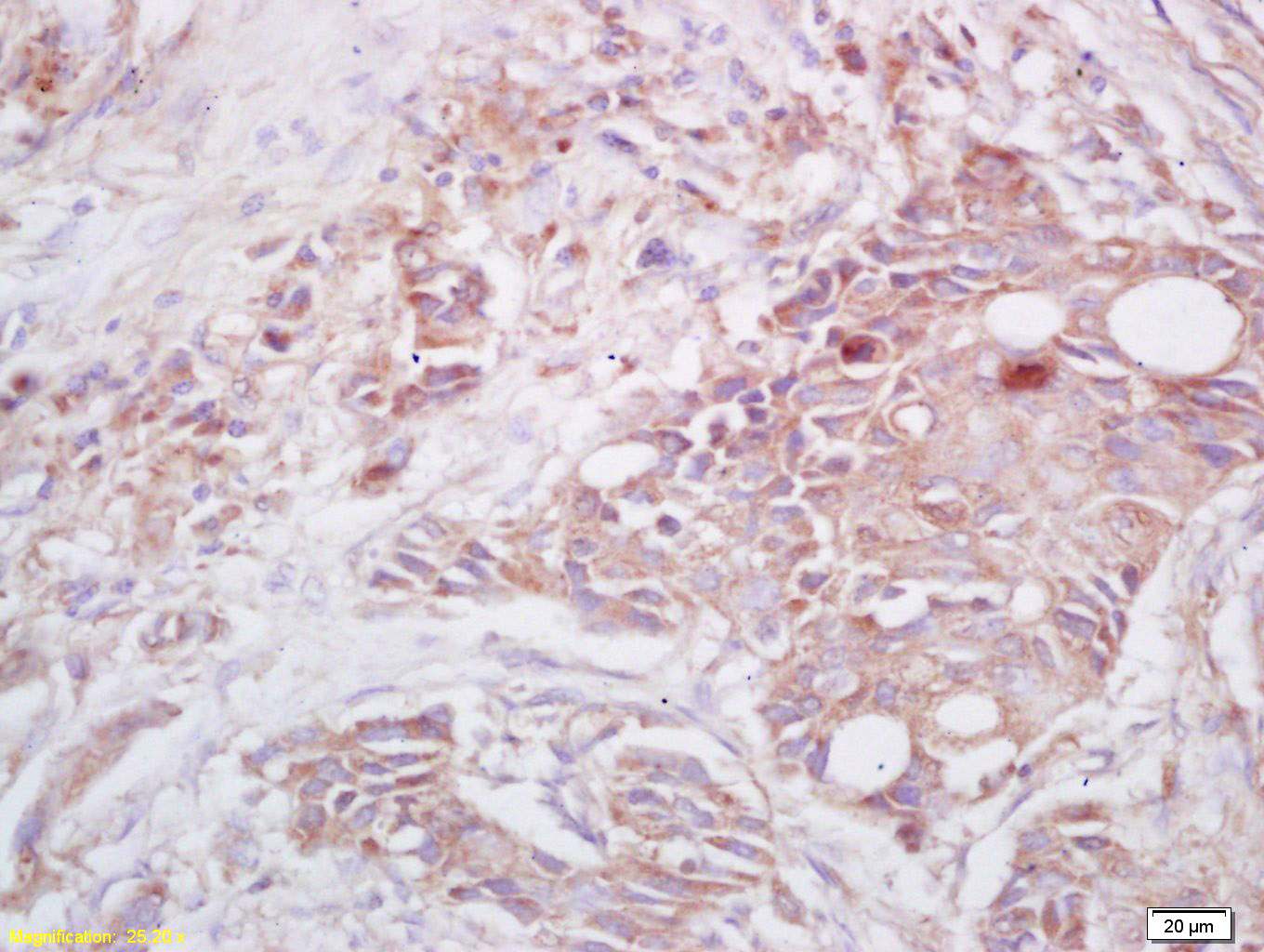 Tissue/cell: human lung carcinoma; 4% Paraformaldehyde-fixed and paraffin-embedded;
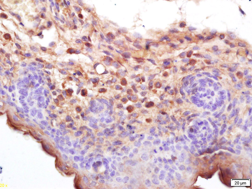
Antigen retrieval: citrate buffer ( 0.01M, pH 6.0 ), Boiling bathing for 15min; Block endogenous peroxidase by 3% Hydrogen peroxide for 30min; Blocking buffer (normal goat serum,C-0005) at 37℃ for 20 min;
Incubation: Anti-Cryopyrin/CIAS1/NALP3 Polyclonal Antibody, Unconjugated(SL10021R) 1:200, overnight at 4°C, followed by conjugation to the secondary antibody(SP-0023) and DAB(C-0010) staining
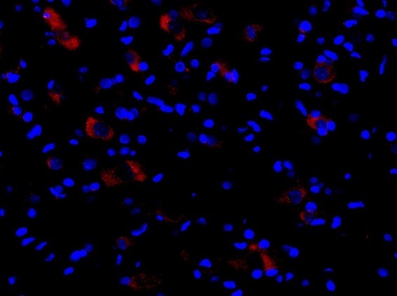 This image was generously provided by Adib Zendedel, PhD from RWTH Aachen University. Formalin-fixed and paraffin embedded rat spinal cord tissue labeled with Rabbit Anti-Cryopyrin Polyclonal Antibody, Unconjugated (SL10021R) at 1:300 followed by conjugation to a secondary antibody
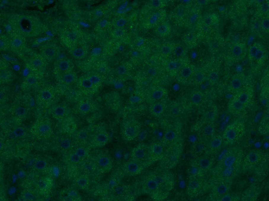 Paraformaldehyde-fixed, paraffin embedded (Human liver cancer); Antigen retrieval by boiling in sodium citrate buffer (pH6.0) for 15min; Block endogenous peroxidase by 3% hydrogen peroxide for 20 minutes; Blocking buffer (normal goat serum) at 37°C for 30min; Antibody incubation with (NALP3/CIAS1) Polyclonal Antibody, Unconjugated (SL10021R) at 1:400 overnight at 4°C, followed by a conjugated Goat Anti-Rabbit IgG antibody (SL0295G-AF488) for 90 minutes, and DAPI for nuclei staining.
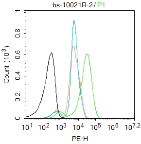 Blank control: Jurkat.
Primary Antibody (green line): Rabbit Anti-NALP3/CIAS1 antibody (SL10021R)
Dilution: 2μg /10^6 cells;
Isotype Control Antibody (orange line): Rabbit IgG .
Secondary Antibody : Goat anti-rabbit IgG-FITC
Dilution: 1μg /test.
Protocol
The cells were fixed with 4% PFA (10min at room temperature)and then permeabilized with 90% ice-cold methanol for 20 min at-20℃.The cells were then incubated in 5%BSA to block non-specific protein-protein interactions for 30 min at room temperature .Cells stained with Primary Antibody for 30 min at room temperature. The secondary antibody used for 40 min at room temperature. Acquisition of 20,000 events was performed.
Cartpieces
Totalgoods,subtotals:¥Checkout
Bought notes(bought amounts latest0)
No one bought this product
User Comment(Total0User Comment Num)
- No comment


 +86 571 56623320
 +86 18668110335

