
Rabbit Anti-Occludin antibody
Occludin, Occludin-1; Occludin1; Occludin 1; FLJ08163; BLCPMG; FLJ18079; FLJ77961; FLJ94056; MGC34277; Occludin; OCLN; OCLN_HUMAN; Tight junction protein occludin.
View History [Clear]
Details
Product Name Occludin Chinese Name 紧密连接蛋白抗体 Alias Occludin, Occludin-1; Occludin1; Occludin 1; FLJ08163; BLCPMG; FLJ18079; FLJ77961; FLJ94056; MGC34277; Occludin; OCLN; OCLN_HUMAN; Tight junction protein occludin. literatures Immunogen Species Rabbit Clonality Polyclonal React Species Human, Mouse, Rat, (predicted: Dog, Pig, Cow, Sheep, ) Applications WB=1:500-2000 ELISA=1:5000-10000 IHC-P=1:100-500 IHC-F=1:100-500 Flow-Cyt=1μg/Test ICC=1:100 IF=1:100-500 (Paraffin sections need antigen repair)
not yet tested in other applications.
optimal dilutions/concentrations should be determined by the end user.Theoretical molecular weight 59kDa Cellular localization The cell membrane Form Liquid Concentration 1mg/ml immunogen KLH conjugated synthetic peptide derived from human Occludin: 151-250/522 <Extracellular> Lsotype IgG Purification affinity purified by Protein A Buffer Solution 0.01M TBS(pH7.4) with 1% BSA, 0.03% Proclin300 and 50% Glycerol. Storage Shipped at 4℃. Store at -20 °C for one year. Avoid repeated freeze/thaw cycles. Attention This product as supplied is intended for research use only, not for use in human, therapeutic or diagnostic applications. PubMed PubMed Product Detail This gene encodes an integral membrane protein which is located at tight junctions. This protein may be involved in the formation and maintenance of the tight junction. The possibility of several alternatively spliced products has been suggested but the full nature of these products has not been described. [provided by RefSeq].
Function:
May play a role in the formation and regulation of the tight junction (TJ) paracellular permeability barrier.
Subunit:
Interacts with TJP1/ZO1 and with VAPA.
Subcellular Location:
Membrane; Multi-pass membrane protein. Cell junction, tight junction.
Tissue Specificity:
Localized at tight junctions of both epithelial and endothelial cells. Highly expressed in kidney. Not detected in testis.
Post-translational modifications:
Phosphorylated upon DNA damage, probably by ATM or ATR. Dephosphorylated by PTPRJ. May be phosphorylated by PKC during translocation to cell-cell contacts.
Similarity:
Belongs to the ELL/occludin family.
Contains 1 MARVEL domain.
SWISS:
Q16625
Gene ID:
100506658
Database links:Entrez Gene: 18260 Mouse
Entrez Gene: 100506658 Human
Omim: 602876 Human
SwissProt: Q16625 Human
SwissProt: Q61146 Mouse
Unigene: 592605 Human
Unigene: 4807 Mouse
Unigene: 31429 Rat
Product Picture 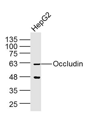 Sample: HepG2 Cell (Human) Lysate at 40 ug
Primary: Anti-Occludin (SL10011R) at 1/300 dilution
Secondary: IRDye800CW Goat Anti-Rabbit IgG at 1/20000 dilution
Predicted band size: 59 kD
Observed band size: 60 kD
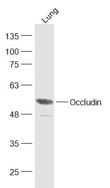 Sample:
Lung (Mouse) Lysate at 40 ug
Primary: Anti-Occludin (SL10011R) at 1/300 dilution
Secondary: IRDye800CW Goat Anti-Rabbit IgG at 1/20000 dilution
Predicted band size: 59 kD
Observed band size: 59 kD
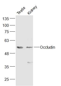 Sample:
Testis (Mouse) Lysate at 40 ug
Kidney (Mouse) Lysate at 40 ug
Primary: Anti-Occludin (SL10011R) at 1/300 dilution
Secondary: IRDye800CW Goat Anti-Rabbit IgG at 1/20000 dilution
Predicted band size: 59 kD
Observed band size: 59 kD
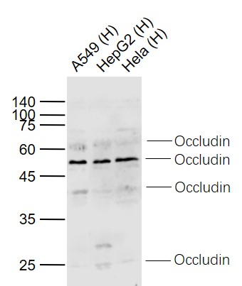 Sample:
Lane 1: A549 (Human) Cell Lysate at 30 ug
Lane 2: HepG2 (Human) Cell Lysate at 30 ug
Lane 3: Hela (Human) Cell Lysate at 30 ug
Primary: Anti-Occludin (SL10011R) at 1/1000 dilution
Secondary: IRDye800CW Goat Anti-Rabbit IgG at 1/20000 dilution
Predicted band size: 65/55/38/28 kD
Observed band size: 62/52/40/25 kD
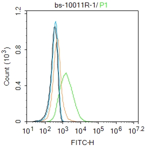
 Blank control(blue): 293T(fixed with 2% paraformaldehyde (10 min) and then permeabilized with ice-cold 90% methanol for 30 min on ice).
Primary Antibody: Rabbit Anti-Occludin/FITC Conjugated antibody (SL10011R /FITC), Dilution: 1μg in 100 μL 1X PBS containing 0.5% BSA;
Isotype Control Antibody: Rabbit IgG/FITC(orange) ,used under the same conditions.
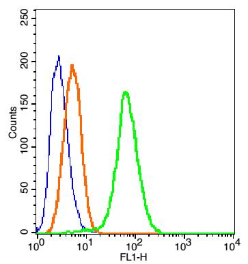
 Blank control: MCF7.
Primary Antibody (green line): Rabbit Anti-Occludin antibody (SL10011R)
Dilution: 1μg /10^6 cells;
Isotype Control Antibody (orange line): Rabbit IgG .
Secondary Antibody : Goat anti-rabbit IgG-FITC
Dilution: 1μg /test.
Protocol
The cells were incubated in 5%BSA to block non-specific protein-protein interactions for 30 min at room temperature .Cells stained with Primary Antibody for 30 min at room temperature. The secondary antibody used for 40 min at room temperature. Acquisition of 20,000 events was performed.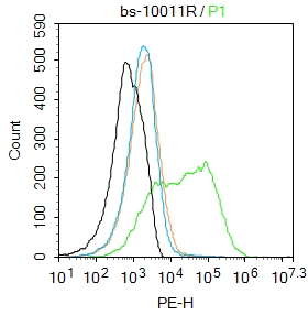 Blank control: Mouse kidney.
Primary Antibody (green line): Rabbit Anti-Occludin antibody (SL10011R)
Dilution: 3μg /10^6 cells;
Isotype Control Antibody (orange line): Rabbit IgG .
Secondary Antibody : Goat anti-rabbit IgG-PE
Dilution: 1μg /test.
Protocol
The cells were incubated in 5%BSA to block non-specific protein-protein interactions for 30 min at at room temperature .Cells stained with Primary Antibody for 30 min at room temperature. The secondary antibody used for 40 min at room temperature. Acquisition of 20,000 events was performed.
Cartpieces
Totalgoods,subtotals:¥Checkout
Bought notes(bought amounts latest0)
No one bought this product
User Comment(Total0User Comment Num)
- No comment


 +86 571 56623320
 +86 18668110335

