
Rabbit Anti-CD38 antibody
ADP-ribosyl cyclase/cyclic ADP-ribose hydrolase 1; 2'-phospho-ADP-ribosyl cyclase; 2'-phospho-ADP-ribosyl cyclase/2'-phospho-cyclic-ADP-ribose transferase; 2'-phospho-cyclic-ADP-ribose transferase; ADP-ribosyl cyclase 1; ADPRC 1; Cyclic ADP-ribose hydrola
View History [Clear]
Details
Product Name CD38 Chinese Name CD38抗体 Alias ADP-ribosyl cyclase/cyclic ADP-ribose hydrolase 1; 2'-phospho-ADP-ribosyl cyclase; 2'-phospho-ADP-ribosyl cyclase/2'-phospho-cyclic-ADP-ribose transferase; 2'-phospho-cyclic-ADP-ribose transferase; ADP-ribosyl cyclase 1; ADPRC 1; Cyclic ADP-ribose hydrolase 1; cADPr hydrolase 1; T10; CD38_HUMAN; CD38_MOUSE; I-19; NIM-R5 antigen. literatures Research Area Tumour immunology Stem cells Diabetes Cell Surface Molecule Natural killer cells lymphocyte t-lymphocyte b-lymphocyte Immunogen Species Rabbit Clonality Polyclonal React Species Human, Mouse, Rat, Applications WB=1:500-2000 ELISA=1:5000-10000 IHC-P=1:100-500 IHC-F=1:100-500 IF=1:100-500 (Paraffin sections need antigen repair)
not yet tested in other applications.
optimal dilutions/concentrations should be determined by the end user.Theoretical molecular weight 34kDa Detection molecular weight 45kDa Cellular localization The cell membrane Form Liquid Concentration 1mg/ml immunogen KLH conjugated synthetic peptide derived from mouse CD38: 101-200/304 <Extracellular> Lsotype IgG Purification affinity purified by Protein A Buffer Solution 0.01M TBS(pH7.4) with 1% BSA, 0.03% Proclin300 and 50% Glycerol. Storage Shipped at 4℃. Store at -20 °C for one year. Avoid repeated freeze/thaw cycles. Attention This product as supplied is intended for research use only, not for use in human, therapeutic or diagnostic applications. PubMed PubMed Product Detail The protein encoded by this gene is a non-lineage-restricted, type II transmembrane glycoprotein that synthesizes and hydrolyzes cyclic adenosine 5'-diphosphate-ribose, an intracellular calcium ion mobilizing messenger. The release of soluble protein and the ability of membrane-bound protein to become internalized indicate both extracellular and intracellular functions for the protein. This protein has an N-terminal cytoplasmic tail, a single membrane-spanning domain, and a C-terminal extracellular region with four N-glycosylation sites. Crystal structure analysis demonstrates that the functional molecule is a dimer, with the central portion containing the catalytic site. It is used as a prognostic marker for patients with chronic lymphocytic leukemia. Alternative splicing results in multiple transcript variants. [provided by RefSeq, Sep 2015]
Function:
Synthesizes cyclic ADP-ribose, a second messenger for glucose-induced insulin secretion. Also has cADPr hydrolase activity. Also moonlights as a receptor in cells of the immune system.
Subcellular Location:
Membrane; Single-pass type II membrane protein.
Tissue Specificity:
Expressed at high levels in pancreas, liver, kidney, brain, testis, ovary, placenta, malignant lymphoma and neuroblastoma.
Similarity:
Belongs to the ADP-ribosyl cyclase family.
SWISS:
P56528
Gene ID:
12494
Database links:Entrez Gene: 952 Human
Entrez Gene: 12494 Mouse
Omim: 107270 Human
SwissProt: P28907 Human
SwissProt: P56528 Mouse
Unigene: 479214 Human
Unigene: 249873 Mouse
CD38分子是单链Ⅱ型跨膜glycoprotein,存在于很多免疫细胞表面上,CD38与无脊椎动物卵的授精、HIV感染、某些癌症以及Diabetes等有关,CD38具有许多复杂而又独特的生物学特性及功能,有调控细胞钙释放的机制。主要表达于胸腺细胞、前T和前B细胞、活化的T和B细胞、单核细胞、NK细胞、浆细胞等,可用于急性白血病的分型和活化的T细胞在自身免疫和免疫缺陷中的作用等方面的研究,也可作为生发中心B细胞的一种选择性标记。Product Picture 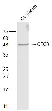 Sample:
Cerebrum (Rat) Lysate at 40 ug
Primary: Anti-CD38 (SL0979R) at 1/1000 dilution
Secondary: IRDye800CW Goat Anti-Rabbit IgG at 1/20000 dilution
Predicted band size: 34 kD
Observed band size: 48 kD
 Sample:
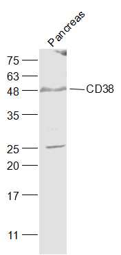
Pancreas (Mouse) Lysate at 40 ug
Primary: Anti-CD38 (SL0979R) at 1/1000 dilution
Secondary: IRDye800CW Goat Anti-Rabbit IgG at 1/20000 dilution
Predicted band size: 34 kD
Observed band size: 48 kD
 Sample:
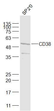
SP2/0(Mouse) Cell Lysate at 30 ug
Primary: Anti-CD38 (SL0979R) at 1/1000 dilution
Secondary: IRDye800CW Goat Anti-Rabbit IgG at 1/20000 dilution
Predicted band size: 34 kD
Observed band size: 54 kD
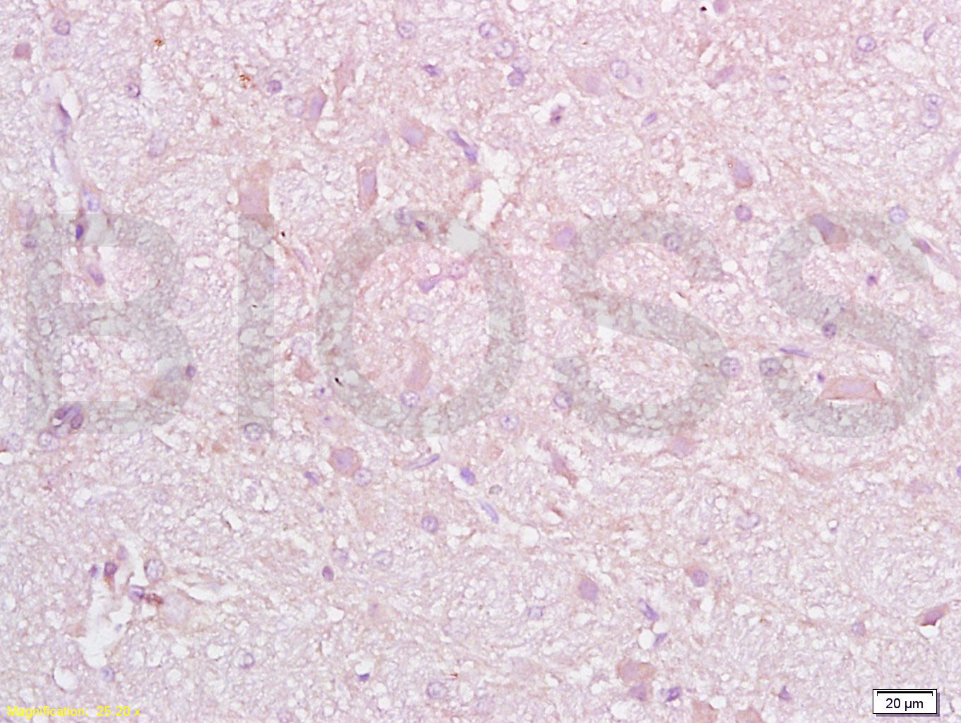 Tissue/cell: rat brain tissue; 4% Paraformaldehyde-fixed and paraffin-embedded;
Antigen retrieval: citrate buffer ( 0.01M, pH 6.0 ), Boiling bathing for 15min; Block endogenous peroxidase by 3% Hydrogen peroxide for 30min; Blocking buffer (normal goat serum,C-0005) at 37℃ for 20 min;
Incubation: Anti-CD38 Polyclonal Antibody, Unconjugated(SL0979R) 1:200, overnight at 4°C, followed by conjugation to the secondary antibody(SP-0023) and DAB(C-0010) staining
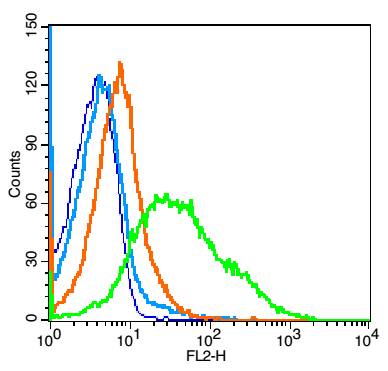 Blank control: mouse spleen cells (blue).
Primary Antibody: Rabbit Anti- CD38 antibody(SL0979R), Dilution: 1μg in 100 μL 1X PBS containing 0.5% BSA;
Isotype Control Antibody: Rabbit IgG(orange) ,used under the same conditions );
Secondary Antibody: Goat anti-rabbit IgG-PE(white blue), Dilution: 1:200 in 1 X PBS containing 0.5% BSA.
Protocol
The cells were fixed with 2% paraformaldehyde (10 min) , then permeabilized with 90% ice-cold methanol for 30 min on ice. Primary antibody (SL0979R, 1μg /1x10^6 cells) were incubated for 30 min on the ice, followed by 1 X PBS containing 0.5% BSA + 1 0% goat serum (15 min) to block non-specific protein-protein interactions. Then the Goat Anti-rabbit IgG/PE antibody was added into the blocking buffer mentioned above to react with the primary antibody at 1/200 dilution for 30 min on ice. Acquisition of 20,000 events was performed.
Cartpieces
Totalgoods,subtotals:¥Checkout
Bought notes(bought amounts latest0)
No one bought this product
User Comment(Total0User Comment Num)
- No comment


 +86 571 56623320
 +86 18668110335

