
Rabbit Anti-PEDF antibody
pigment epithelium-derived factor; Serpin-F1; Stromal cell-derived factor 3; SDF-3; Caspin; Alpha 2 antiplasmin; EPC 1; EPC1; PIG 35; PIG35; Pigment epithelium derived factor; Proliferation inducing protein 35; SERPIN F1; Serpin peptidase inhibitor clade
View History [Clear]
Details
Product Name PEDF Chinese Name 色素上皮源性因子抗体 Alias pigment epithelium-derived factor; Serpin-F1; Stromal cell-derived factor 3; SDF-3; Caspin; Alpha 2 antiplasmin; EPC 1; EPC1; PIG 35; PIG35; Pigment epithelium derived factor; Proliferation inducing protein 35; SERPIN F1; Serpin peptidase inhibitor clade F member 1; SERPINF 1; SERPINF1; PEDF_HUMAN. literatures Research Area Tumour Cell biology immunology Neurobiology Growth factors and hormones Immunogen Species Rabbit Clonality Polyclonal React Species Human, Rat, (predicted: Mouse, Dog, Pig, Cow, ) Applications ELISA=1:5000-10000 IHC-P=1:100-500 IHC-F=1:100-500 IF=1:100-500 (Paraffin sections need antigen repair)
not yet tested in other applications.
optimal dilutions/concentrations should be determined by the end user.Theoretical molecular weight 46kDa Cellular localization Secretory protein Form Liquid Concentration 1mg/ml immunogen KLH conjugated synthetic peptide derived from human PEDF: 201-300/418 Lsotype IgG Purification affinity purified by Protein A Buffer Solution 0.01M TBS(pH7.4) with 1% BSA, 0.03% Proclin300 and 50% Glycerol. Storage Shipped at 4℃. Store at -20 °C for one year. Avoid repeated freeze/thaw cycles. Attention This product as supplied is intended for research use only, not for use in human, therapeutic or diagnostic applications. PubMed PubMed Product Detail Pigment epithelium derived factor, originally identified in conditioned medium of cultured human fetal retinal pigment epithelial (RPE) cells, is a neurotrophic protein that induces extensive neuronal differentiation in human Y79 retinoblastoma cells, a neoplastic counterpart of normal retinoblasts. It has been suggested that PEDF is synthesized by RPE cells and secreted into the retina interphotoreceptor matrix where it may influence development/differentiation of the neural retina. PEDF is a potent inhibitor of angiogenesis. As it does not undergo the S (stressed) to R (relaxed) conformational transition characteristic of active serpins, it exhibits no serine protease inhibitory activity. The PEDF gene is a member of the serpin gene family. Serpins are a group of serine protease inhibitors, some of which have also been reported to exhibit neurotrophic activity.
Function:
Neurotrophic protein; induces extensive neuronal differentiation in retinoblastoma cells. Potent inhibitor of angiogenesis. As it does not undergo the S (stressed) to R (relaxed) conformational transition characteristic of active serpins, it exhibits no serine protease inhibitory activity.
Subcellular Location:
Secreted. Melanosome. Enriched in stage I melanosomes.
Tissue Specificity:
Retinal pigment epithelial cells and blood plasma.
Post-translational modifications:
The N-terminus is blocked. Extracellular phosphorylation enhances antiangiogenic activity.
N- and O-glycosylated. O-glycosylated with a core 1 or possibly core 8 glycan.
DISEASE:
Defects in SERPINF1 are the cause of osteogenesis imperfecta type 12 (OI12) [MIM:613982]. OI12 is a connective tissue disorder characterized by bone fragility, low bone mass, and recurrent fractures. OI12 is characterized by features compatible with osteogenesis imperfecta type III in the Sillence classification. Patients have normal grayish sclerae and fractures of long bones and severe vertebral compression fractures, with resulting deformities observed as early as the first year of life.
Similarity:
Belongs to the serpin family.
SWISS:
P97298
Gene ID:
5176
Database links:Entrez Gene: 5176 Human
Entrez Gene: 20317 Mouse
Omim: 172860 Human
SwissProt: P36955 Human
SwissProt: P97298 Mouse
Unigene: 532768 Human
Unigene: 2044 Mouse
Product Picture 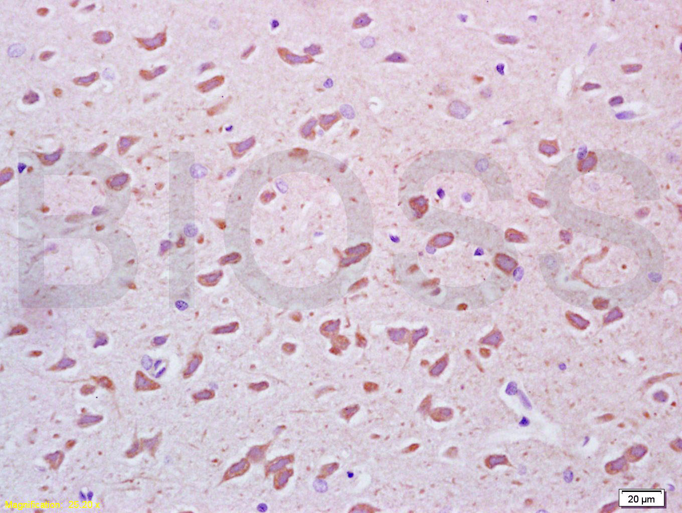 Tissue/cell: rat brain tissue; 4% Paraformaldehyde-fixed and paraffin-embedded;
Antigen retrieval: citrate buffer ( 0.01M, pH 6.0 ), Boiling bathing for 15min; Block endogenous peroxidase by 3% Hydrogen peroxide for 30min; Blocking buffer (normal goat serum,C-0005) at 37℃ for 20 min;
Incubation: Anti-PEGF Polyclonal Antibody, Unconjugated(SL0731R) 1:200, overnight at 4°C, followed by conjugation to the secondary antibody(SP-0023) and DAB(C-0010) staining
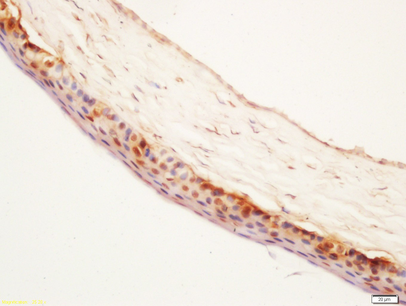 Tissue/cell: chorioid of rat eyes; 4% Paraformaldehyde-fixed and paraffin-embedded;
Antigen retrieval: citrate buffer ( 0.01M, pH 6.0 ), Boiling bathing for 15min; Block endogenous peroxidase by 3% Hydrogen peroxide for 30min; Blocking buffer (normal goat serum,C-0005) at 37℃ for 20 min;
Incubation: Anti-PEGF Polyclonal Antibody, Unconjugated(SL0731R) 1:200, overnight at 4°C, followed by conjugation to the secondary antibody(SP-0023) and DAB(C-0010) staining
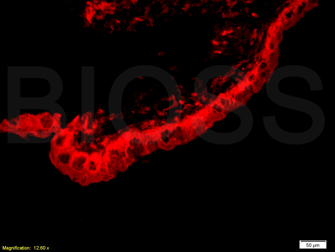 Tissue/cell: ciliary body of rat eye;4% Paraformaldehyde-fixed and paraffin-embedded;
Antigen retrieval: citrate buffer ( 0.01M, pH 6.0 ), Boiling bathing for 15min; Blocking buffer (normal goat serum,C-0005) at 37℃ for 20 min;
Incubation: Anti-PEGF Polyclonal Antibody, Unconjugated(SL0731R) 1:800, overnight at 4°C; The secondary antibody was Goat Anti-Rabbit IgG, Cy3 conjugated(SL0295G-Cy3)used at 1:200 dilution for 40 minutes at 37°C.
Cartpieces
Totalgoods,subtotals:¥Checkout
Bought notes(bought amounts latest0)
No one bought this product
User Comment(Total0User Comment Num)
- No comment


 +86 571 56623320
 +86 18668110335

