
Rabbit Anti-LDLR antibody
LDLR_HUMAN; Low-density lipoprotein receptor; LDL receptor; FH; FHC; FHCL1; LDLCQ2; low density lipoprotein receptor;
View History [Clear]
Details
Product Name LDLR Chinese Name 低密度Lipoprotein受体抗体 Alias LDLR_HUMAN; Low-density lipoprotein receptor; LDL receptor; FH; FHC; FHCL1; LDLCQ2; low density lipoprotein receptor; literatures Research Area Tumour immunology Immunogen Species Rabbit Clonality Polyclonal React Species Human, Mouse, (predicted: Rat, Dog, Pig, Cow, Horse, Rabbit, Guinea Pig, ) Applications WB=1:500-2000 ELISA=1:5000-10000 Flow-Cyt=1μg/Test ICC=1:100
not yet tested in other applications.
optimal dilutions/concentrations should be determined by the end user.Theoretical molecular weight 92kDa Cellular localization cytoplasmic The cell membrane Form Liquid Concentration 1mg/ml immunogen KLH conjugated synthetic peptide derived from human LDL-R: 781-860/860 <Cytoplasmic> Lsotype IgG Purification affinity purified by Protein A Buffer Solution 0.01M TBS(pH7.4) with 1% BSA, 0.03% Proclin300 and 50% Glycerol. Storage Shipped at 4℃. Store at -20 °C for one year. Avoid repeated freeze/thaw cycles. Attention This product as supplied is intended for research use only, not for use in human, therapeutic or diagnostic applications. PubMed PubMed Product Detail The low density lipoprotein receptor (LDLR) gene family consists of cell surface proteins involved in receptor-mediated endocytosis of specific ligands. The encoded protein is normally bound at the cell membrane, where it binds low density lipoprotein/cholesterol and is taken into the cell. Lysosomes release the cholesterol, which is made available for repression of microsomal enzyme 3-hydroxy-3-methylglutaryl coenzyme A (HMG CoA) reductase, the rate-limiting step in cholesterol synthesis. At the same time, a reciprocal stimulation of cholesterol ester synthesis takes place. Mutations in this gene cause the autosomal dominant disorder, familial hypercholesterolemia. Alternate splicing results in multiple transcript variants.[provided by RefSeq, May 2022]
Function:
Binds LDL, the major cholesterol-carrying lipoprotein of plasma, and transports it into cells by endocytosis. In order to be internalized, the receptor-ligand complexes must first cluster into clathrin-coated pits. In case of HIV-1 infection, functions as a receptor for extracellular Tat in neurons, mediating its internalization in uninfected cells.
Subunit:
Interacts with LDLRAP1. Interacts with SNX17. Interacts with HCV E1/E2 heterodimer. Interacts with HIV-1 Tat.
Subcellular Location:
Cell membrane; Single-pass type I membrane protein. Endomembrane system; Single-pass type I membrane protein. Membrane, clathrin-coated pit; Single-pass type I membrane protein. Note=Found distributed from the plasma membrane to intracellular compartments.
Tissue Specificity:
Binds LDL, the major cholesterol-carrying lipoprotein of plasma, and transports it into cells by endocytosis. In order to be internalized, the receptor-ligand complexes must first cluster into clathrin-coated pits. In case of HIV-1 infection, functions as a receptor for extracellular Tat in neurons, mediating its internalization in uninfected cells.
Post-translational modifications:
N- and O-glycosylated.
Ubiquitinated by MYLIP leading to degradation.
Similarity:
Belongs to the LDLR family.
Contains 3 EGF-like domains.
Contains 7 LDL-receptor class A domains.
Contains 6 LDL-receptor class B repeats.
SWISS:
P01130
Gene ID:
3949
Database links:Entrez Gene: 3949 Human
Entrez Gene: 16835 Mouse
Omim: 606945 Human
SwissProt: P01130 Human
SwissProt: P35951 Mouse
Unigene: 213289 Human
Unigene: 728190 Human
Unigene: 3213 Mouse
Unigene: 10483 Rat
低密度Lipoprotein受体 LDLR是一种存在于细胞表面的、可识别多种配体的Lipoprotein受体,在体内对于富含甘油三酯的Lipoprotein代谢非常重要;
LDL R目前主要用于代谢及Tumour方面的研究。Product Picture 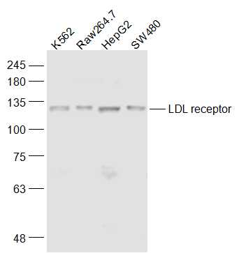 Sample:
K562(Human) Cell Lysate at 30 ug
Raw264.7(Mouse) Cell Lysate at 30 ug
HepG2(Human) Cell Lysate at 30 ug
SW480(Human) Cell Lysate at 30 ug
Primary: Anti-LDL receptor (SL0705R) at 1/1000 dilution
Secondary: IRDye800CW Goat Anti-Rabbit IgG at 1/20000 dilution
Predicted band size: 92 kD
Observed band size: 120 kD
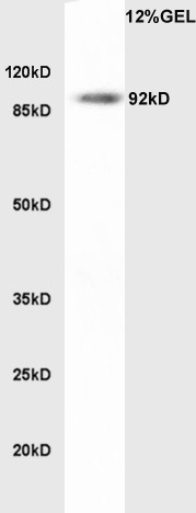 Sample:Human colon Lysate at 45ug; Primary: Anti-LDL receptor (SL0705R) at 1:300 dilution; Secondary: HRP conjugated Goat Anti-Rabbit IgG(SL0295G-HRP) at 1: 5000 dilution; Predicted band size : 92kD Observed band size : 92kD
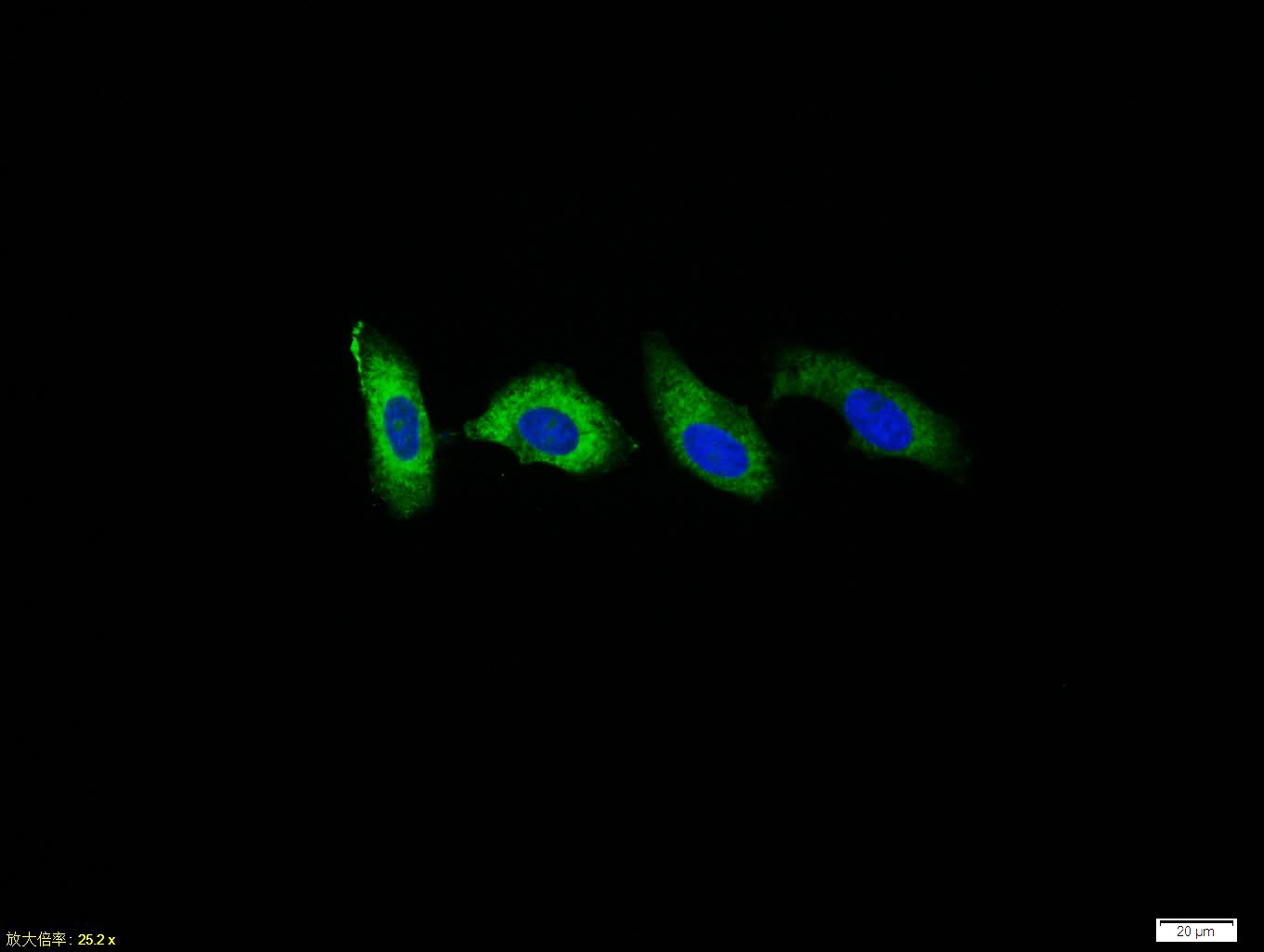 HepG2 cell; 4% Paraformaldehyde-fixed; Triton X-100 at room temperature for 20 min; Blocking buffer (normal goat serum, C-0005) at 37°C for 20 min; Antibody incubation with (LDL receptor) polyclonal Antibody, Unconjugated (SL0705R) 1:100, 90 minutes at 37°C; followed by a conjugated Goat Anti-Rabbit IgG antibody at 37°C for 90 minutes, DAPI (blue, C02-04002) was used to stain the cell nuclei.
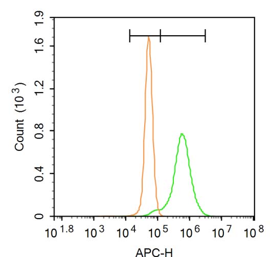 Blank control:A431.
Primary Antibody (green line): Rabbit Anti-LDL receptor antibody (SL0705R),Dilution: 1μg /10^6 cells.
Isotype Control Antibody (orange line): Rabbit IgG.
Secondary Antibody:Goat anti-rabbit IgG-AF647,Dilution: 1μg /test.
Protocol
A431 cells were fixed with 4% PFA (10min at room temperature)and then permeabilized with 0.1% PBST for 20 min at room temperature. The cells were then incubated in 5%BSA to block non-specific protein-protein interactions for 30 min at room temperature .Cells stained with Primary Antibody for 30 min at room temperature. The secondary antibody used for 40 min at room temperature. Acquisition of 20,000 events was performed.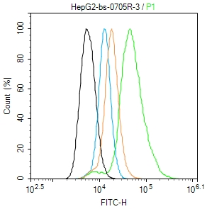 Blank control (black line): HepG2 (black) (The cells were fixed with 2% paraformaldehyde (10 min) , then permeabilized with PBST for 30 min on room temperature)
Primary Antibody (green line): Rabbit Anti-LDLreceptor antibody (SL0705R) ; Dilution: 1μg /10^6 cells;
Isotype Control Antibody (orange line): Rabbit IgG .
Secondary Antibody (white blue line): Goat anti-rabbit IgG-FITC;Dilution: 1μg /test.
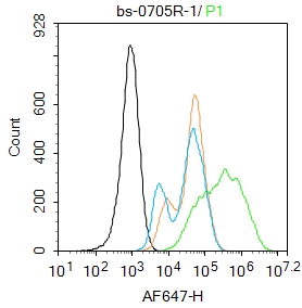 Blank control: Raw264.7.
Primary Antibody (green line): Rabbit Anti-LDL receptor antibody (SL0705R)
Dilution: 1μg /10^6 cells;
Isotype Control Antibody (orange line): Rabbit IgG .
Secondary Antibody : Goat anti-rabbit IgG-AF647
Dilution: 1μg /test.
Protocol
The cells were fixed with 4% PFA (10min at room temperature)and then permeabilized with 0.1% PBST for 20 min at room temperature. The cells were then incubated in 5%BSA to block non-specific protein-protein interactions for 30 min at room temperature .Cells stained with Primary Antibody for 30 min at room temperature. The secondary antibody used for 40 min at room temperature. Acquisition of 20,000 events was performed.
Cartpieces
Totalgoods,subtotals:¥Checkout
Bought notes(bought amounts latest0)
No one bought this product
User Comment(Total0User Comment Num)
- No comment


 +86 571 56623320
 +86 18668110335

