
Rabbit Anti-CD147 antibody
TCSF; Emmprin; 5A11 antigen; 5F7; Basigin (Ok blood group); Basigin; Blood brain barrier HT7 antigen; BSG; CD 147; CD147 antigen; Collagenase stimulatory factor; Extracellular matrix metalloproteinase inducer; Leukocyte activation antigen M6; M6; M6 leuko
View History [Clear]
Details
Product Name CD147 Chinese Name Extracellular matrix金属蛋白酶诱导因子抗体 Alias TCSF; Emmprin; 5A11 antigen; 5F7; Basigin (Ok blood group); Basigin; Blood brain barrier HT7 antigen; BSG; CD 147; CD147 antigen; Collagenase stimulatory factor; Extracellular matrix metalloproteinase inducer; Leukocyte activation antigen M6; M6; M6 leukocyte activation antigen; neurothelin; OK; OK blood group; OK blood group antigen; TCSF; Tumor cell derived collagenase stimulatory factor; BASI_HUMAN. literatures Research Area Tumour immunology Synthesis and Degradation Cell Surface Molecule Immunogen Species Rabbit Clonality Polyclonal React Species Human, Mouse, Rat, (predicted: Chicken, Dog, Pig, Cow, Rabbit, Guinea Pig, ) Applications WB=1:500-2000 ELISA=1:5000-10000 IHC-P=1:100-500 IHC-F=1:100-500 Flow-Cyt=1μg /test IF=1:100-500 (Paraffin sections need antigen repair)
not yet tested in other applications.
optimal dilutions/concentrations should be determined by the end user.Theoretical molecular weight 40kDa Detection molecular weight 55-65 kDa Cellular localization The cell membrane Form Liquid Concentration 1mg/ml immunogen KLH conjugated synthetic peptide derived from human TCSF: 301-385/385 <Cytoplasmic> Lsotype IgG Purification affinity purified by Protein A Buffer Solution 0.01M TBS(pH7.4) with 1% BSA, 0.03% Proclin300 and 50% Glycerol. Storage Shipped at 4℃. Store at -20 °C for one year. Avoid repeated freeze/thaw cycles. Attention This product as supplied is intended for research use only, not for use in human, therapeutic or diagnostic applications. PubMed PubMed Product Detail The protein encoded by this gene is a plasma membrane protein that is important in spermatogenesis, embryo implantation, neural network formation, and tumor progression. The encoded protein is also a member of the immunoglobulin superfamily. Multiple transcript variants encoding different isoforms have been found for this gene. [provided by RefSeq, Jul 2008]
Function:
Plays pivotal roles in spermatogenesis, embryo implantation, neural network formation and tumor progression. Stimulates adjacent fibroblasts to produce matrix metalloproteinases (MMPS). May target monocarboxylate transporters SLC16A1, SLC16A3 and SLC16A8 to plasma membranes of retinal pigment epithelium and neural retina. Seems to be a receptor for oligomannosidic glycans. In vitro, promotes outgrowth of astrocytic processes.
Subunit:
Forms homooligomers in a cis-dependent manner on the plasma membrane. Forms heterooligomers of isoform 2 and isoform 3. Forms a complex with MMP1 at the tumor cell surface. Interacts with SLC16A1 and SLC1A3; probably a BSG dimer is associated with a monocarboxylate transporter dimer. Interacts with ATP1B2, MAG and L1CAM. Interacts with AJAP1.
Subcellular Location:
Cell membrane; Single-pass type I membrane protein. Melanosome.
Tissue Specificity:
Present only in vascular endothelium in non-neoplastic regions of the brain, whereas it is present in tumor cells but not in proliferating blood vessels in malignant gliomas.
Post-translational modifications:
N-glycosylated.
Similarity:
Contains 1 Ig-like C2-type (immunoglobulin-like) domain.
Contains 1 Ig-like V-type (immunoglobulin-like) domain.
SWISS:
P35613
Gene ID:
682
Database links:Entrez Gene: 682 Human
Entrez Gene: 12215 Mouse
Omim: 109480 Human
SwissProt: P35613 Human
SwissProt: P18572 Mouse
Unigene: 501293 Human
Synthesis and Degradation(Synthesis and Degradation)
主要由Tumour细胞合成,刺激纤维母细胞产生MMPProduct Picture 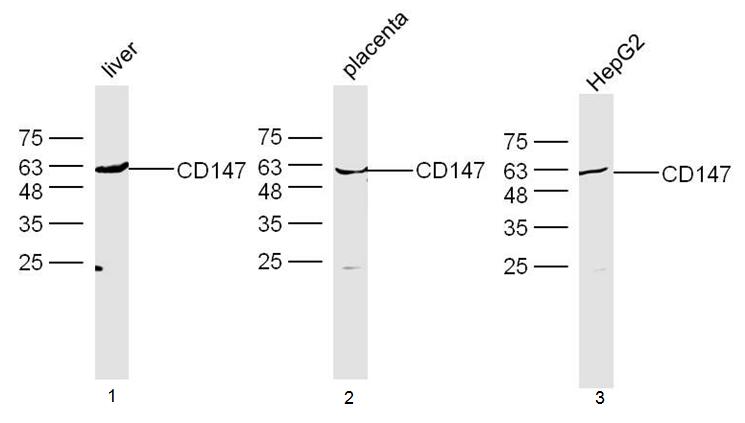 Sample:
Liver(Mouse) Lysate at 40 ug
Placenta(Mouse) Lysate at 40 ug
HepG2 Cell Lysate at 40 ug
Primary: Anti-CD147 (SL0684R) at 1/300 dilution
Secondary: IRDye800CW Goat Anti-Rabbit IgG at 1/20000 dilution
Predicted band size: 40 kD
Observed band size: 60 kD
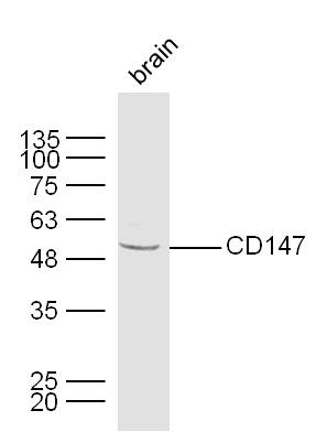 Sample:
Brain (Mouse) Lysate at 40 ug
Primary: Anti-CD147 (Bs-0684R) at 1/300 dilution
Secondary: IRDye800CW Goat Anti-Rabbit IgG at 1/20000 dilution
Predicted band size: 40 kD
Observed band size: 50 kD
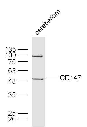 Sample:
Cerebellum (Mouse) Lysate at 40 ug
Primary: Anti-CD147 (Bs-0684R) at 1/300 dilution
Secondary: IRDye800CW Goat Anti-Rabbit IgG at 1/20000 dilution
Predicted band size: 40 kD
Observed band size: 50 kD
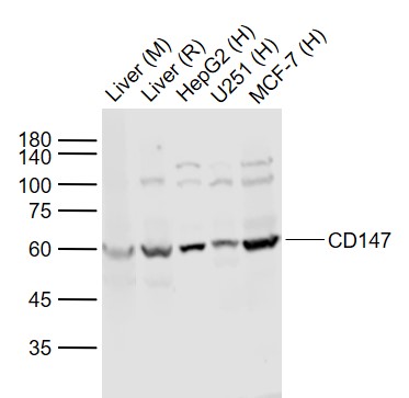 Sample:
Lane 1: Liver (Mouse) Lysate at 40 ug
Lane 2: Liver (Rat) Lysate at 40 ug
Lane 3: HepG2 (Human) Cell Lysate at 30 ug
Lane 4: U251 (Human) Cell Lysate at 30 ug
Lane 5: MCF-7 (Human) Cell Lysate at 30 ug
Primary: Anti-CD147 (SL0684R) at 1/1000 dilution
Secondary: IRDye800CW Goat Anti-Rabbit IgG at 1/20000 dilution
Predicted band size: 50-60 kD
Observed band size: 60 kD
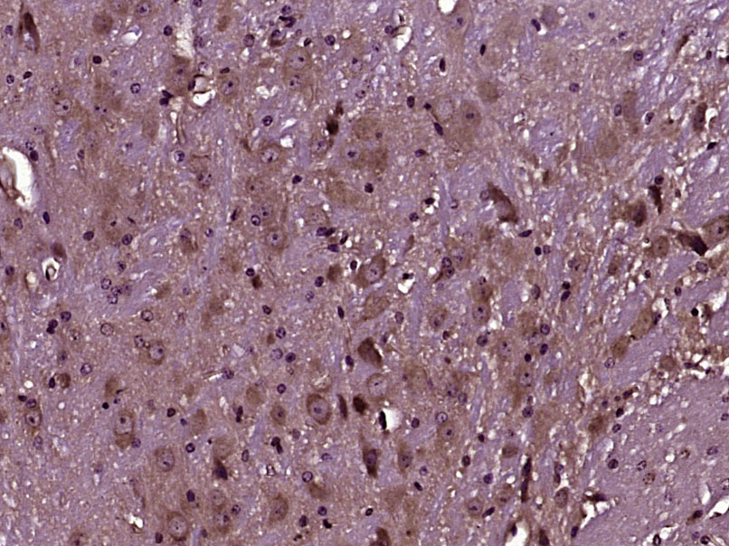 Paraformaldehyde-fixed, paraffin embedded (Mouse cerebellum); Antigen retrieval by boiling in sodium citrate buffer (pH6.0) for 15min; Block endogenous peroxidase by 3% hydrogen peroxide for 20 minutes; Blocking buffer (normal goat serum) at 37°C for 30min; Antibody incubation with (CD147) Polyclonal Antibody, Unconjugated (SL0684R) at 1:400 overnight at 4°C, followed by operating according to SP Kit(Rabbit) (sp-0023) instructionsand DAB staining.
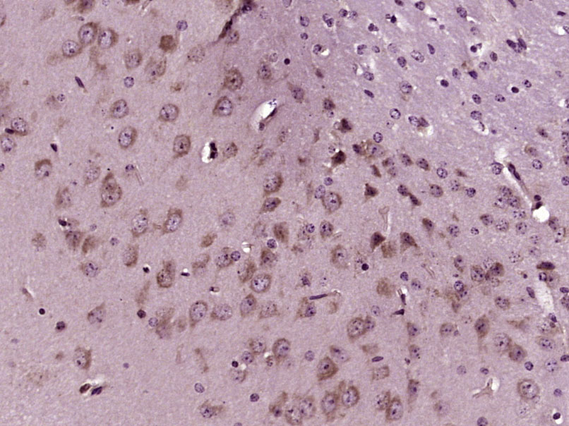 Paraformaldehyde-fixed, paraffin embedded (Mouse brain); Antigen retrieval by boiling in sodium citrate buffer (pH6.0) for 15min; Block endogenous peroxidase by 3% hydrogen peroxide for 20 minutes; Blocking buffer (normal goat serum) at 37°C for 30min; Antibody incubation with (CD147) Polyclonal Antibody, Unconjugated (SL0684R) at 1:400 overnight at 4°C, followed by operating according to SP Kit(Rabbit) (sp-0023) instructionsand DAB staining.
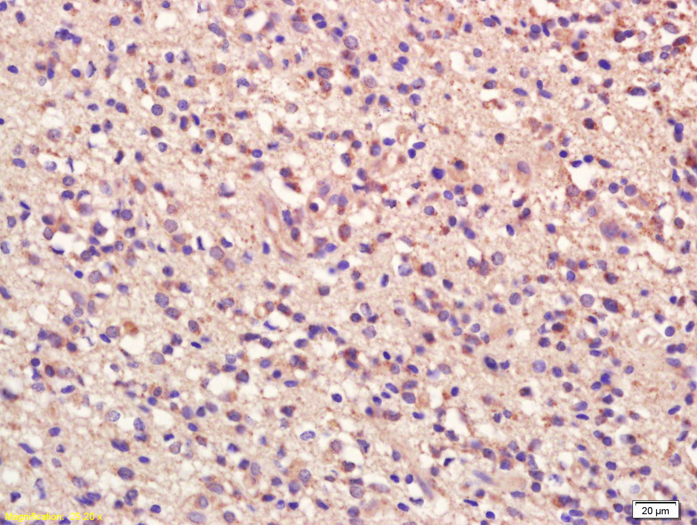 Tissue/cell: human glioma tissue; 4% Paraformaldehyde-fixed and paraffin-embedded;
Antigen retrieval: citrate buffer ( 0.01M, pH 6.0 ), Boiling bathing for 15min; Block endogenous peroxidase by 3% Hydrogen peroxide for 30min; Blocking buffer (normal goat serum,C-0005) at 37℃ for 20 min;
Incubation: Anti-CD147/TCSF/Emmprin Polyclonal Antibody, Unconjugated(SL0684R) 1:200, overnight at 4°C, followed by conjugation to the secondary antibody(SP-0023) and DAB(C-0010) staining
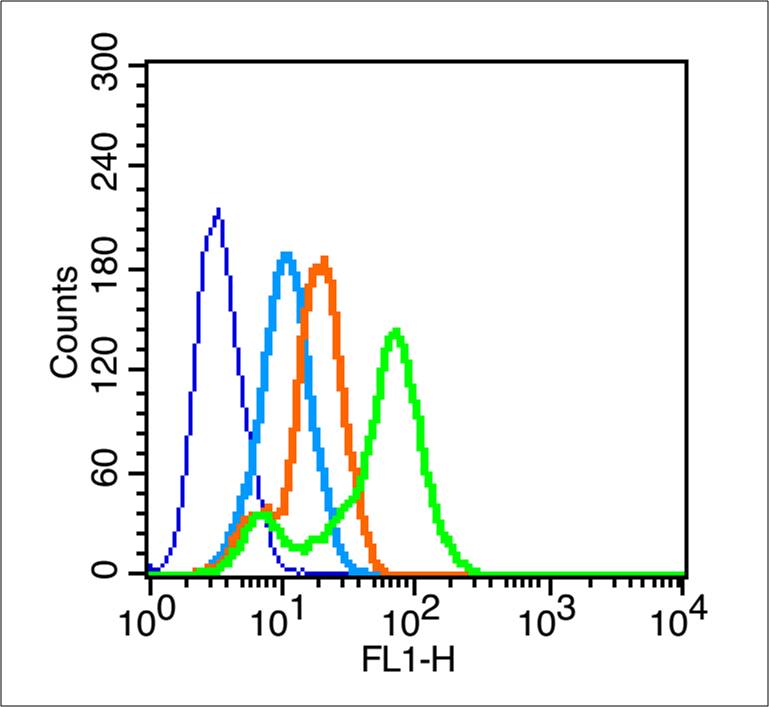 Blank control (blue line): MCF7 (blue).
Primary Antibody (green line):Rabbit Anti-CD147 antibody(SL0684R)
Dilution: 1μg /10^6 cells;
Isotype Control Antibody (orange line): Rabbit IgG .
Secondary Antibody (white blue line): F(ab’)2 fragment goat anti-rabbit IgG-FITC
Dilution: 1μg /test.
Protocol
The cells were fixed with 2% paraformaldehyde for 10 min at room temperature. Cells stained with Primary Antibody for 30 min at room temperature. The cells were then incubated in 1 X PBS/2%BSA/10% goat serum to block non-specific protein-protein interactions followed by the antibody for 15 min at room temperature. The secondary antibody used for 40 min at room temperature. Acquisition of 20,000 events was performed.
Cartpieces
Totalgoods,subtotals:¥Checkout
Bought notes(bought amounts latest0)
No one bought this product
User Comment(Total0User Comment Num)
- No comment


 +86 571 56623320
 +86 18668110335

