
Rabbit Anti-SYVN1 antibody
1200010C09Rik; HRD1; KIAA1810; MGC40372; Synovial apoptosis inhibitor 1 synoviolin; Synoviolin 1 isoform b; SYVN1_HUMAN; DER3; E3 ubiquitin-protein ligase synoviolin; HMG coA reductase degradation 1 homolog; OTTHUMP00000230429; OTTHUMP00000230430; OTTHUMP
View History [Clear]
Details
Product Name SYVN1 Chinese Name 滑膜Apoptosis抑制物1抗体 Alias 1200010C09Rik; HRD1; KIAA1810; MGC40372; Synovial apoptosis inhibitor 1 synoviolin; Synoviolin 1 isoform b; SYVN1_HUMAN; DER3; E3 ubiquitin-protein ligase synoviolin; HMG coA reductase degradation 1 homolog; OTTHUMP00000230429; OTTHUMP00000230430; OTTHUMP00000230431; OTTHUMP00000230432; Synovial apoptosis inhibitor 1; Synoviolin 1 isoform b; SYNOVIOLIN; SYVN1. literatures Research Area Cell biology immunology Apoptosis Immunogen Species Rabbit Clonality Polyclonal React Species Human, Mouse, (predicted: Rat, Dog, Cow, ) Applications WB=1:500-2000 ELISA=1:5000-10000 IHC-P=1:100-500 IHC-F=1:100-500 Flow-Cyt=3μg/Test IF=1:100-500 (Paraffin sections need antigen repair)
not yet tested in other applications.
optimal dilutions/concentrations should be determined by the end user.Theoretical molecular weight 65kDa Cellular localization cytoplasmic The cell membrane Form Liquid Concentration 1mg/ml immunogen KLH conjugated synthetic peptide derived from human SYVN1: 531-617/617 Lsotype IgG Purification affinity purified by Protein A Buffer Solution 0.01M TBS(pH7.4) with 1% BSA, 0.03% Proclin300 and 50% Glycerol. Storage Shipped at 4℃. Store at -20 °C for one year. Avoid repeated freeze/thaw cycles. Attention This product as supplied is intended for research use only, not for use in human, therapeutic or diagnostic applications. PubMed PubMed Product Detail This gene encodes a protein involved in endoplasmic reticulum (ER)-associated degradation. The encoded protein removes unfolded proteins, accumulated during ER stress, by retrograde transport to the cytosol from the ER. This protein also uses the ubiquitin-proteasome system for additional degradation of unfolded proteins. Sequence analysis identified two transcript variants that encode different isoforms. [provided by RefSeq, May 2011]
Function:
Acts as an E3 ubiquitin-protein ligase which accepts ubiquitin specifically from endoplasmic reticulum-associated UBC7 E2 ligase and transfers it to substrates, promoting their degradation. Component of the endoplasmic reticulum quality control (ERQC) system also called ER-associated degradation (ERAD) involved in ubiquitin-dependent degradation of misfolded endoplasmic reticulum proteins. Also promotes the degradation of normal but naturally short-lived proteins such as SGK. Protects cells from ER stress-induced apoptosis. Protects neurons from apoptosis induced by polyglutamine-expanded huntingtin (HTT) or unfolded GPR37 by promoting their degradation. Sequesters p53/TP53 in the cytoplasm and promotes its degradation, thereby negatively regulating its biological function in transcription, cell cycle regulation and apoptosis.
Subunit:
Homodimer. Interacts with p53/TP53 and HTT. Interacts with VCP, HERPUD1 and DERL1. Part of a complex containing SYVN1, HERPUD1, SELS and DERL1; which probably transfer misfolded proteins from the ER to VCP. Part of a complex containing SYVN1, SEL1L and DERL2. Interacts with UBXN6. Interacts with SEL1L; recruits ERLEC1 and OS9. May form a complex with ERLEC1; HSPA5; OS9 AND SEL1L.
Subcellular Location:
Endoplasmic reticulum membrane; Multi-pass membrane protein.
Tissue Specificity:
Ubiquitously expressed, with highest levels in liver and kidney (at protein level). Up-regulated in synovial tissues from patients with rheumatoid arthritis (at protein level).
Post-translational modifications:
Not N-glycosylated.
Auto-ubiquitinated.
Similarity:
Belongs to the HRD1 family.
Contains 1 RING-type zinc finger.
SWISS:
Q86TM6
Gene ID:
84447
Database links:Entrez Gene: 84447 Human
Entrez Gene: 74126 Mouse
Omim: 608046 Human
SwissProt: Q86TM6 Human
SwissProt: Q9DBY1 Mouse
Unigene: 75859 Human
Unigene: 149870 Mouse
与关节滑膜的损伤有关。Product Picture 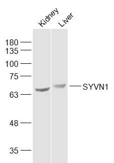 Sample:
Kidney (Mouse) Lysate at 40 ug
Liver (Mouse) Lysate at 40 ug
Primary: Anti-SYVN1 (SL0679R) at 1/1000 dilution
Secondary: IRDye800CW Goat Anti-Rabbit IgG at 1/20000 dilution
Predicted band size: 65 kD
Observed band size: 65/70 kD
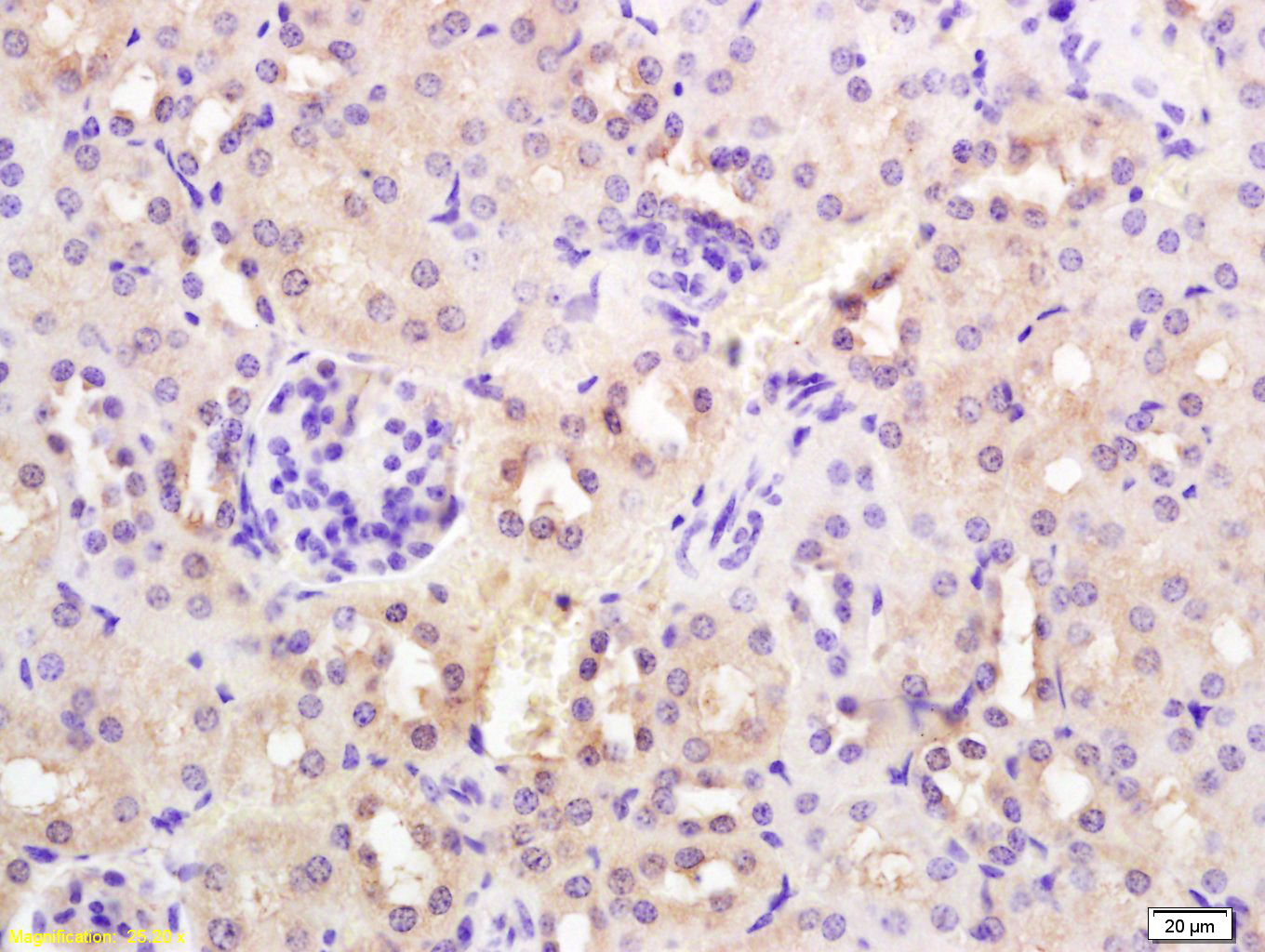 Tissue/cell: mouse kidney tissue; 4% Paraformaldehyde-fixed and paraffin-embedded;
Antigen retrieval: citrate buffer ( 0.01M, pH 6.0 ), Boiling bathing for 15min; Block endogenous peroxidase by 3% Hydrogen peroxide for 30min; Blocking buffer (normal goat serum,C-0005) at 37℃ for 20 min;
Incubation: Anti-SYVN1/HRD1 Polyclonal Antibody, Unconjugated(SL0679R) 1:200, overnight at 4°C, followed by conjugation to the secondary antibody(SP-0023) and DAB(C-0010) staining
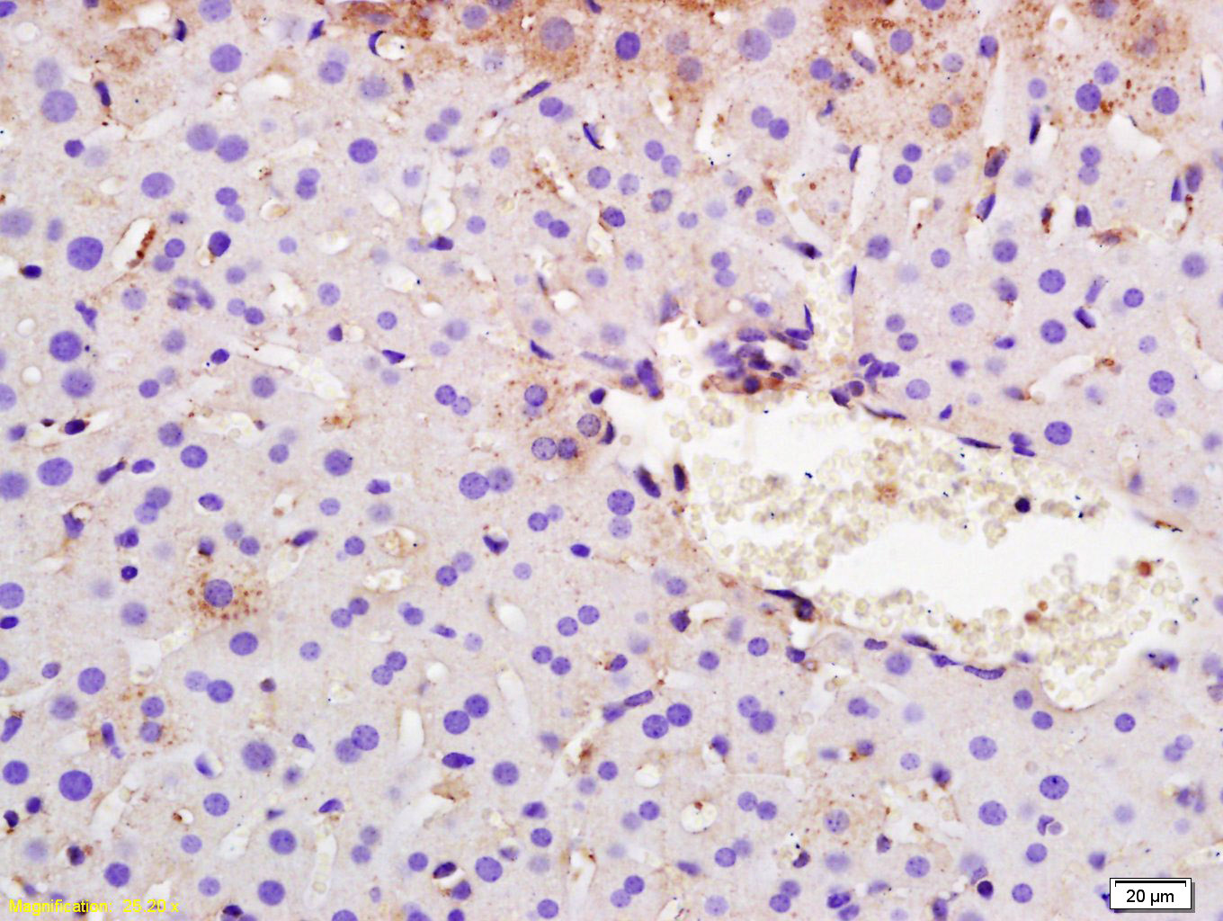 Tissue/cell: mouse liver tissue; 4% Paraformaldehyde-fixed and paraffin-embedded;
Antigen retrieval: citrate buffer ( 0.01M, pH 6.0 ), Boiling bathing for 15min; Block endogenous peroxidase by 3% Hydrogen peroxide for 30min; Blocking buffer (normal goat serum,C-0005) at 37℃ for 20 min;
Incubation: Anti-SYVN1/HRD1 Polyclonal Antibody, Unconjugated(SL0679R) 1:200, overnight at 4°C, followed by conjugation to the secondary antibody(SP-0023) and DAB(C-0010) staining
 The figure annotation:
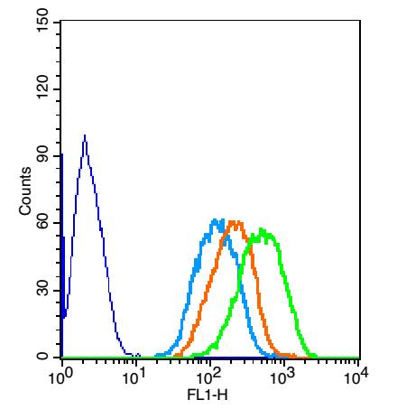
The blue histogram is unstained cells.
The Wathet Blue histogram is cells stained with secondary antibody (SL0295G-FITC) alone.
The Orange histogram is cells stained with rabbit IgG isotype control antibody(SL0295P)plus secondary antibody.
The green histogram is cells stained with Rabbit Anti-SYVN1 antibody (SL0679R) plus secondary antibody.
Positive control: Hepg2 cells
Concentration: 1:50
Cartpieces
Totalgoods,subtotals:¥Checkout
Bought notes(bought amounts latest0)
No one bought this product
User Comment(Total0User Comment Num)
- No comment


 +86 571 56623320
 +86 18668110335

