
Rabbit Anti-C-jun antibody
Transcription factor AP-1; Jun oncogene; JUN; AP 1; AP1; AP-1; Enhancer Binding Protein AP1; Jun Activation Domain Binding Protein; JUN protein; JUNC; p39; Proto oncogene cJun; Transcription Factor AP1; V jun avian sarcoma virus 17 oncogene homolog; vJun
View History [Clear]
Details
Product Name C-jun Chinese Name 原癌基因蛋白/活化蛋白1抗体 Alias Transcription factor AP-1; Jun oncogene; JUN; AP 1; AP1; AP-1; Enhancer Binding Protein AP1; Jun Activation Domain Binding Protein; JUN protein; JUNC; p39; Proto oncogene cJun; Transcription Factor AP1; V jun avian sarcoma virus 17 oncogene homolog; vJun Avian Sarcoma Virus 17 Oncogene Homolog; JUN_HUMAN; Activator 1; Proto-oncogene c-Jun; V-jun avian sarcoma virus 17 oncogene homolog. literatures Research Area Tumour Cell biology Signal transduction transcriptional regulatory factor Kinases and Phosphatases Immunogen Species Rabbit Clonality Polyclonal React Species Human, Mouse, Rat, (predicted: Chicken, Dog, Pig, Cow, Sheep, ) Applications WB=1:500-2000 ELISA=1:5000-10000 IHC-P=1:100-500 IHC-F=1:100-500 Flow-Cyt=1μg/Test IF=1:100-500 (Paraffin sections need antigen repair)
not yet tested in other applications.
optimal dilutions/concentrations should be determined by the end user.Theoretical molecular weight 36kDa Detection molecular weight 43/36 kDa Cellular localization The nucleus Form Liquid Concentration 1mg/ml immunogen KLH conjugated synthetic peptide derived from human Transcription factor AP-1: 31-331/331 Lsotype IgG Purification affinity purified by Protein A Buffer Solution 0.01M TBS(pH7.4) with 1% BSA, 0.03% Proclin300 and 50% Glycerol. Storage Shipped at 4℃. Store at -20 °C for one year. Avoid repeated freeze/thaw cycles. Attention This product as supplied is intended for research use only, not for use in human, therapeutic or diagnostic applications. PubMed PubMed Product Detail The human protooncogene JUN is the putative transforming gene of avian sarcoma virus 17, and it encodes a protein which is highly homologous to the viral protein. cJun (previously known as the Fos binding protein p39) and c Fos form a complex in the nucleus. AP 1 (activating protein 1) is a collective term referring to these dimeric transcription factors composed of Jun, Fos or ATF subunits that bind to a common DNA site, the AP1 binding site. AP 1 proteins, mostly the Jun group, regulate the expression and function of cell cycle regulators such as Cyclin D1, p53, p21 (cip1/waf1), p19 (ARF) and p16. Fos and Jun proto oncogene expression is induced transiently by a variety of extracellular stimuli associated with mitogenesis, differentiation processes or depolarization of neurons. JUN has been mapped to 1p32 to p31, a chromosomal region involved in both translocations and deletions in human malignancies.
Function:
Transcription factor that recognizes and binds to the enhancer heptamer motif 5'-TGA[CG]TCA-3'. Promotes activity of NR5A1 when phosphorylated by HIPK3 leading to increased steroidogenic gene expression upon cAMP signaling pathway stimulation.
Subunit:
Heterodimer with either FOS or BATF3 or ATF7. The ATF7/JUN heterodimer is essential for ATF7 transactivation activity. Interacts with DSIPI; the interaction inhibits the binding of active AP1 to its target DNA. Interacts with HIVEP3 and MYBBP1A. Interacts with SP1, SPIB and TCF20. Interacts with COPS5; the interaction leads indirectly to its phosphorylation. Component of the SMAD3/SMAD4/JUN/FOS/complex which forms at the AP1 promoter site. The SMAD3/SMAD4 heterodimer acts syngernistically with the JUN/FOS heterodimer to activate transcription in response to TGF-beta. Interacts (via its basic DNA binding and leucine zipper domains) with SMAD3 (via an N-terminal domain); the interaction is required for TGF-beta-mediated transactivation of the SMAD3/SMAD4/JUN/FOS/complex. Interacts with RNF187. Binds to HIPK3.
Subcellular Location:
Nucleus.
Post-translational modifications:
Phosphorylated by CaMK4 and PRKDC; phosphorylation enhances the transcriptional activity. Phosphorylated by HIPK3. [PTM] Phosphorylated at Thr-239, Ser-243 and Ser-249 by GSK3B; phosphorylation reduces its ability to bind DNA.
Phosphorylated by PAK2 at Thr-2, Thr-8, Thr-89, Thr-93 and Thr-286 thereby promoting JUN-mediated cell proliferation andtransformation.
Similarity:
Belongs to the bZIP family. Jun subfamily.
Contains 1 bZIP domain.
SWISS:
P05412
Gene ID:
3725
Database links:Entrez Gene: 3725 Human
Entrez Gene: 16476 Mouse
Omim: 165160 Human
SwissProt: P05412 Human
SwissProt: P05627 Mouse
Unigene: 525704 Human
Unigene: 696684 Human
Unigene: 275071 Mouse
Unigene: 93714 Rat
transcriptional regulatory factor(Transcriptin Regulators)
C-jun(Oncoprotein C-jun:active protein 1)基因与鸟类肉瘤病毒17的转化基因具有同源性,是早期应答基因家族成员之一。主要用于各种恶性Tumour的研究。C-jun又称应激活化蛋白激酶.Product Picture 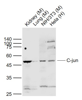 Sample:
Lane 1: Kidney (Mouse) Lysate at 40 ug
Lane 2: Lung (Mouse) Lysate at 40 ug
Lane 3: NIH/3T3 (Mouse) Cell Lysate at 30 ug
Lane 4: Hela (Human) Cell Lysate at 30 ug
Primary: Anti-C-jun (SL0670R) at 1/1000 dilution
Secondary: IRDye800CW Goat Anti-Rabbit IgG at 1/20000 dilution
Predicted band size: 43/36 kD
Observed band size: 45 kD
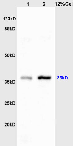 Sample:
Liver(Rat)lysate at 30ug;
Brain(Rat) lysates at 30ug;
Primary: Anti-C-jun/AP-1 (SL0670R) at 1:200;
Secondary: HRP conjugated Goat-Anti-Rabbit IgG(SL0295G-HRP) at 1: 3000;
Predicted band size : 36kD
Observed band size : 36kD
 Paraformaldehyde-fixed, paraffin embedded (Mouse brain); Antigen retrieval by boiling in sodium citrate buffer (pH6.0) for 15min; Block endogenous peroxidase by 3% hydrogen peroxide for 20 minutes; Blocking buffer (normal goat serum) at 37°C for 30min; Antibody incubation with (C-jun) Polyclonal Antibody, Unconjugated (SL0670R) at 1:400 overnight at 4°C, followed by a conjugated secondary antibody (sp-0023) for 20 minutes and DAB staining.
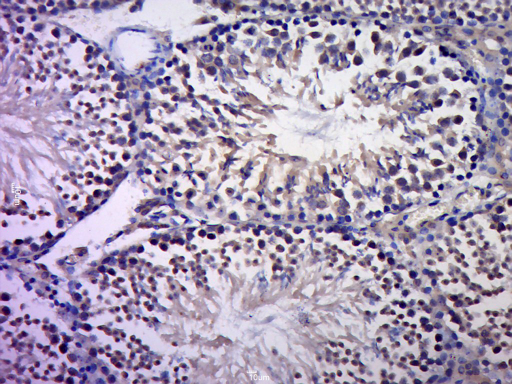 Paraformaldehyde-fixed, paraffin embedded (Rat testis); Antigen retrieval by boiling in sodium citrate buffer (pH6.0) for 15min; Block endogenous peroxidase by 3% hydrogen peroxide for 20 minutes; Blocking buffer (normal goat serum) at 37°C for 30min; Antibody incubation with (C-jun) Polyclonal Antibody, Unconjugated (SL0670R) at 1:400 overnight at 4°C, followed by a conjugated secondary (sp-0023) for 20 minutes and DAB staining.
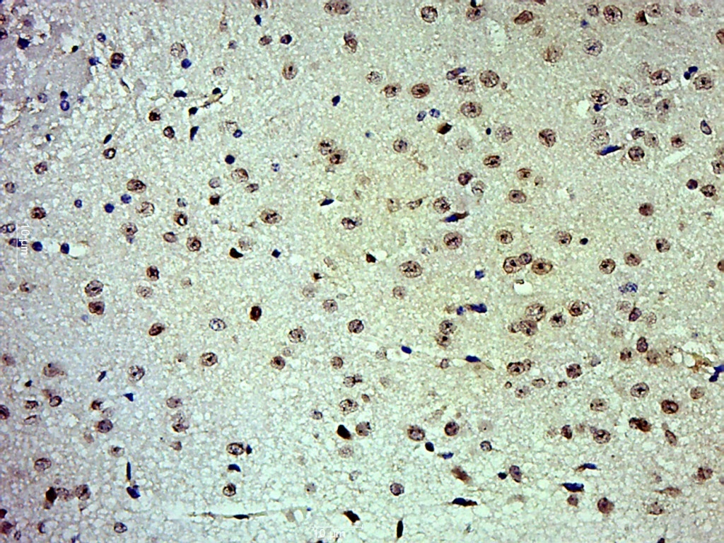 Paraformaldehyde-fixed, paraffin embedded (Mouse brain); Antigen retrieval by boiling in sodium citrate buffer (pH6.0) for 15min; Block endogenous peroxidase by 3% hydrogen peroxide for 20 minutes; Blocking buffer (normal goat serum) at 37°C for 30min; Antibody incubation with (C-jun) Polyclonal Antibody, Unconjugated (SL0670R) at 1:500 overnight at 4°C, followed by a conjugated secondary (sp-0023) for 20 minutes and DAB staining.
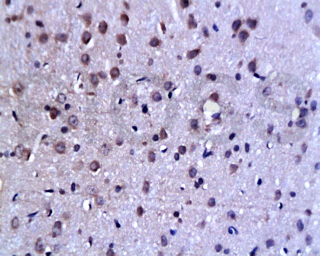 Tissue/cell: rat brain tissue; 4% Paraformaldehyde-fixed and paraffin-embedded;
Antigen retrieval: citrate buffer ( 0.01M, pH 6.0 ), Boiling bathing for 15min; Block endogenous peroxidase by 3% Hydrogen peroxide for 30min; Blocking buffer (normal goat serum,C-0005) at 37℃ for 20 min;
Incubation: Anti-C-Jun Polyclonal Antibody, Unconjugated(SL0670R) 1:200, overnight at 4°C, followed by conjugation to the secondary antibody(SP-0023) and DAB(C-0010) staining
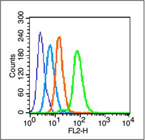 Blank control (blue line): HepG2 (blue).
Primary Antibody (green line): Rabbit Anti-C-jun antibody (SL4601R)
Dilution: 1μg /10^6 cells;
Isotype Control Antibody (orange line): Rabbit IgG .
Secondary Antibody (white blue line): Goat anti-rabbit IgG-PE
Dilution: 1μg /test.
Protocol
The cells were fixed with 70% methanol (Overnight at 4℃) and then permeabilized with 90% ice-cold methanol for 20 min at -20℃. Cells stained with Primary Antibody for 30 min at room temperature. The cells were then incubated in 1 X PBS/2%BSA/10% goat serum to block non-specific protein-protein interactions followed by the antibody for 15 min at room temperature. The secondary antibody used for 40 min at room temperature. Acquisition of 20,000 events was performed.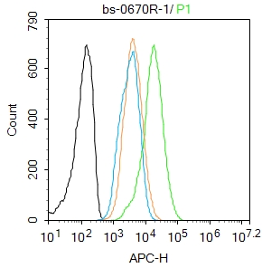 Blank control: Hela.
Primary Antibody (green line): Rabbit Anti-C-jun antibody (SL0670R)
Dilution: 1μg /10^6 cells;
Isotype Control Antibody (orange line): Rabbit IgG .
Secondary Antibody : Goat anti-rabbit IgG-AF647
Dilution: 1μg /test.
Protocol
The cells were fixed with 4% PFA (10min at room temperature)and then permeabilized with 90% ice-cold methanol for 20 min at -20℃. The cells were then incubated in 5%BSA to block non-specific protein-protein interactions for 30 min at room temperature .Cells stained with Primary Antibody for 30 min at room temperature. The secondary antibody used for 40 min at room temperature. Acquisition of 20,000 events was performed.
Cartpieces
Totalgoods,subtotals:¥Checkout
Bought notes(bought amounts latest0)
No one bought this product
User Comment(Total0User Comment Num)
- No comment


 +86 571 56623320
 +86 18668110335

