
Rabbit Anti-Fibronectin/FN1 antibody
CIG; Cold insoluble globulin; DKFZp686F10164; DKFZp686H0342; DKFZp686I1370; DKFZp686O13149; ED B; ED-B; fibronectin 1; FINC; FN 1; FN; FN1; FNZ; GFND; GFND2; LETS; Migration stimulating factor; MSF; Transformation sensitive protein; FINC_HUMAN; Fibronecti
View History [Clear]
Details
Product Name Fibronectin/FN1 Chinese Name 纤维连接蛋白抗体 Alias CIG; Cold insoluble globulin; DKFZp686F10164; DKFZp686H0342; DKFZp686I1370; DKFZp686O13149; ED B; ED-B; fibronectin 1; FINC; FN 1; FN; FN1; FNZ; GFND; GFND2; LETS; Migration stimulating factor; MSF; Transformation sensitive protein; FINC_HUMAN; Fibronectin; Cold-insoluble globulin; Ugl-Y3. literatures Research Area Tumour Cell biology immunology Signal transduction transcriptional regulatory factor Cell adhesion molecule Immunogen Species Rabbit Clonality Polyclonal React Species Human, Mouse, Rat, Cow, (predicted: Dog, ) Applications ELISA=1:5000-10000 IHC-P=1:100-500 IHC-F=1:100-500 IF=1:200-800 (Paraffin sections need antigen repair)
not yet tested in other applications.
optimal dilutions/concentrations should be determined by the end user.Theoretical molecular weight 259kDa Cellular localization Extracellular matrix Secretory protein Form Liquid Concentration 1mg/ml immunogen KLH conjugated synthetic peptide derived from human Fibronectin/Ugl-Y3: 1201-1300/2386 Lsotype IgG Purification affinity purified by Protein A Buffer Solution 0.01M TBS(pH7.4) with 1% BSA, 0.03% Proclin300 and 50% Glycerol. Storage Shipped at 4℃. Store at -20 °C for one year. Avoid repeated freeze/thaw cycles. Attention This product as supplied is intended for research use only, not for use in human, therapeutic or diagnostic applications. PubMed PubMed Product Detail This gene encodes fibronectin, a glycoprotein present in a soluble dimeric form in plasma, and in a dimeric or multimeric form at the cell surface and in extracellular matrix. Fibronectin is involved in cell adhesion and migration processes including embryogenesis, wound healing, blood coagulation, host defense, and metastasis. The gene has three regions subject to alternative splicing, with the potential to produce 20 different transcript variants. However, the full-length nature of some variants has not been determined. [provided by RefSeq, Jul 2008].
Function:
Fibronectins bind cell surfaces and various compounds including collagen, fibrin, heparin, DNA, and actin. Fibronectins are involved in cell adhesion, cell motility, opsonization, wound healing, and maintenance of cell shape.
Anastellin binds fibronectin and induces fibril formation. This fibronectin polymer, named superfibronectin, exhibits enhanced adhesive properties. Both anastellin and superfibronectin inhibit tumor growth, angiogenesis and metastasis. Anastellin activates p38 MAPK and inhibits lysophospholipid signaling.
Subunit:
Mostly heterodimers or multimers of alternatively spliced variants, connected by 2 disulfide bonds near the carboxyl ends; to a lesser extent homodimers. Interacts with FBLN1, AMBP, TNR, LGALS3BP and COL13A1. Interacts with FBLN7. Interacts with COMP. Interacts with S.aureus fnbA. Interacts with TNR; the interaction inhibits cell adhesion and neurite outgrowth. Interacts with FST3.
Subcellular Location:
Secreted, extracellular space, extracellular matrix.
Tissue Specificity:
Plasma FN (soluble dimeric form) is secreted by hepatocytes. Cellular FN (dimeric or cross-linked multimeric forms), made by fibroblasts, epithelial and other cell types, is deposited as fibrils in the extracellular matrix. Ugl-Y1, Ugl-Y2 and Ugl-Y3 are found in urine.
Post-translational modifications:
Sulfated.
It is not known whether both or only one of Thr-2064 and Thr-2065 are/is glycosylated.
Forms covalent cross-links mediated by a transglutaminase, such as F13A or TGM2, between a glutamine and the epsilon-amino group of a lysine residue, forming homopolymers and heteropolymers (e.g. fibrinogen-fibronectin, collagen-fibronectin heteropolymers).
Phosphorylation sites are present in the extracellular medium.
Proteolytic processing produces the C-terminal NC1 peptide, anastellin.
DISEASE:
Defects in FN1 are the cause of glomerulopathy with fibronectin deposits type 2 (GFND2) [MIM:601894]; also known as familial glomerular nephritis with fibronectin deposits or fibronectin glomerulopathy. GFND is a genetically heterogeneous autosomal dominant disorder characterized clinically by proteinuria, microscopic hematuria, and hypertension that leads to end-stage renal failure in the second to fifth decade of life.
Similarity:
Contains 12 fibronectin type-I domains. Contains 2 fibronectin type-II domains. Contains 16 fibronectin type-III domains.
SWISS:
P11276
Gene ID:
2335
Database links:
Entrez Gene: 2335 Human
Entrez Gene: 14268 Mouse
Omim: 135600 Human
SwissProt: P02751 Human
SwissProt: P11276 Mouse
细胞粘附蛋白(Call Adhesion Protein)
FaN(Fibronectin)是一种细胞粘附蛋白,参与细胞粘附,分化,移动,伤口愈合,凝血和机体防卫。主要分布于结缔组织及大部分基底膜上,作为判断良恶性上皮源性Tumour(如乳腺癌等)的一个主要参考指标,亦可作为恶性Tumour转移和病人预后的判断指标
Product Picture 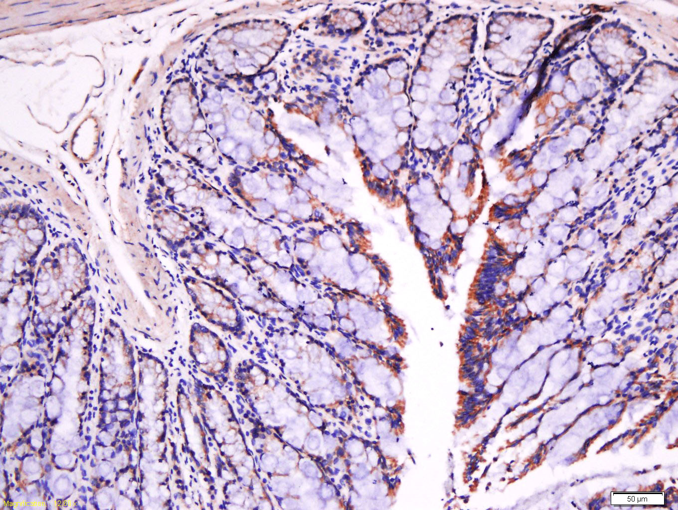 Tissue/cell: rat intestine tissue; 4% Paraformaldehyde-fixed and paraffin-embedded;
Antigen retrieval: citrate buffer ( 0.01M, pH 6.0 ), Boiling bathing for 15min; Block endogenous peroxidase by 3% Hydrogen peroxide for 30min; Blocking buffer (normal goat serum,C-0005) at 37℃ for 20 min;
Incubation: Anti-Fibronectin/FN1 Polyclonal Antibody, Unconjugated(SL0666R) 1:500, overnight at 4°C, followed by conjugation to the secondary antibody(SP-0023) and DAB(C-0010) staining
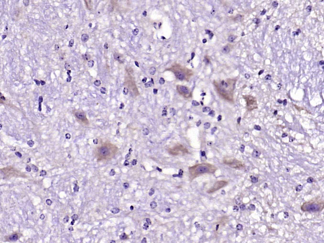 Paraformaldehyde-fixed, paraffin embedded (mouse cerebellum); Antigen retrieval by boiling in sodium citrate buffer (pH6.0) for 15min; Block endogenous peroxidase by 3% hydrogen peroxide for 20 minutes; Blocking buffer (normal goat serum) at 37°C for 30min; Antibody incubation with (Fibronectin) Polyclonal Antibody, Unconjugated (SL0666R) at 1:200 overnight at 4°C, followed by operating according to SP Kit(Rabbit) (sp-0023) instructionsand DAB staining.
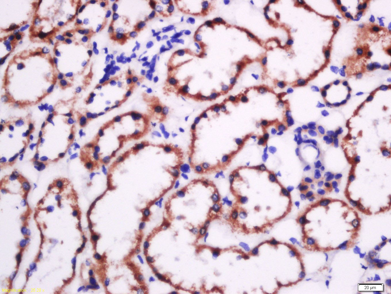 Tissue/cell: human kidney tissue; 4% Paraformaldehyde-fixed and paraffin-embedded;
Antigen retrieval: citrate buffer ( 0.01M, pH 6.0 ), Boiling bathing for 15min; Block endogenous peroxidase by 3% Hydrogen peroxide for 30min; Blocking buffer (normal goat serum,C-0005) at 37℃ for 20 min;
Incubation: Anti-Fibronectin/FN1 Polyclonal Antibody, Unconjugated(SL0666R) 1:500, overnight at 4°C, followed by conjugation to the secondary antibody(SP-0023) and DAB(C-0010) staining
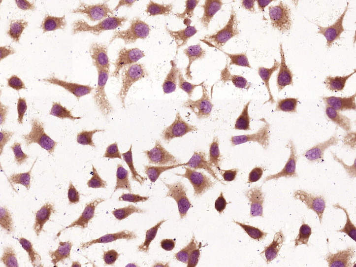 Tissue/cell: HepG2 cell; 4% Paraformaldehyde-fixed; Triton X-100 at room temperature for 20 min; Block endogenous peroxidase by 3% hydrogen peroxide for 20 minutes; Blocking buffer (normal goat serum) at 37°C for 30min; Antibody incubation with (Fibronectin/FN1) Polyclonal Antibody, Unconjugated (SL0666R) at 1:400 overnight at 4°C, followed by operating according to SP Kit(Rabbit) (sp-0023) instructionsand DAB staining.
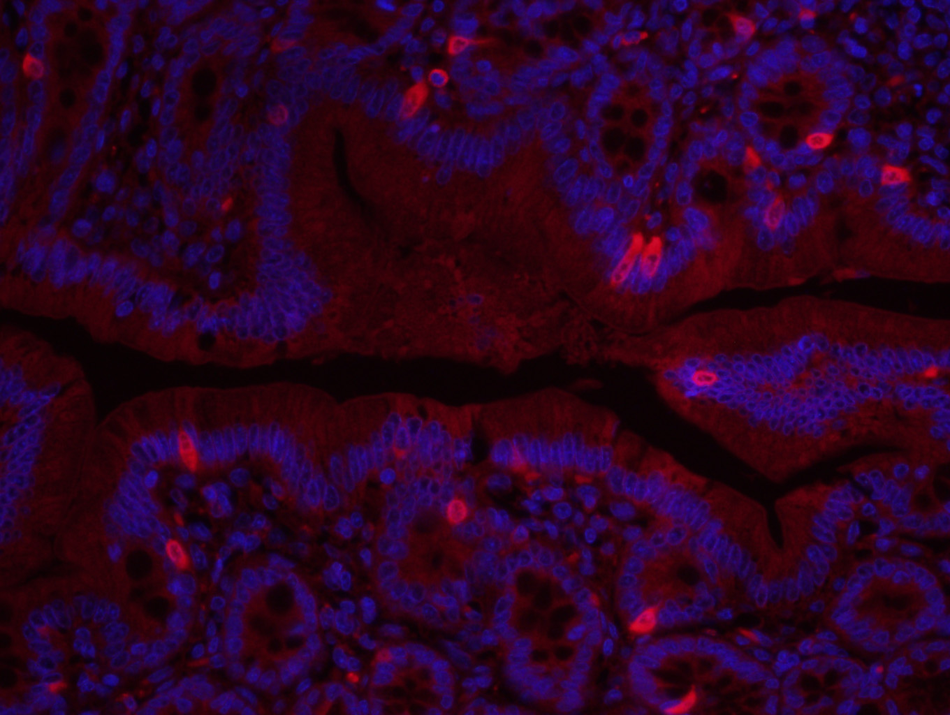 Paraformaldehyde-fixed, paraffin embedded (Rat colon); Antigen retrieval by boiling in sodium citrate buffer (pH6.0) for 15min; Block endogenous peroxidase by 3% hydrogen peroxide for 20 minutes; Blocking buffer (normal goat serum) at 37°C for 30min; Incubation: Anti-Fibronectin Polyclonal Antibody, conjugated (SL0666R-AF647) 1:400, 1.5 hours at 37°C; DAPI (5ug/ml, blue, C-0033) was used to stain the cell nuclei.
Cartpieces
Totalgoods,subtotals:¥Checkout
Bought notes(bought amounts latest0)
No one bought this product
User Comment(Total0User Comment Num)
- No comment


 +86 571 56623320
 +86 18668110335

