
Rabbit Anti-HMGB1 antibody
High mobility group protein B1; Amphoterin; High mobility group 1; High Mobility Group Box 1; High mobility group protein 1; HMG3; HMGB 1; HMGB-1; Hmgb1 protein; Nonhistone chromosomal protein HMG1; SBP 1; SBP-1; Sulfoglucuronyl carbohydrate binding prote
View History [Clear]
Details
Product Name HMGB1 Chinese Name 高迁移率族蛋白B1抗体 Alias High mobility group protein B1; Amphoterin; High mobility group 1; High Mobility Group Box 1; High mobility group protein 1; HMG3; HMGB 1; HMGB-1; Hmgb1 protein; Nonhistone chromosomal protein HMG1; SBP 1; SBP-1; Sulfoglucuronyl carbohydrate binding protein; HMGB1_HUMAN. literatures Research Area Tumour Cell biology immunology transcriptional regulatory factor Binding protein Immunogen Species Rabbit Clonality Polyclonal React Species Human, Mouse, Rat, (predicted: Cow, ) Applications WB=1:500-2000 ELISA=1:5000-10000 IHC-P=1:100-500 IHC-F=1:100-500 Flow-Cyt=1μg/Test ICC=1:100 IF=1:100-500 (Paraffin sections need antigen repair)
not yet tested in other applications.
optimal dilutions/concentrations should be determined by the end user.Theoretical molecular weight 25kDa Cellular localization The nucleus Form Liquid Concentration 1mg/ml immunogen KLH conjugated synthetic peptide derived from human HMGB1: 75-170/215 Lsotype IgG Purification affinity purified by Protein A Buffer Solution 0.01M TBS(pH7.4) with 1% BSA, 0.03% Proclin300 and 50% Glycerol. Storage Shipped at 4℃. Store at -20 °C for one year. Avoid repeated freeze/thaw cycles. Attention This product as supplied is intended for research use only, not for use in human, therapeutic or diagnostic applications. PubMed PubMed Product Detail High Mobility Group Box-1 (HMGB1) is a cytokine implicated in the pathogenesis of rheumatoid arthritis (RA) and other inflammatory diseases. The cholinergic anti-inflammatory pathway, a vagus nerve dependent mechanism, inhibits HMGB1 release in experimental disease models
Function:
DNA binding proteins that associates with chromatin and has the ability to bend DNA. Binds preferentially single-stranded DNA. Involved in V(D)J recombination by acting as a cofactor of the RAG complex. Acts by stimulating cleavage and RAG protein binding at the 23 bp spacer of conserved recombination signal sequences (RSS). Heparin-binding protein that has a role in the extension of neurite-type cytoplasmic processes in developing cells.
Subunit:
Component of the RAG complex composed of core components RAG1 and RAG2, and associated component HMGB1 or HMGB2.
Subcellular Location:
Nucleus. Chromosome.
Similarity:
Belongs to the HMGB family.
Contains 2 HMG box DNA-binding domains.
SWISS:
P09429
Gene ID:
3146
Database links:
Entrez Gene: 3146 Human
Entrez Gene: 100862258 Mouse
Entrez Gene: 15289 Mouse
Omim: 163905 Human
SwissProt: P09429 Human
近来的研究表明称之为高迁移率族蛋白B-1(HMG-B1)的核内结构蛋白在核外表达时是一种有效的早期炎症介质。
高迁移性B1组蛋白(HMGB1): 是一种核Binding protein,在DNA重组、修复、复制和基因转录中起作用。HMGB1也是巨噬细胞分泌的一种介质。
此外,高迁移性B1组蛋白亦被受刺激的巨噬细胞或单核细胞主动分泌。在这一主动分泌过程中,HMGB1首先经乙酰化并由核内转移至溶酶体内,继而在ATP和溶血磷脂胆碱两种分泌信号指导下转移至细胞外。由坏死细胞被动释放的HMGB1和炎症细胞主动分泌的HMGB1存在分子上的差异。胞外的HMGB1可作为cell factor参与信号传导,因为它既可识别Toll 样受体(TLR)家族的一些成员,又能与识别晚期糖基化终末产物受体(RAGE)相作用。
HMGB1能启动炎症反应,包括产生多种cell factor、对某些Stem cells产生趋化作用、诱导血管粘附分子、削弱肠epithelial cells的功能等。Product Picture 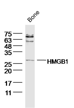 Sample: bone (mouse) Lysate at 40 ug
Primary: Anti- HMGB1(SL0664R)at 1/300 dilution
Secondary: IRDye800CW Goat Anti-Rabbit IgG at 1/20000 dilution
Predicted band size: 25 kD
Observed band size: 27kD
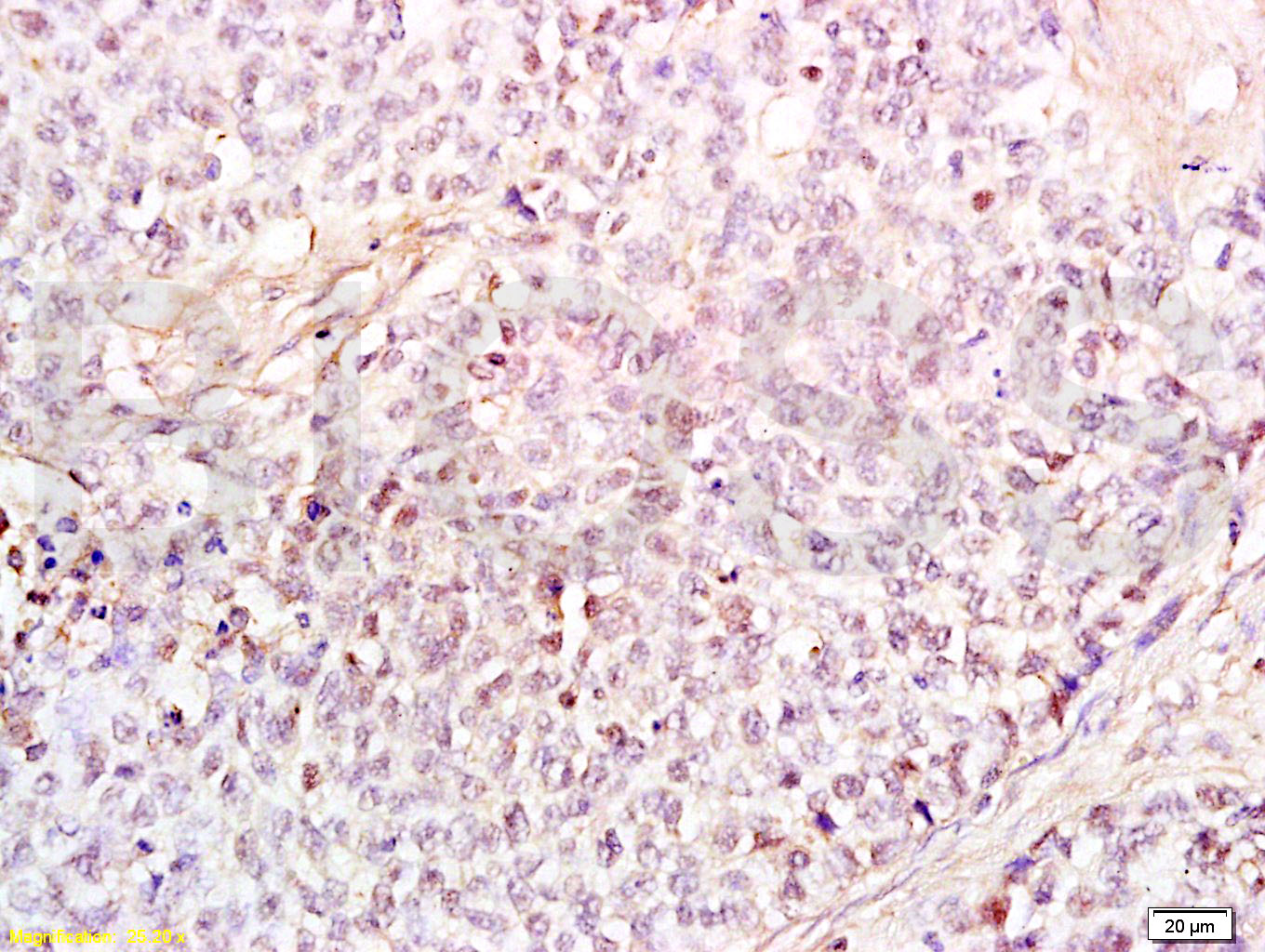 Tissue/cell: human endometrium carcinoma; 4% Paraformaldehyde-fixed and paraffin-embedded;
Antigen retrieval: citrate buffer ( 0.01M, pH 6.0 ), Boiling bathing for 15min; Block endogenous peroxidase by 3% Hydrogen peroxide for 30min; Blocking buffer (normal goat serum,C-0005) at 37℃ for 20 min;
Incubation: Anti-HMGB1 Polyclonal Antibody, Unconjugated(SL0664R) 1:200, overnight at 4癈, followed by conjugation to the secondary antibody(SP-0023) and DAB(C-0010) staining
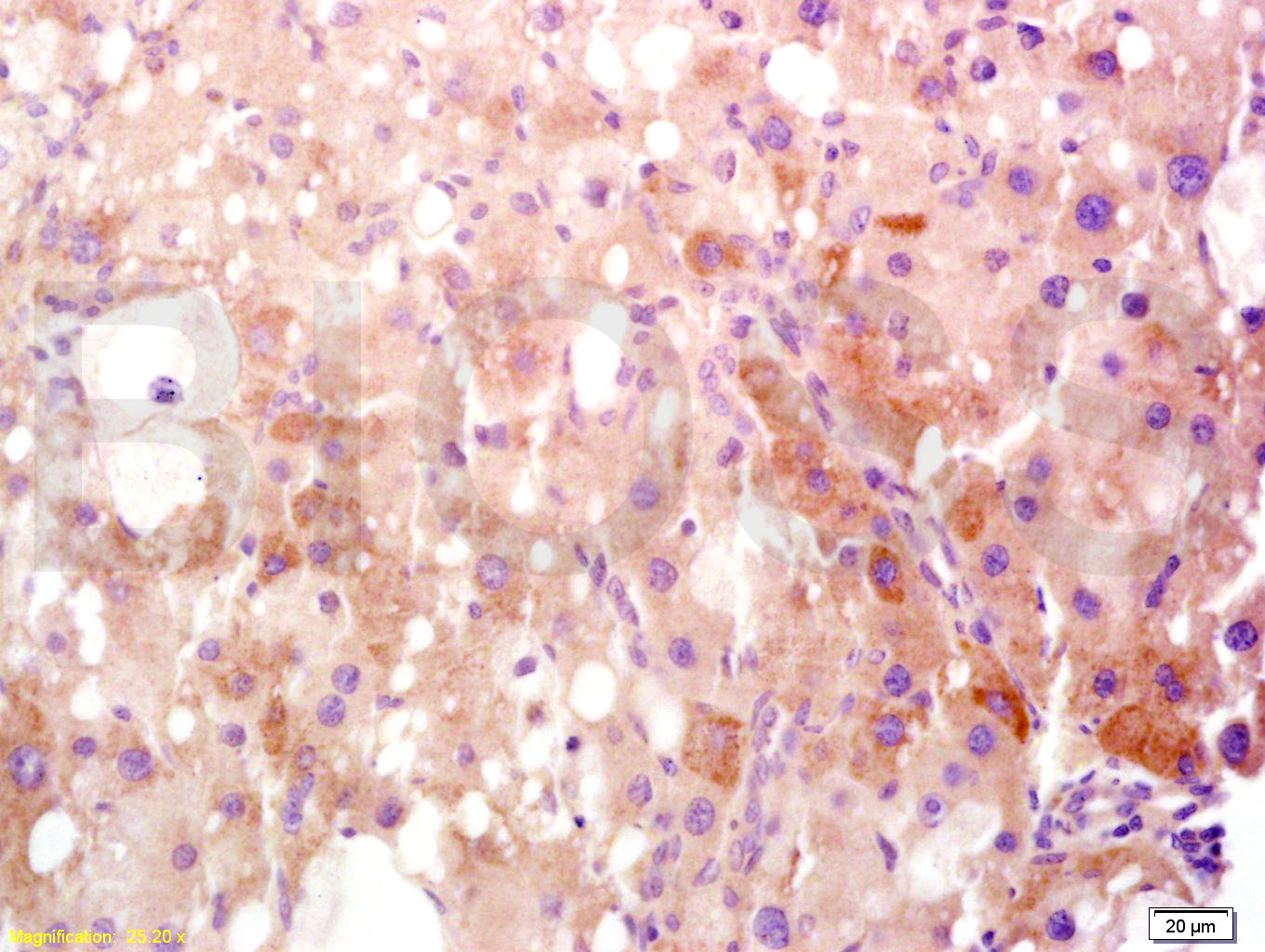 Tissue/cell: rat liver tissue; 4% Paraformaldehyde-fixed and paraffin-embedded;
Antigen retrieval: citrate buffer ( 0.01M, pH 6.0 ), Boiling bathing for 15min; Block endogenous peroxidase by 3% Hydrogen peroxide for 30min; Blocking buffer (normal goat serum,C-0005) at 37℃ for 20 min;
Incubation: Anti-HMGB1 Polyclonal Antibody, Unconjugated(SL0664R) 1:400, overnight at 4癈, followed by conjugation to the secondary antibody(SP-0023) and DAB(C-0010) staining
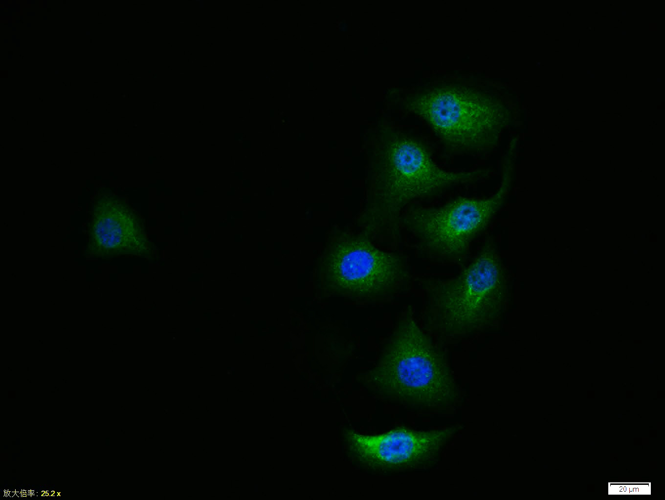 HepG2 cell; 4% Paraformaldehyde-fixed; Triton X-100 at room temperature for 20 min; Blocking buffer (normal goat serum, C-0005) at 37°C for 20 min; Antibody incubation with (HMGB1) polyclonal Antibody, Unconjugated (SL0664R) 1:100, 90 minutes at 37°C; followed by a conjugated Goat Anti-Rabbit IgG antibody at 37°C for 90 minutes, DAPI (blue, C02-04002) was used to stain the cell nuclei.
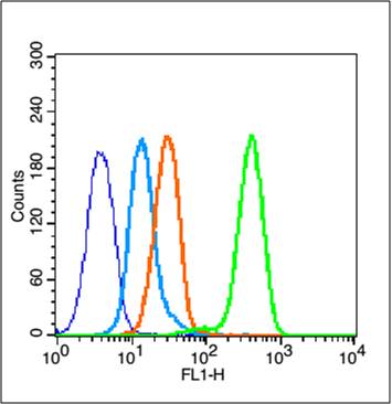 Blank control (blue line): MCF7 (blue).
Primary Antibody (green line): Rabbit Anti-HMGB1 antibody (SL0664R)
Dilution: 1μg /10^6 cells;
Isotype Control Antibody (orange line): Rabbit IgG .
Secondary Antibody (white blue line): Goat anti-rabbit IgG-FITC
Dilution: 1μg /test.
Protocol
The cells were fixed with 70% ethanol (Overnight at 4℃) and then permeabilized with 90% ice-cold methanol for 30 min on ice. Cells stained with Primary Antibody for 30 min at room temperature. The cells were then incubated in 1 X PBS/2%BSA/10% goat serum to block non-specific protein-protein interactions followed by the antibody for 15 min at room temperature. The secondary antibody used for 40 min at room temperature. Acquisition of 20,000 events was performed.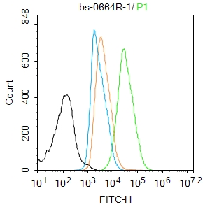 Blank control:HL-60.
Primary Antibody (green line): Rabbit Anti-HMGB1 antibody (SL0664R)
Dilution: 1μg /10^6 cells;
Isotype Control Antibody (orange line): Rabbit IgG .
Secondary Antibody : Goat anti-rabbit IgG-AF488
Dilution: 1μg /test.
Protocol
The cells were fixed with 4% PFA (10min at room temperature)and then permeabilized with 90% ice-cold methanol for 20 min at-20℃. The cells were then incubated in 5%BSA to block non-specific protein-protein interactions for 30 min at room temperature .Cells stained with Primary Antibody for 30 min at room temperature. The secondary antibody used for 40 min at room temperature. Acquisition of 20,000 events was performed.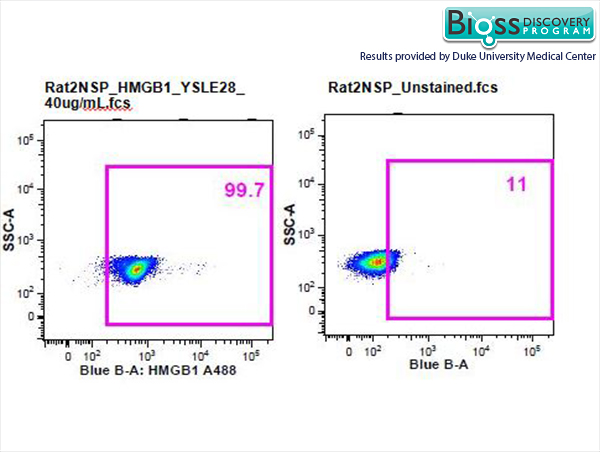 Rat splenocytes stained with Anti- HMGB1 Polyclonal Antibody, A488 Conjugated (SL0664R-A488) at 1:50.
 The figure annotation:
The blue histogram is unstained cells. The Orange histogram is cells stained with Rabbit IgG/FITC (SL0295P-FITC) The green histogram is cells stained with Rabbit Anti-HMGB1/FITC Conjugated antibody (SL0664R-FITC).
Controls
Positive control: HepG 2 cells Isotype control: Cell lines treated with Rabbit IgG/FITC (SL0295P-FITC). instead of the primary antibody to confirm that primary antibody binding is specific. 5μgin 100 μL 1 X PBS containing 0.5% BSA.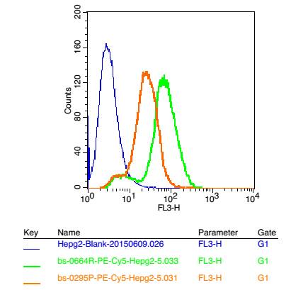 Positive control: HepG2 cells
Concebtration: 5μg/10^6 cells
Incubation conditions: Avoid light , 30 minutes on the ice.
Cartpieces
Totalgoods,subtotals:¥Checkout
Bought notes(bought amounts latest0)
No one bought this product
User Comment(Total0User Comment Num)
- No comment


 +86 571 56623320
 +86 18668110335

