
Rabbit Anti-ADRA1A antibody
ADA1A_HUMAN; Adrenergic alpha 1A receptor; Adrenergic alpha 1C receptor; Adrenergic alpha 1D receptor; alpha 1 Adrenergic Receptor; Alpha 1A adrenergic receptor; Alpha-1A adrenergic receptor; Alpha-1A adrenoreceptor; Alpha-1C adrenergic receptor; Alpha-ad
View History [Clear]
Details
Product Name ADRA1A Chinese Name alpha 1肾上腺素能受体A抗体 Alias ADA1A_HUMAN; Adrenergic alpha 1A receptor; Adrenergic alpha 1C receptor; Adrenergic alpha 1D receptor; alpha 1 Adrenergic Receptor; Alpha 1A adrenergic receptor; Alpha-1A adrenergic receptor; Alpha-1A adrenoreceptor; Alpha-1C adrenergic receptor; Alpha-adrenergic receptor 1c; ADRA1A; ADRA1C; Alpha 1A adrenoceptor; alpha-1A adrenergic receptor isoform 1; adrenergic, alpha-1A-, receptor variant 1; adrenergic, alpha-1A-, receptor variant 3; adrenergic, alpha-1A-, receptor variant 5; adrenergic, alpha-1A-, receptor variant 8; G protein coupled receptor; alpha-1A adrenoceptor; ADRA1L1; ALPHA1AAR. literatures Research Area Cell biology Neurobiology The cell membrane受体 Immunogen Species Rabbit Clonality Polyclonal React Species Human, Mouse, Rat, (predicted: Chicken, Dog, Pig, Cow, Horse, Rabbit, Sheep, Guinea Pig, ) Applications WB=1:500-2000 ELISA=1:5000-10000 IHC-P=1:100-500 IHC-F=1:100-500 IF=1:100-500 (Paraffin sections need antigen repair)
not yet tested in other applications.
optimal dilutions/concentrations should be determined by the end user.Theoretical molecular weight 51kDa Cellular localization The nucleus The cell membrane Form Liquid Concentration 1mg/ml immunogen KLH conjugated synthetic peptide derived from human Alpha-1A adrenergic receptor: 201-300/466 Lsotype IgG Purification affinity purified by Protein A Buffer Solution 0.01M TBS(pH7.4) with 1% BSA, 0.03% Proclin300 and 50% Glycerol. Storage Shipped at 4℃. Store at -20 °C for one year. Avoid repeated freeze/thaw cycles. Attention This product as supplied is intended for research use only, not for use in human, therapeutic or diagnostic applications. PubMed PubMed Product Detail Alpha-1-adrenergic receptors (alpha-1-ARs) are members of the G protein-coupled receptor superfamily. They activate mitogenic responses and regulate growth and proliferation of many cells. There are 3 alpha-1-AR subtypes: alpha-1A, -1B and -1D, all of which signal through the Gq/11 family of G-proteins and different subtypes show different patterns of activation. This gene encodes alpha-1A-adrenergic receptor. Alternative splicing of this gene generates four transcript variants, which encode four different isoforms with distinct C-termini but having similar ligand binding properties. [provided by RefSeq, Jul 2008].
Function:
This alpha-adrenergic receptor mediates its action by association with G proteins that activate a phosphatidylinositol-calcium second messenger system. Its effect is mediated by G(q) and G(11) proteins. Nuclear ADRA1A-ADRA1B heterooligomers regulate phenylephrine(PE)-stimulated ERK signaling in cardiac myocytes.
Subunit:
Homo- and heterooligomer. Heterooligomerizes with ADRA1B homooligomers in cardiac myocytes.
Subcellular Location:
Nucleus membrane; Multi-pass membrane protein. Cell membrane; Multi-pass membrane protein. Note=Location at the nuclear membrane facilitates heterooligomerization and regulates ERK-mediated signaling in cardiac myocytes. Colocalizes with GNAQ, PLCB1 as well as LAP2 at the nuclear membrane of cardiac myocytes.
Tissue Specificity:
Expressed in heart, brain, liver and prostate, but not in kidney, lung, adrenal, aorta and pituitary. Within the prostate, expressed in the apex, base, periurethral and lateral lobe. Isoform 4 is the most abundant isoform expressed in the prostate with high levels also detected in liver and heart.
Post-translational modifications:
C-terminal Ser or Thr residues may be phosphorylated.
Similarity:
Belongs to the G-protein coupled receptor 1 family. Adrenergic receptor subfamily. ADRA1A sub-subfamily.
SWISS:
P35348
Gene ID:
148
Database links:Entrez Gene: 148 Human
Entrez Gene: 11549 Mouse
Omim: 104221 Human
SwissProt: P35348 Human
SwissProt: P97718 Mouse
Unigene: 52931 Human
Unigene: 709175 Human
Unigene: 57064 Mouse
Unigene: 9991 Rat
ADRA1肾上腺素能α1受体位于突触后,在血管平滑肌上,兴奋时可使血管收缩;
alpha 1-adrenergic receptor有兴奋效应也有抑制效应。肾上腺素能受体又可分为α和β两种。alpha 受体与儿茶酚胺结合后,主要是兴奋平滑肌,如血管收缩、子宫收缩和瞳孔开张肌收缩等;但也有抑制作用,如使小肠平滑肌舒张。β受体又可分为β1和β2两个亚型.Product Picture 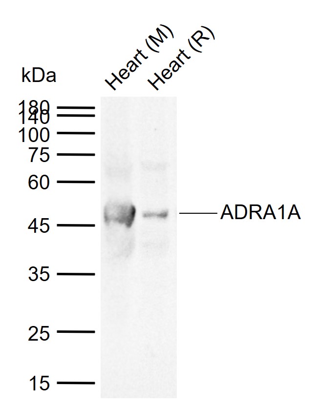 Sample:
Lane 1: Mouse Heart tissue lysates
Lane 2: Rat Heart tissue lysates
Primary: Anti-ADRA1A (SL0600R) at 1/1000 dilution
Secondary: IRDye800CW Goat Anti-Rabbit IgG at 1/20000 dilution
Predicted band size: 51 kDa
Observed band size: 47 kDa
 Sample:
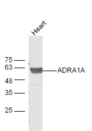
Heart (Mouse) Lysate at 40 ug
Primary: Anti-ADRA1A (SL0600R) at 1/300 dilution
Secondary: IRDye800CW Goat Anti-Rabbit IgG at 1/20000 dilution
Predicted band size: 51 kD
Observed band size: 62 kD
 Sample:
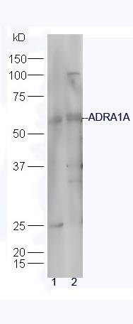
U937 Cell (Human) Lysate at 30 ug
Raji Cell (Human) Lysate at 30 ug
Primary: Anti-ADRA1A (SL0600R) at 1/300 dilution
Secondary: IRDye800CW Goat Anti-Rabbit IgG at 1/20000 dilution
Predicted band size: 51 kD
Observed band size: 55 kD
 Sample:
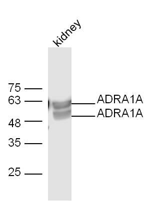
kidney (Mouse) Lysate at 40 ug
Primary: Anti-ADRA1A (SL0600R) at 1/300 dilution
Secondary: IRDye800CW Goat Anti-Rabbit IgG at 1/20000 dilution
Predicted band size: 51 kD
Observed band size: 62 kD
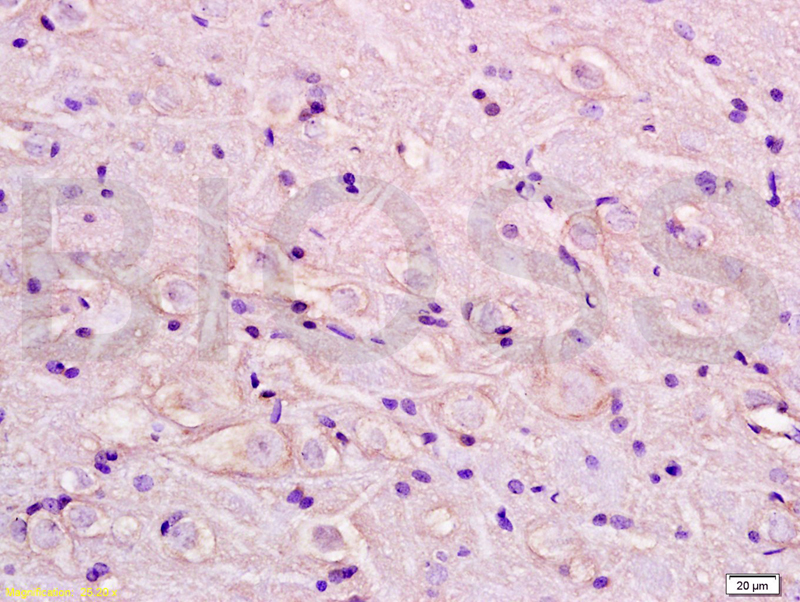 Tissue/cell: rat brain tissue; 4% Paraformaldehyde-fixed and paraffin-embedded;
Antigen retrieval: citrate buffer ( 0.01M, pH 6.0 ), Boiling bathing for 15min; Block endogenous peroxidase by 3% Hydrogen peroxide for 30min; Blocking buffer (normal goat serum,C-0005) at 37℃ for 20 min;
Incubation: Anti-ADRA1/ADRA1B/alpha 1 Adrenergic Receptor Polyclonal Antibody, Unconjugated (SL0600R) 1:200, overnight at 4°C, followed by conjugation to the secondary antibody(SP-0023) and DAB(C-0010) staining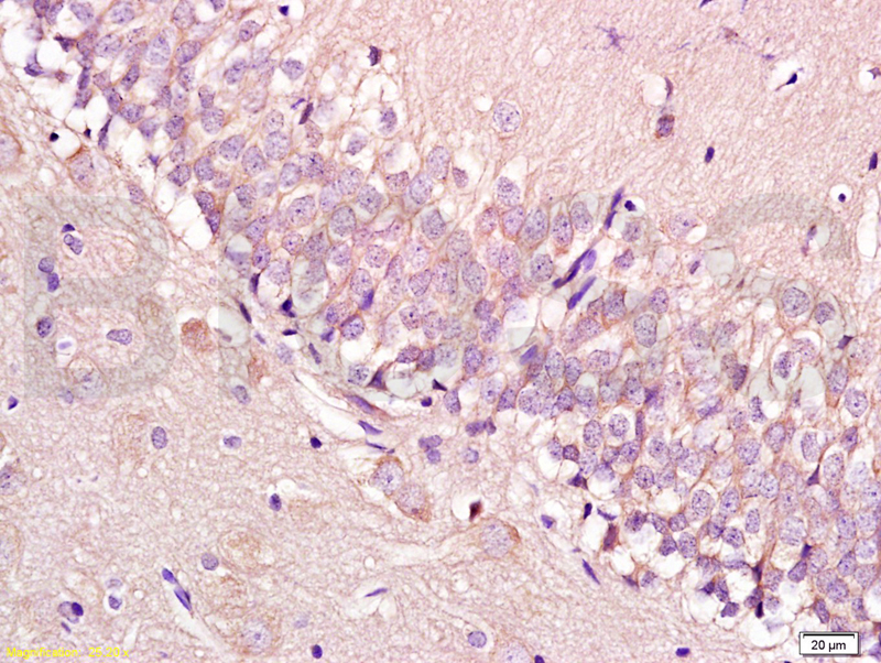
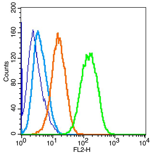 Blank control: U-87MG(blue).
Primary Antibody:Rabbit Anti-ADRA1A antibody(SL0600R), Dilution: 1μg in 100 μL 1X PBS containing 0.5% BSA;
Isotype Control Antibody: Rabbit IgG(orange) ,used under the same conditions );
Secondary Antibody: Goat anti-rabbit IgG-PE(white blue), Dilution: 1:200 in 1 X PBS containing 0.5% BSA.
Protocol
The cells were fixed with 2% paraformaldehyde (10 min) , then permeabilized with 90% ice-cold methanol for 30 min on ice. Primary antibody (SL0600R,1μg /1x10^6 cells) were incubated for 30 min on the ice, followed by 1 X PBS containing 0.5% BSA + 1 0% goat serum (15 min) to block non-specific protein-protein interactions. Then the Goat Anti-rabbit IgG/PE antibody was added into the blocking buffer mentioned above to react with the primary antibody at 1/200 dilution for 30 min on ice. Acquisition of 20,000 events was performed.
Cartpieces
Totalgoods,subtotals:¥Checkout
Bought notes(bought amounts latest0)
No one bought this product
User Comment(Total0User Comment Num)
- No comment


 +86 571 56623320
 +86 18668110335

