
Rabbit Anti-DVL1 antibody
Dishevelled 1; Dishevelled; Dishevelled dsh homolog 1; Dishevelled1; DSH homolog 1; Dvl; MGC54245; Segment polarity protein dishevelled homolog DVL 1; Segment polarity protein dishevelled homolog DVL1; DVL1_HUMAN.
View History [Clear]
Details
Product Name DVL1 Chinese Name 蓬乱蛋白1抗体 Alias Dishevelled 1; Dishevelled; Dishevelled dsh homolog 1; Dishevelled1; DSH homolog 1; Dvl; MGC54245; Segment polarity protein dishevelled homolog DVL 1; Segment polarity protein dishevelled homolog DVL1; DVL1_HUMAN. literatures Research Area immunology Neurobiology Immunogen Species Rabbit Clonality Polyclonal React Species Human, Mouse, Rat, Applications WB=1:500-2000 ELISA=1:5000-10000 IHC-P=1:100-500 IHC-F=1:100-500 IF=1:100-500 (Paraffin sections need antigen repair)
not yet tested in other applications.
optimal dilutions/concentrations should be determined by the end user.Theoretical molecular weight 76kDa Cellular localization cytoplasmic The cell membrane Form Liquid Concentration 1mg/ml immunogen KLH conjugated synthetic peptide derived from human DVL1: 21-100/695 Lsotype IgG Purification affinity purified by Protein A Buffer Solution 0.01M TBS(pH7.4) with 1% BSA, 0.03% Proclin300 and 50% Glycerol. Storage Shipped at 4℃. Store at -20 °C for one year. Avoid repeated freeze/thaw cycles. Attention This product as supplied is intended for research use only, not for use in human, therapeutic or diagnostic applications. PubMed PubMed Product Detail DVL1 may play a role in the signal transduction pathway mediated by multiple Wnt genes. [Subunit] Interacts with BRD7 and INVS. Interacts through its PDZ domain with the C-terminal regions of VANGL1, VANGL2 and CCDC88C/DAPLE. Interacts (via PDZ domain) with NXN (By similarity). Interacts with CXXC4. [Subcellular Location] Cytoplasm (Potential). Belongs to the DSH family.
Function:
Participates in Wnt signaling by binding to the cytoplasmic C-terminus of frizzled family members and transducing the Wnt signal to down-stream effectors. Plays a role both in canonical and non-canonical Wnt signaling. Plays a role in the signal transduction pathways mediated by multiple Wnt genes. Required for LEF1 activation upon WNT1 and WNT3A signaling. DVL1 and PAK1 form a ternary complex with MUSK which is important for MUSK-dependent regulation of AChR clustering during the formation of the neuromuscular junction (NMJ).
Subunit:
Interacts with CXXC4. Interacts (via PDZ domain) with NXN (By similarity). Interacts with BRD7 and INVS. Interacts through its PDZ domain with the C-terminal regions of VANGL1, VANGL2 and CCDC88C/DAPLE. Interacts with ARRB1; the interaction is enhanced by phosphorylation of DVL1. Interacts with CYLD (By similarity). Interacts (via PDZ domain) with RYK. Self-associates (via DIX domain) and forms higher homooligomers. Interacts (via PDZ domain) with DACT1 and FZD7, where DACT1 and FZD7 compete for the same binding site (By similarity). Interacts (via DEP domain) with MUSK; the interaction is direct and mediates the formation a DVL1, MUSK and PAK1 ternary complex involved in AChR clustering (By similarity).
Subcellular Location:
Cell membrane. Cytoplasm, cytosol. Cytoplasmic vesicle. Localizes at the cell membrane upon interaction with frizzled family members.
Post-translational modifications:
Ubiquitinated; undergoes both 'Lys-48'-linked ubiquitination, leading to its subsequent degradation by the ubiquitin-proteasome pathway, and 'Lys-63'-linked ubiquitination. The interaction with INVS is required for ubiquitination. Deubiquitinated by CYLD, which acts on 'Lys-63'-linked ubiquitin chains.
Similarity:
Belongs to the DSH family.
Contains 1 DEP domain.
Contains 1 DIX domain.
Contains 1 PDZ (DHR) domain.
SWISS:
O14640
Gene ID:
1855
Database links:Entrez Gene: 1855 Human
Entrez Gene: 13542 Mouse
Omim: 601365 Human
SwissProt: Q5IS48 Chimpanzee
SwissProt: O14640 Human
SwissProt: P51141 Mouse
Unigene: 741165 Human
Unigene: 3400 Mouse
Unigene: 144610 Rat
dishevelled-1蓬乱蛋白-1主要用于神经退行性改变-AD病方面的研究.Product Picture 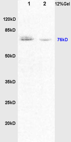 Protein:
Brain(Rat) lysate at 30ug;
Lung(Rat) lysate at 30ug;
Primary: Anti-DVL1/Dishevelled (SL0598R) at 1:200 dilution;
Secondary: HRP conjugated Goat-Anti-Rabbit IgG(SL0295G-HRP) at 1: 3000 dilution;
Predicted band size : 76kD
Observed band size : 76kD
 Sample:
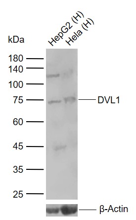
Lane 1: Human HepG2 cell lysates
Lane 2: Human Hela cell lysates
Primary: Anti-DVL1 (SL0598R) at 1/1000 dilution
Secondary: IRDye800CW Goat Anti-Rabbit IgG at 1/20000 dilution
Predicted band size: 76 kDa
Observed band size: 75 kDa
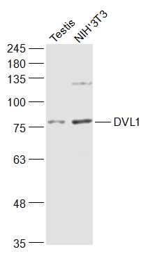 Sample:
Testis (Mouse) Lysate at 40 ug
NIH/3T3 (Mouse) Cell Lysate at 30 ug
Primary: Anti-DVL1 (SL0598R) at 1/1000 dilution
Secondary: IRDye800CW Goat Anti-Rabbit IgG at 1/20000 dilution
Predicted band size: 76 kD
Observed band size: 77 kD
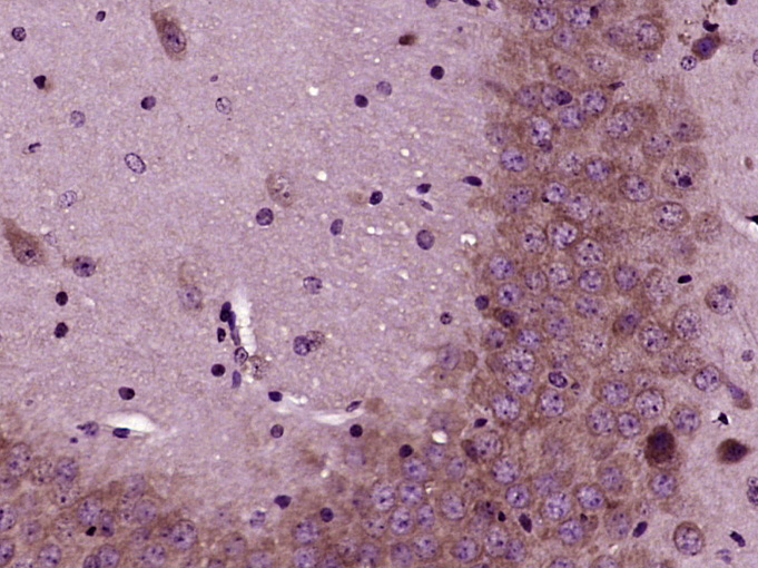 Paraformaldehyde-fixed, paraffin embedded (Mouse brain); Antigen retrieval by boiling in sodium citrate buffer (pH6.0) for 15min; Block endogenous peroxidase by 3% hydrogen peroxide for 20 minutes; Blocking buffer (normal goat serum) at 37°C for 30min; Antibody incubation with (DVL1) Polyclonal Antibody, Unconjugated (SL0598R) at 1:400 overnight at 4°C, followed by operating according to SP Kit(Rabbit) (sp-0023) instructionsand DAB staining.
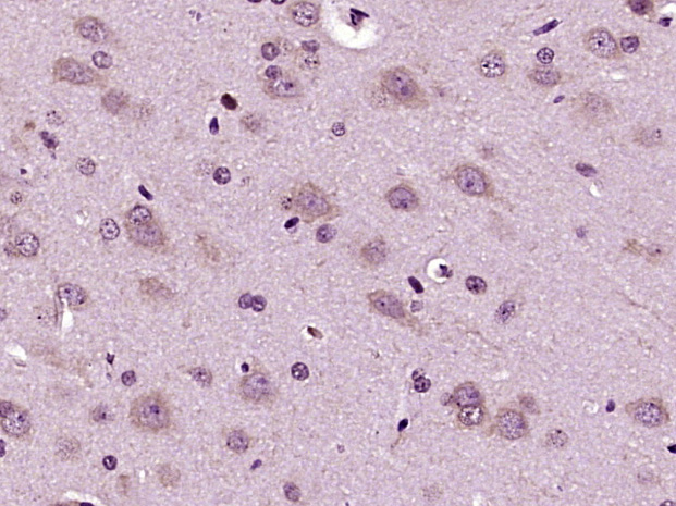 Paraformaldehyde-fixed, paraffin embedded (Rat brain); Antigen retrieval by boiling in sodium citrate buffer (pH6.0) for 15min; Block endogenous peroxidase by 3% hydrogen peroxide for 20 minutes; Blocking buffer (normal goat serum) at 37°C for 30min; Antibody incubation with (DVL1) Polyclonal Antibody, Unconjugated (SL0598R) at 1:400 overnight at 4°C, followed by operating according to SP Kit(Rabbit) (sp-0023) instructionsand DAB staining.
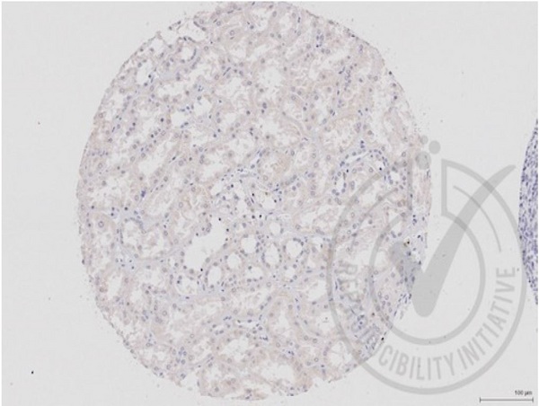 Images provided the Independent Validation Program (badge number 029657)Formalin-fixed and paraffin embedded human kidney labeled with Rabbit Anti-DVL1 Polyclonal Antibody (SL0598R) at 1:250 overnight at room temperature followed by conjugation to secondary antibody.
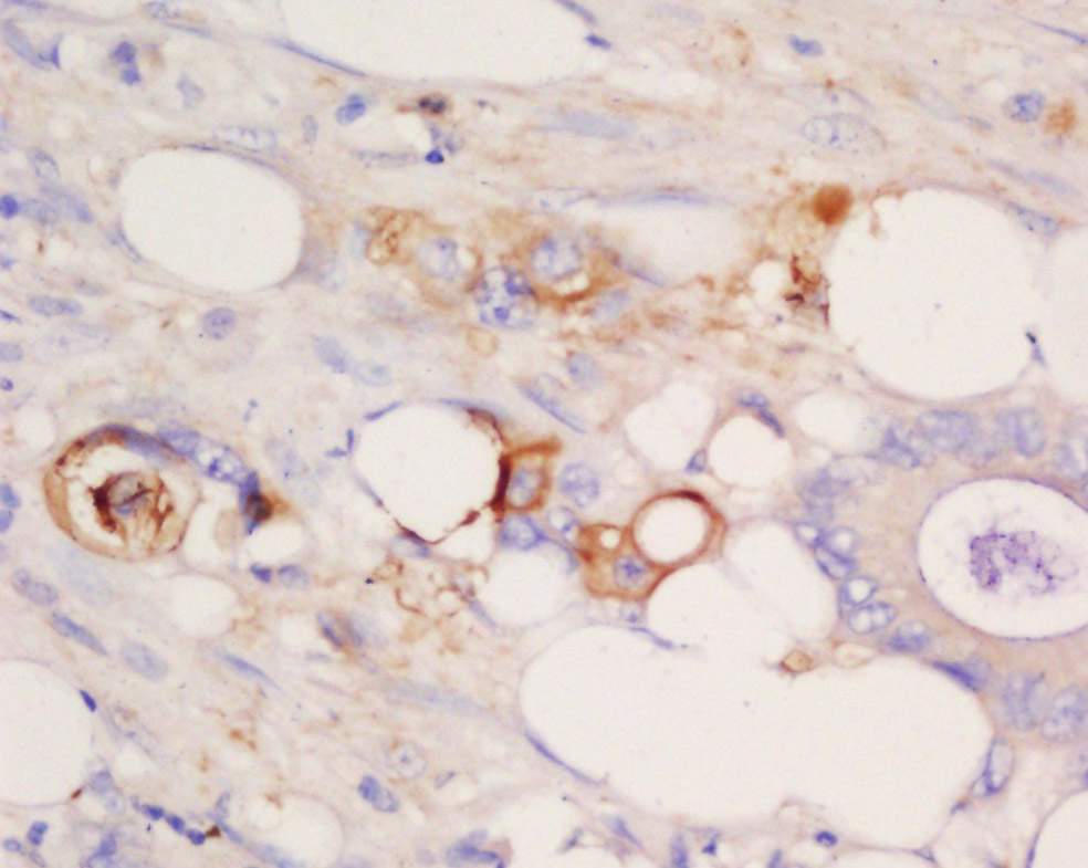 Tissue/cell: human cervical carcinoma; 4% Paraformaldehyde-fixed and paraffin-embedded;
Antigen retrieval: citrate buffer ( 0.01M, pH 6.0 ), Boiling bathing for 15min; Block endogenous peroxidase by 3% Hydrogen peroxide for 30min; Blocking buffer (normal goat serum,C-0005) at 37℃ for 20 min;
Incubation: Anti-DVL1/Dishevelled Polyclonal Antibody, Unconjugated(SL0598R) 1:200, overnight at 4°C, followed by conjugation to the secondary antibody(SP-0023) and DAB(C-0010) staining
 Blank control: A431.
Primary Antibody (green line): Rabbit Anti-DVL1 antibody (SL0598R)
Dilution: 1μg /10^6 cells;
Isotype Control Antibody (orange line): Rabbit IgG .
Secondary Antibody: Goat anti-rabbit IgG-AF647
Dilution: 1μg /test.
Protocol
The cells were fixed with 4% PFA (10min at room temperature)and then permeabilized with 0.1% PBST for 20 min at room temperature. The cells were then incubated in 5%BSA to block non-specific protein-protein interactions for 30 min at room temperature.Cells stained with Primary Antibody for 30 min at room temperature. The secondary antibody used for 40 min at room temperature. Acquisition of 20,000 events was performed.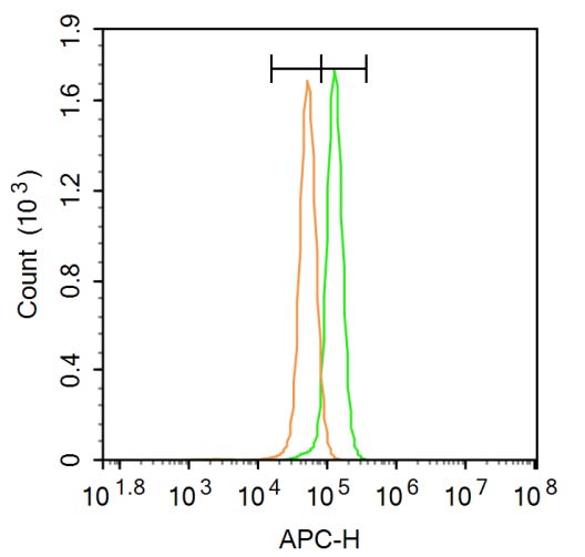 Blank control: A431.
Primary Antibody (green line): Rabbit Anti-DVL1 antibody (SL0598R)
Dilution: 3μg /10^6 cells;
Isotype Control Antibody (orange line): Rabbit IgG .
Secondary Antibody : Goat anti-rabbit IgG-AF647
Dilution: 3μg /test.
Protocol
The cells were fixed with 4% PFA (10min at room temperature)and then permeabilized with 20% PBST for 20 min at room temperature. The cells were then incubated in 5%BSA to block non-specific protein-protein interactions for 30 min at room temperature .Cells stained with Primary Antibody for 30 min at room temperature. The secondary antibody used for 40 min at room temperature. Acquisition of 20,000 events was performed.
Cartpieces
Totalgoods,subtotals:¥Checkout
Bought notes(bought amounts latest0)
No one bought this product
User Comment(Total0User Comment Num)
- No comment


 +86 571 56623320
 +86 18668110335

