
Rabbit Anti-CDK7 antibody
CDK7_HUMAN; Cyclin-dependent kinase 7; EC:2.7.11.22; EC:2.7.11.23; CDK7; CAK; CAK1; CDKN7; MO15; STK1; 39 kDa protein kinase (p39 Mo15); CDK-activating kinase 1; Cell division protein kinase 7; Serine/threonine-protein kinase 1; TFIIH basal transcription
View History [Clear]
Details
Product Name CDK7 Chinese Name 周期素依赖性激酶7抗体 Alias CDK7_HUMAN; Cyclin-dependent kinase 7; EC:2.7.11.22; EC:2.7.11.23; CDK7; CAK; CAK1; CDKN7; MO15; STK1; 39 kDa protein kinase (p39 Mo15); CDK-activating kinase 1; Cell division protein kinase 7; Serine/threonine-protein kinase 1; TFIIH basal transcription factor complex kinase subunit; Research Area Tumour Cell biology Chromatin and nuclear signals Signal transduction Cyclin Kinases and Phosphatases Immunogen Species Rabbit Clonality Polyclonal React Species Human, Mouse, Rat, (predicted: Cow, ) Applications WB=1:500-2000 ELISA=1:5000-10000 IHC-P=1:100-500 IHC-F=1:100-500 ICC=1:100 IF=1:100-500 (Paraffin sections need antigen repair)
not yet tested in other applications.
optimal dilutions/concentrations should be determined by the end user.Theoretical molecular weight 40kDa Cellular localization The nucleus cytoplasmic Form Liquid Concentration 1mg/ml immunogen KLH conjugated synthetic peptide derived from human CDK7: 1-80/346 Lsotype IgG Purification affinity purified by Protein A Buffer Solution 0.01M TBS(pH7.4) with 1% BSA, 0.03% Proclin300 and 50% Glycerol. Storage Shipped at 4℃. Store at -20 °C for one year. Avoid repeated freeze/thaw cycles. Attention This product as supplied is intended for research use only, not for use in human, therapeutic or diagnostic applications. PubMed PubMed Product Detail The protein encoded by this gene is a member of the cyclin-dependent protein kinase (CDK) family. CDK family members are highly similar to the gene products of Saccharomyces cerevisiae cdc28, and Schizosaccharomyces pombe cdc2, and are known to be important regulators of cell cycle progression. This protein forms a trimeric complex with cyclin H and MAT1, which functions as a Cdk-activating kinase (CAK). It is an essential component of the transcription factor TFIIH, that is involved in transcription initiation and DNA repair. This protein is thought to serve as a direct link between the regulation of transcription and the cell cycle. [provided by RefSeq, Jul 2008]
Function:
Serine/threonine kinase involved in cell cycle control and in RNA polymerase II-mediated RNA transcription. Cyclin-dependent kinases (CDKs) are activated by the binding to a cyclin and mediate the progression through the cell cycle. Each different complex controls a specific transition between 2 subsequent phases in the cell cycle. Required for both activation and complex formation of CDK1/cyclin-B during G2-M transition, and for activation of CDK2/cyclins during G1-S transition (but not complex formation). CDK7 is the catalytic subunit of the CDK-activating kinase (CAK) complex. Phosphorylates SPT5/SUPT5H, SF1/NR5A1, POLR2A, p53/TP53, CDK1, CDK2, CDK4, CDK6 and CDK11B/CDK11. CAK activates the cyclin-associated kinases CDK1, CDK2, CDK4 and CDK6 by threonine phosphorylation, thus regulating cell cycle progression. CAK complexed to the core-TFIIH basal transcription factor activates RNA polymerase II by serine phosphorylation of the repetitive C-terminus domain (CTD) of its large subunit (POLR2A), allowing its escape from the promoter and elongation of the transcripts. Phosphorylation of POLR2A in complex with DNA promotes transcription initiation by triggering dissociation from DNA. Its expression and activity are constant throughout the cell cycle. Upon DNA damage, triggers p53/TP53 activation by phosphorylation, but is inactivated in turn by p53/TP53; this feedback loop may lead to an arrest of the cell cycle and of the transcription, helping in cell recovery, or to apoptosis. Required for DNA-bound peptides-mediated transcription and cellular growth inhibition.
Subunit:
Associates primarily with cyclin-H (CCNH) and MAT1 to form the CAK complex. CAK can further associate with the core-TFIIH to form the TFIIH basal transcription factor; this complex is sensitive to UV light. The CAK complex binds to p53/TP53 in response to DNA damage. Interacts with CDK2, SF1/NR5A1, PUF60 and PRKCI.
Subcellular Location:
Nucleus. Cytoplasm. Cytoplasm, perinuclear region. Note=Colocalizes with PRKCI in the cytoplasm and nucleus. Translocates from the nucleus to cytoplasm and perinuclear region in response to DNA-bound peptides.
Tissue Specificity:
Ubiquitous.
Post-translational modifications:
Phosphorylation of Ser-164 during mitosis inactivates the enzyme. Phosphorylation of Thr-170 is required for activity. Phosphorylated at Ser-164 and Thr-170 by CDK2.
Similarity:
Belongs to the protein kinase superfamily. CMGC Ser/Thr protein kinase family. CDC2/CDKX subfamily.
Contains 1 protein kinase domain.
SWISS:
P50613
Gene ID:
1022
Database links:Entrez Gene: 1022 Human
Entrez Gene: 12572 Mouse
Omim: 601955 Human
SwissProt: P50613 Human
SwissProt: Q03147 Mouse
Unigene: 184298 Human
Unigene: 259718 Mouse
Unigene: 98896 Rat
Product Picture 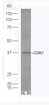 Sample:HL-60 Cell lysate;
Primary:Anti-CDK7 (SL0569R) at 1:300;
Secondary: HRP conjugated Goat-Anti-rabbit IgG(SL0295G-HRP) at 1: 5000;
Predicted band size:40 kD
Observed band size:37 kD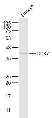 Sample:
Embryo (Mouse) Lysate at 40 ug
Primary: Anti-CDK7 (SL0569R) at 1/1000 dilution
Secondary: IRDye800CW Goat Anti-Rabbit IgG at 1/20000 dilution
Predicted band size: 40 kD
Observed band size: 40 kD
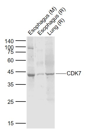 Sample:
Lane 1: Esophagus (Mouse) Lysate at 40 ug
Lane 2: Esophagus (Rat) Lysate at 40 ug
Lane 3: Lung (Rat) Lysate at 40 ug
Primary: Anti-CDK7 (SL0569R) at 1/1000 dilution
Secondary: IRDye800CW Goat Anti-Rabbit IgG at 1/20000 dilution
Predicted band size: 43 kD
Observed band size: 43 kD
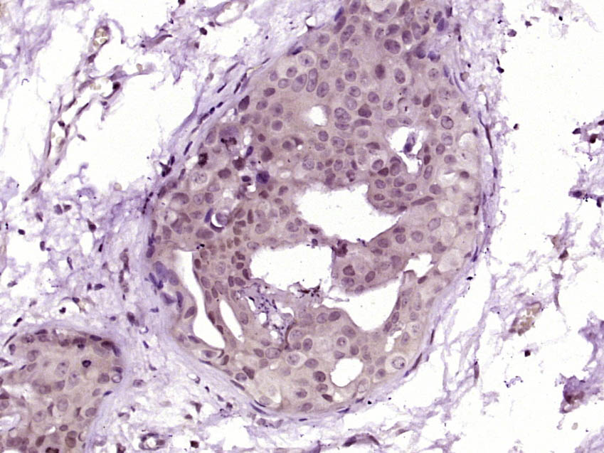 Paraformaldehyde-fixed, paraffin embedded (Human breast carcinoma); Antigen retrieval by boiling in sodium citrate buffer (pH6.0) for 15min; Block endogenous peroxidase by 3% hydrogen peroxide for 20 minutes; Blocking buffer (normal goat serum) at 37°C for 30min; Antibody incubation with (CDK7) Polyclonal Antibody, Unconjugated (SL0569R) at 1:400 overnight at 4°C, followed by operating according to SP Kit(Rabbit) (sp-0023) instructionsand DAB staining.
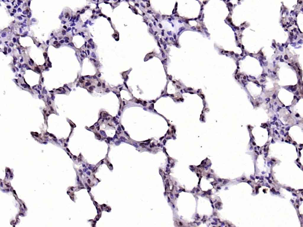 Paraformaldehyde-fixed, paraffin embedded (rat lung); Antigen retrieval by boiling in sodium citrate buffer (pH6.0) for 15min; Block endogenous peroxidase by 3% hydrogen peroxide for 20 minutes; Blocking buffer (normal goat serum) at 37°C for 30min; Antibody incubation with (CDK7) Polyclonal Antibody, Unconjugated (SL0569R) at 1:200 overnight at 4°C, followed by operating according to SP Kit(Rabbit) (sp-0023) instructionsand DAB staining.
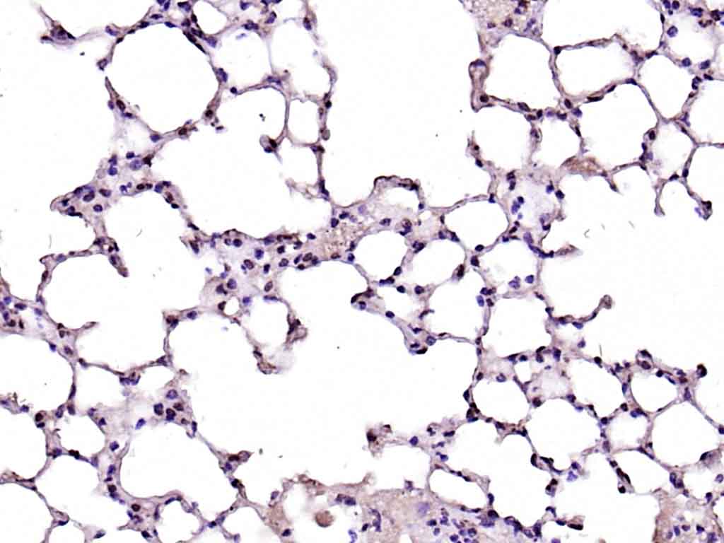 Paraformaldehyde-fixed, paraffin embedded (mouse lung); Antigen retrieval by boiling in sodium citrate buffer (pH6.0) for 15min; Block endogenous peroxidase by 3% hydrogen peroxide for 20 minutes; Blocking buffer (normal goat serum) at 37°C for 30min; Antibody incubation with (CDK7) Polyclonal Antibody, Unconjugated (SL0569R) at 1:200 overnight at 4°C, followed by operating according to SP Kit(Rabbit) (sp-0023) instructionsand DAB staining.
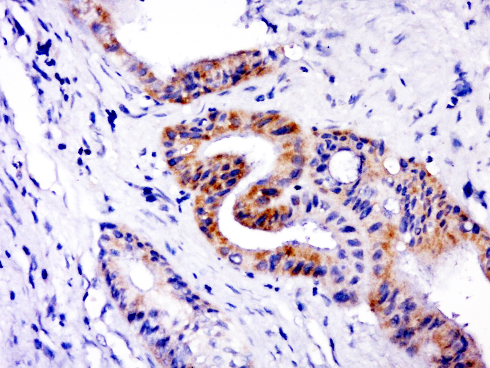 Paraformaldehyde-fixed, paraffin embedded (human cervical carcinoma); Antigen retrieval by boiling in sodium citrate buffer (pH6.0) for 15min; Block endogenous peroxidase by 3% hydrogen peroxide for 20 minutes; Blocking buffer (normal goat serum) at 37°C for 30min; Antibody incubation with (CDK7) Polyclonal Antibody, Unconjugated (SL0569R) at 1:400 overnight at 4°C, followed by a conjugated secondary (sp-0023) for 20 minutes and DAB staining.
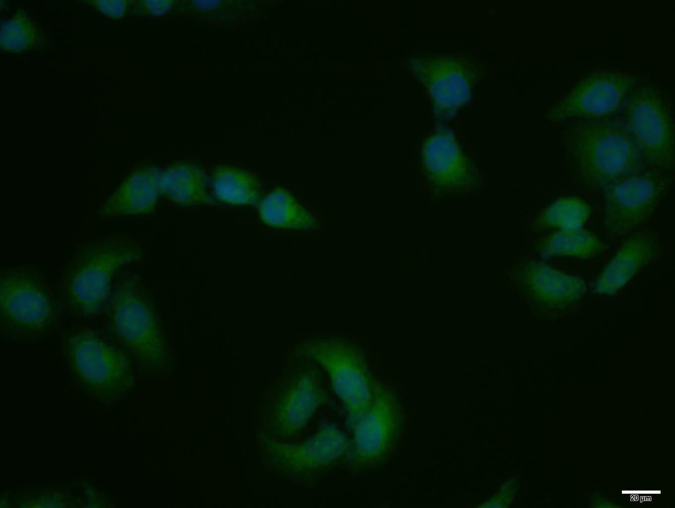 Hela cell; 4% Paraformaldehyde-fixed; Triton X-100 at room temperature for 20 min; Blocking buffer (normal goat serum, C-0005) at 37°C for 20 min; Antibody incubation with (CDK7) polyclonal Antibody, Unconjugated (SL0569R) 1:100, 90 minutes at 37°C; followed by a conjugated Goat Anti-Rabbit IgG antibody at 37°C for 90 minutes, DAPI (blue, C02-04002) was used to stain the cell nuclei.
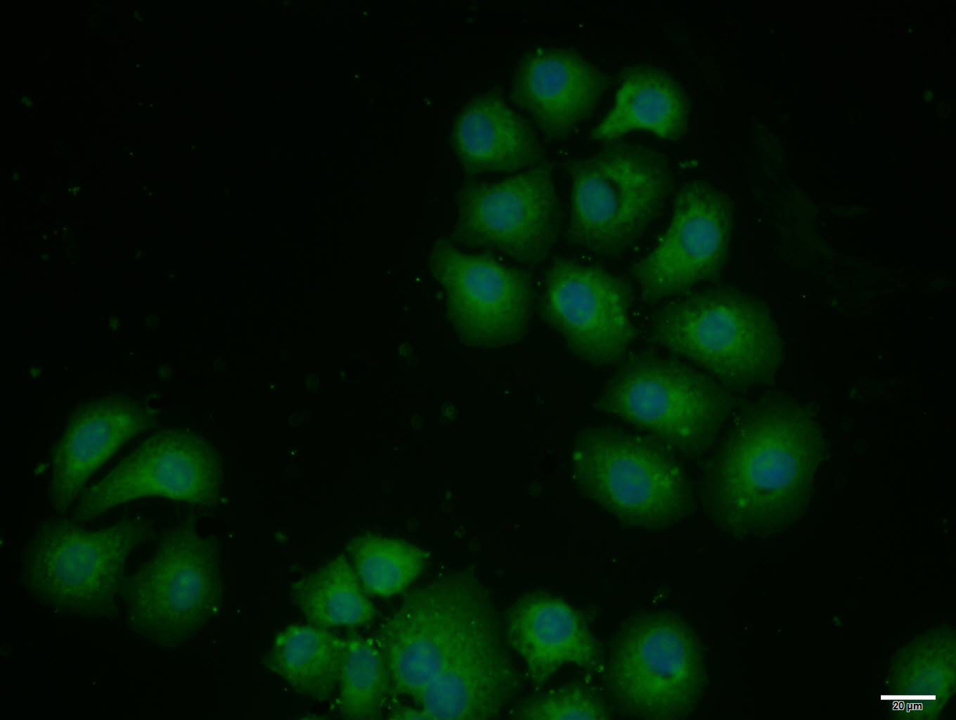 HepG2 cell; 4% Paraformaldehyde-fixed; Triton X-100 at room temperature for 20 min; Blocking buffer (normal goat serum, C-0005) at 37°C for 20 min; Antibody incubation with (CDK7) polyclonal Antibody, Unconjugated (SL0569R) 1:100, 90 minutes at 37°C; followed by a conjugated Goat Anti-Rabbit IgG antibody at 37°C for 90 minutes, DAPI (blue, C02-04002) was used to stain the cell nuclei.
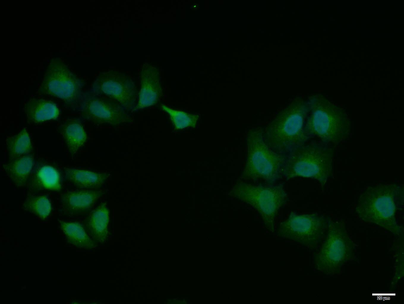 Hela cell; 4% Paraformaldehyde-fixed; Triton X-100 at room temperature for 20 min; Blocking buffer (normal goat serum, C-0005) at 37°C for 20 min; Antibody incubation with (CDK7) polyclonal Antibody, Unconjugated (SL0569R) 1:100, 90 minutes at 37°C; followed by a conjugated Goat Anti-Rabbit IgG antibody at 37°C for 90 minutes, DAPI (blue, C02-04002) was used to stain the cell nuclei.
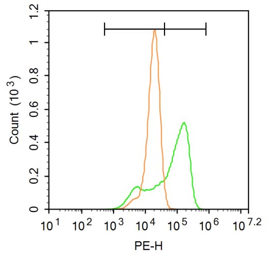 Blank control:A549. Primary Antibody (green line): Rabbit Anti-CDK7 antibody (SL0659R) Dilution: 3μg /10^6 cells; Isotype Control Antibody (orange line): Rabbit IgG . Secondary Antibody : Goat anti-rabbit IgG-PE Dilution: 1μg /test. Protocol The cells were fixed with 4% PFA (10min at room temperature)and then permeabilized with 90% ice-cold methanol for 20 min at-20℃. The cells were then incubated in 5% BSA to block non-specific protein-protein interactions for 30 min at at room temperature .Cells stained with Primary Antibody for 30 min at room temperature. The secondary antibody used for 40 min at room temperature. Acquisition of 20,000 events was performed.
Cartpieces
Totalgoods,subtotals:¥Checkout
Bought notes(bought amounts latest0)
No one bought this product
User Comment(Total0User Comment Num)
- No comment


 +86 571 56623320
 +86 18668110335

