
Rabbit Anti-MDR1 antibody
P-Glycoprotein; Multi Drug Resistance Associated Protein, ABCB1; ATP-binding cassette sub-family B (MDR/TAP), member 1; ABC20; CD243; CD243 antigen; CLCS; Multidrug resistance protein 1; P glycoprotein 1; p-GP; gp170; P gp; PGY1; MDR1_HUMAN.
View History [Clear]
Details
Product Name MDR1 Chinese Name 多药耐药蛋白/P-glycoprotein抗体 Alias P-Glycoprotein; Multi Drug Resistance Associated Protein, ABCB1; ATP-binding cassette sub-family B (MDR/TAP), member 1; ABC20; CD243; CD243 antigen; CLCS; Multidrug resistance protein 1; P glycoprotein 1; p-GP; gp170; P gp; PGY1; MDR1_HUMAN. literatures Research Area Tumour Cardiovascular Cell biology immunology Signal transduction transcriptional regulatory factor Immunogen Species Rabbit Clonality Polyclonal React Species Human, Mouse, Rat, Applications WB=1:500-2000 ELISA=1:5000-10000 IHC-P=1:100-500 IHC-F=1:100-500 Flow-Cyt=1μg/Test ICC=1:100 IF=1:100-500 (Paraffin sections need antigen repair)
not yet tested in other applications.
optimal dilutions/concentrations should be determined by the end user.Theoretical molecular weight 141kDa Cellular localization The cell membrane Form Liquid Concentration 1mg/ml immunogen KLH conjugated synthetic peptide derived from human MDR1: 21-100/1272 <Cytoplasmic> Lsotype IgG Purification affinity purified by Protein A Buffer Solution 0.01M TBS(pH7.4) with 1% BSA, 0.03% Proclin300 and 50% Glycerol. Storage Shipped at 4℃. Store at -20 °C for one year. Avoid repeated freeze/thaw cycles. Attention This product as supplied is intended for research use only, not for use in human, therapeutic or diagnostic applications. PubMed PubMed Product Detail P Glycoprotein, the product of the MDR1 gene, is expressed in distinct non-malignant cells, typically cells with secretory and excretory functions. It is assumed to function as an ATP-dependent drug efflux pump with broad substrate specificity. The highest expression of P Glycoprotein has been observed in kidney (proximal tubules), liver (bile canaliculi), adrenal gland and intestine, suggesting that the primary role of P Glycoprotein is in the normal secretion of physiological metabolites and ingested chemicals into bile, urine and the lumen of the intestinal tract. Elevated levels of P Glycoprotein have also been reported in multidrug-resistant cell lines and in colon, endometrial, ovarian, and breast tumors, as well as in sarcomas and leukemias / lymphomas.
Function:
Energy-dependent efflux pump responsible for decreased drug accumulation in multidrug-resistant cells.
Subunit:
Interacts with PSMB5.
Subcellular Location:
Cell membrane; Multi-pass membrane protein.
Tissue Specificity:
Expressed in liver, kidney, small intestine and brain.
DISEASE:
Genetic variations in ABCB1 are associated with susceptibility to inflammatory bowel disease type 13 (IBD13) [MIM:612244]. Inflammatory bowel disease is characterized by a chronic relapsing intestinal inflammation. It is subdivided into Crohn disease and ulcerative colitis phenotypes. Crohn disease may involve any part of the gastrointestinal tract, but most frequently the terminal ileum and colon. Bowel inflammation is transmural and discontinuous; it may contain granulomas or be associated with intestinal or perianal fistulas. In contrast, in ulcerative colitis, the inflammation is continuous and limited to rectal and colonic mucosal layers; fistulas and granulomas are not observed. Both diseases include extraintestinal inflammation of the skin, eyes, or joints. Crohn disease and ulcerative colitis are commonly classified as autoimmune diseases.
Similarity:
Belongs to the ABC transporter superfamily. ABCB family. Multidrug resistance exporter (TC 3.A.1.201) subfamily.
Contains 2 ABC transmembrane type-1 domains.
Contains 2 ABC transporter domains.
SWISS:
P21447
Gene ID:
5243
Database links:Entrez Gene: 5243 Human
Entrez Gene: 18669 Mouse
Omim: 171050 Human
SwissProt: P08183 Human
SwissProt: P06795 Mouse
Product Picture 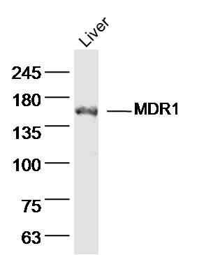 Sample: Liver (mouse) Lysate at 40 ug
Primary: Anti- MDR1 (SL0563R)at 1/300 dilution
Secondary: IRDye800CW Goat Anti-Rabbit IgG at 1/20000 dilution
Predicted band size: 141kD
Observed band size: 150kD
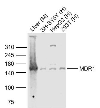 Sample:
Lane 1: Liver (Mouse) Lysate at 40 ug
Lane 2: SH-SY5Y (Human) Cell Lysate at 30 ug
Lane 3: HepG2 (Human) Cell Lysate at 30 ug
Lane 4: 293T (Human) Cell Lysate at 30 ug
Primary: Anti-MDR1 (SL0563R) at 1/1000 dilution
Secondary: IRDye800CW Goat Anti-Rabbit IgG at 1/20000 dilution
Predicted band size: 150-180 kD
Observed band size: 150 kD
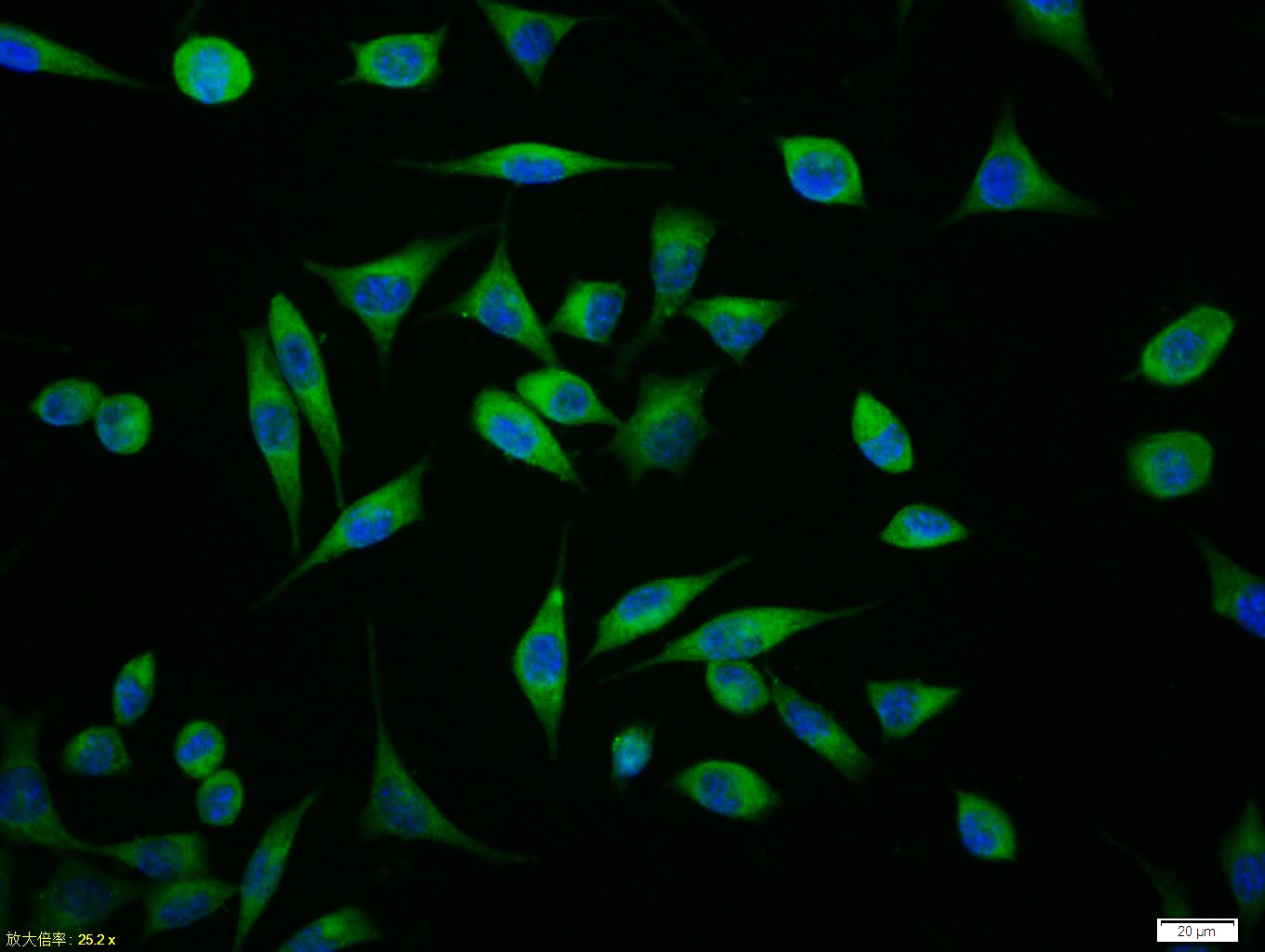 Tissue/cell:SH-SY5Y cell; 4% Paraformaldehyde-fixed; Triton X-100 at room temperature for 20 min; Blocking buffer (normal goat serum,C-0005) at 37°C for 20 min; Antibody incubation with (MDR1) polyclonal Antibody, Unconjugated (SL0563R) 1:100, 90 minutes at 37°C; followed by a FITC conjugated Goat Anti-Rabbit IgG antibody at 37°C for 90 minutes, DAPI (blue, C02-04002) was used to stain the cell nuclei.
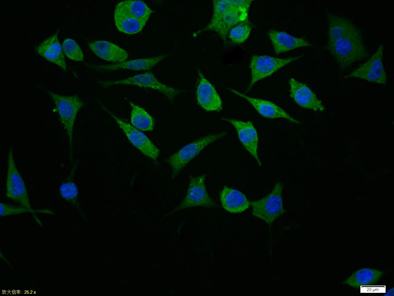 Tissue/cell:SH-SY5Y cell; 4% Paraformaldehyde-fixed; Triton X-100 at room temperature for 20 min; Blocking buffer (normal goat serum,C-0005) at 37°C for 20 min; Antibody incubation with (MDR1) polyclonal Antibody, Unconjugated (SL0563R) 1:100, 90 minutes at 37°C; followed by a FITC conjugated Goat Anti-Rabbit IgG antibody at 37°C for 90 minutes, DAPI (blue, C02-04002) was used to stain the cell nuclei.
 Blank control (blue line): Hela (fixed with 70% methanol (Overnight at -20℃) and then permeabilized with ice-cold 90% methanol for 30 min on ice).
Primary Antibody (green line): Rabbit Anti-MDR1 antibody (SL0563R),Dilution: 1μg /10^6 cells;
Isotype Control Antibody (orange line): Rabbit IgG .
Secondary Antibody (white blue line): Goat anti-rabbit IgG-FITC,Dilution: 1μg /test.
Cartpieces
Totalgoods,subtotals:¥Checkout
Bought notes(bought amounts latest0)
No one bought this product
User Comment(Total0User Comment Num)
- No comment


 +86 571 56623320
 +86 18668110335

