
Rabbit Anti-EMP-1 antibody
CL-20; EMP 1; EMP1; EMP1_HUMAN; Epithelial membrane protein 1; Protein B4B; TMP; Tumor-associated membrane protein.
View History [Clear]
Details
Product Name EMP-1 Chinese Name 表皮膜蛋白1抗体 Alias CL-20; EMP 1; EMP1; EMP1_HUMAN; Epithelial membrane protein 1; Protein B4B; TMP; Tumor-associated membrane protein. literatures Research Area Cell biology immunology Apoptosis The cell membrane受体 Immunogen Species Rabbit Clonality Polyclonal React Species Human, Mouse, Rat, Applications ELISA=1:5000-10000 IHC-P=1:100-500 IHC-F=1:100-500 IF=1:100-500 (Paraffin sections need antigen repair)
not yet tested in other applications.
optimal dilutions/concentrations should be determined by the end user.Theoretical molecular weight 17kDa Cellular localization The cell membrane Form Liquid Concentration 1mg/ml immunogen KLH conjugated synthetic peptide derived from mouse EMP-1: 101-160/160 Lsotype IgG Purification affinity purified by Protein A Buffer Solution 0.01M TBS(pH7.4) with 1% BSA, 0.03% Proclin300 and 50% Glycerol. Storage Shipped at 4℃. Store at -20 °C for one year. Avoid repeated freeze/thaw cycles. Attention This product as supplied is intended for research use only, not for use in human, therapeutic or diagnostic applications. PubMed PubMed Product Detail Epithelial membrane protein-1 (EMP-1) is a four pass transmembrane protein consisting of 160 amino acids. It is a member of a small family of epithelial membrane proteins. EMP-1 is very similar in structure to its close relative, Peripheral Myelin Protein 22 (PMP22). It is most predominantly expressed in tissues of the gastrointestinal tract but has also been found to be a junctional protein in the liver expressed along the intercellular border. EMP-1 directly interacts with the C-terminus of the P2X7 receptor and may be involved in membrane blebbing. EMP-1 may also be an important regulator in cell communication, signaling, and adhesion. When EMP-1 is overexpressed, cell proliferation is inhibited, S phase is arrested and G1 phase is prolonged in esophogeal cancer cells. EMP-1 may play a role in tumorigenesis and has been identified as a biomarker for gefitinib treatment resistance for patients with lung cancer.
Subcellular Location:
Membrane; Multi-pass membrane protein.
Tissue Specificity:
Most prominently found in the gastrointestinal tract, skin, lung, and brain but not in liver.
Similarity:
Belongs to the PMP-22/EMP/MP20 family.
SWISS:
P47801
Gene ID:
13730
Database links:Entrez Gene: 2012 Human
Omim: 602333 Human
SwissProt: P54849 Human
Unigene: 719042 Human
emp-1蛋白主要参与细胞生长发育、细胞增殖、死亡、表皮发育等作用。Product Picture 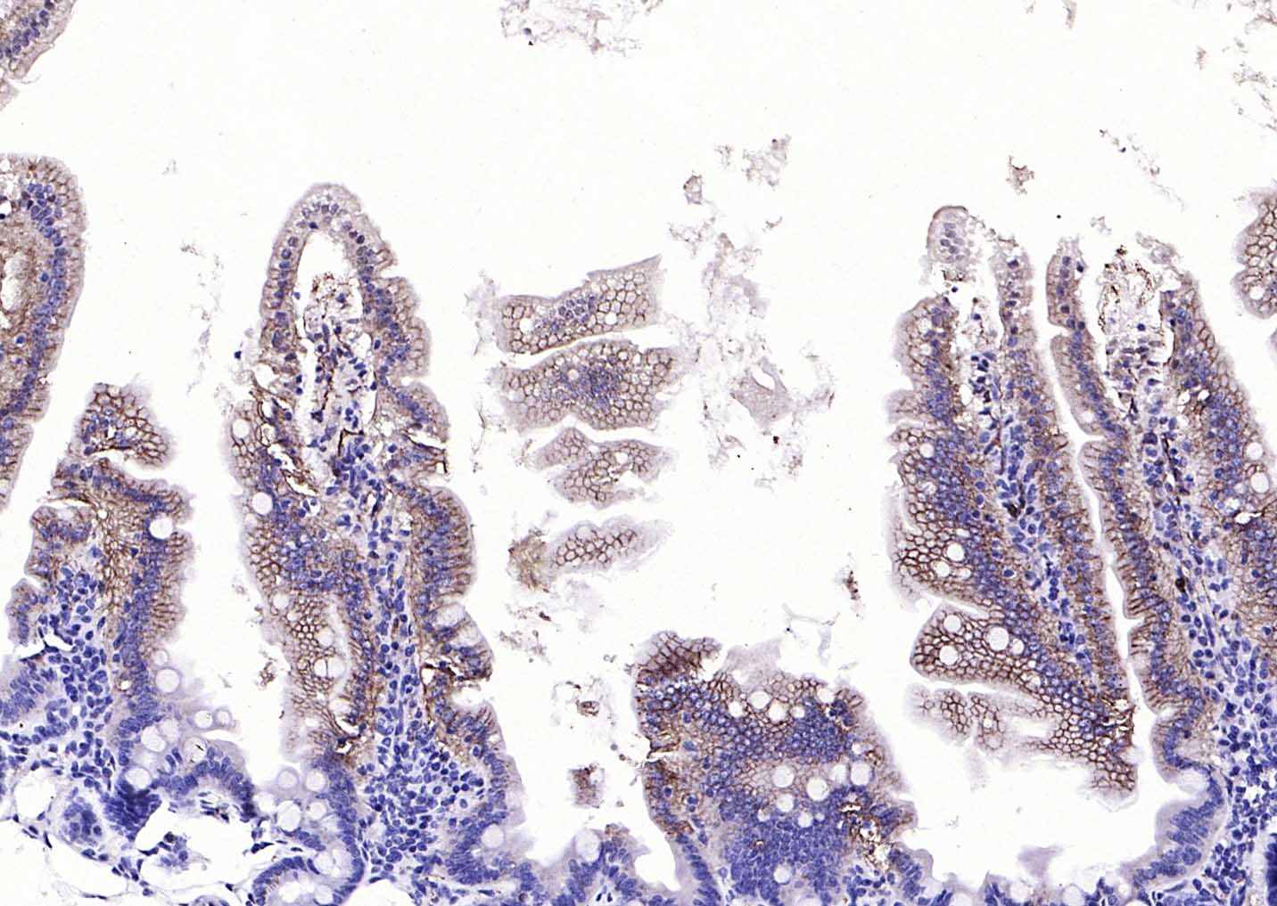 Paraformaldehyde-fixed, paraffin embedded (mouse small intestine); Antigen retrieval by boiling in sodium citrate buffer (pH6.0) for 15min; Block endogenous peroxidase by 3% hydrogen peroxide for 20 minutes; Blocking buffer (normal goat serum) at 37°C for 30min; Antibody incubation with (EMP-1) Polyclonal Antibody, Unconjugated (SL0558R) at 1:200 overnight at 4°C, followed by operating according to SP Kit(Rabbit) (sp-0023) instructionsand DAB staining.
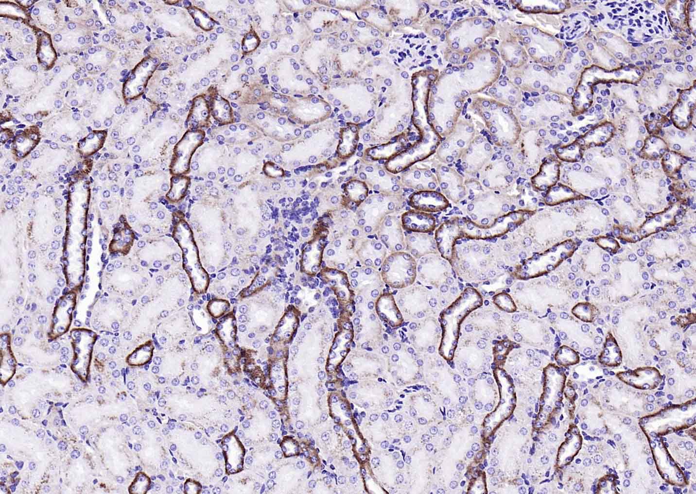 Paraformaldehyde-fixed, paraffin embedded (mouse kidney); Antigen retrieval by boiling in sodium citrate buffer (pH6.0) for 15min; Block endogenous peroxidase by 3% hydrogen peroxide for 20 minutes; Blocking buffer (normal goat serum) at 37°C for 30min; Antibody incubation with (EMP-1) Polyclonal Antibody, Unconjugated (SL0558R) at 1:200 overnight at 4°C, followed by operating according to SP Kit(Rabbit) (sp-0023) instructionsand DAB staining.
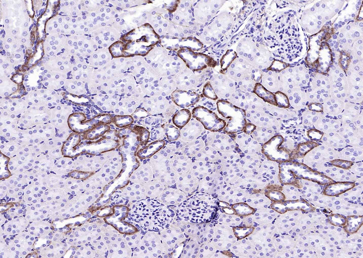 Paraformaldehyde-fixed, paraffin embedded (rat kidney); Antigen retrieval by boiling in sodium citrate buffer (pH6.0) for 15min; Block endogenous peroxidase by 3% hydrogen peroxide for 20 minutes; Blocking buffer (normal goat serum) at 37°C for 30min; Antibody incubation with (EMP-1) Polyclonal Antibody, Unconjugated (SL0558R) at 1:200 overnight at 4°C, followed by operating according to SP Kit(Rabbit) (sp-0023) instructionsand DAB staining.
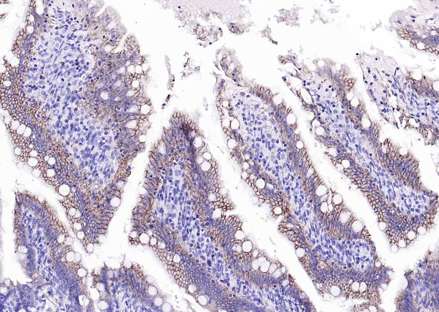 Paraformaldehyde-fixed, paraffin embedded (rat small intestine); Antigen retrieval by boiling in sodium citrate buffer (pH6.0) for 15min; Block endogenous peroxidase by 3% hydrogen peroxide for 20 minutes; Blocking buffer (normal goat serum) at 37°C for 30min; Antibody incubation with (EMP-1) Polyclonal Antibody, Unconjugated (SL0558R) at 1:200 overnight at 4°C, followed by operating according to SP Kit(Rabbit) (sp-0023) instructionsand DAB staining.
 Paraformaldehyde-fixed, paraffin embedded (Human kidney); Antigen retrieval by boiling in sodium citrate buffer (pH6.0) for 15min; Block endogenous peroxidase by 3% hydrogen peroxide for 20 minutes; Blocking buffer (normal goat serum) at 37°C for 30min; Antibody incubation with (EMP-1) Polyclonal Antibody, Unconjugated (SL0558R) at 1:200 overnight at 4°C, followed by operating according to SP Kit(Rabbit) (sp-0023) instructionsand DAB staining.
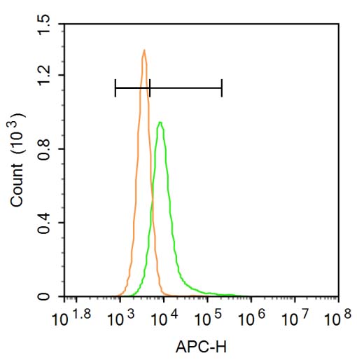 Blank control: A431.
Primary Antibody (green line): Rabbit Anti-EMP-1 antibody (SL0558R)
Dilution: 3μg /10^6 cells;
Isotype Control Antibody (orange line): Rabbit IgG .
Secondary Antibody: Goat anti-rabbit IgG-AF647
Dilution: 3μg /test.
Protocol
The cells were incubated in 5%BSA to block non-specific protein-protein interactions for 30 min at at room temperature .Cells stained with Primary Antibody for 30 min at room temperature. The secondary antibody used for 40 min at room temperature. Acquisition of 20,000 events was performed.
Cartpieces
Totalgoods,subtotals:¥Checkout
Bought notes(bought amounts latest0)
No one bought this product
User Comment(Total0User Comment Num)
- No comment


 +86 571 56623320
 +86 18668110335

