
Rabbit Anti-CD45 antibody
B220; CD 45; CD-45; cd45 antigen; ec3.1.3.48; CD45R; GP180; GP180; GP 180; L CA; LCA; L-CA; Leukocyte common antigen; LY5; Ly-5 glycoprotein; Protein tyrosine phosphatase receptor type C; Protein tyrosine phosphatase receptor type c polypeptide; protein
View History [Clear]
Details
Product Name CD45 Chinese Name 白细胞共同抗原CD45抗体 Alias B220; CD 45; CD-45; cd45 antigen; ec3.1.3.48; CD45R; GP180; GP180; GP 180; L CA; LCA; L-CA; Leukocyte common antigen; LY5; Ly-5 glycoprotein; Protein tyrosine phosphatase receptor type C; Protein tyrosine phosphatase receptor type c polypeptide; protein tyrosine phosphatase, receptor type, C; Receptor-type tyrosine-protein phosphatase C; PTPRC; PTPRC_HUMAN; SCID due to PTPRC deficiency; T200; T200 glycoprotein; T200 leukocyte common antigen; Human homolog of severe combined immunodeficiency due to PTPRC deficiency. literatures Research Area Cell biology immunology Neurobiology Signal transduction Stem cells transcriptional regulatory factor Cell Surface Molecule glycoprotein Cell type markers Natural killer cells lymphocyte t-lymphocyte b-lymphocyte The cell membrane蛋白 Immunogen Species Rabbit Clonality Polyclonal React Species (predicted: Rat, Cow, ) Applications WB=1:500-2000 ELISA=1:5000-10000 Flow-Cyt=1μg/Test
not yet tested in other applications.
optimal dilutions/concentrations should be determined by the end user.Theoretical molecular weight 143kDa Detection molecular weight 220 kDa Cellular localization The cell membrane Form Liquid Concentration 1mg/ml immunogen KLH conjugated synthetic peptide derived from human CD45: 1210-1304/1304 <Cytoplasmic> Lsotype IgG Purification affinity purified by Protein A Buffer Solution 0.01M TBS(pH7.4) with 1% BSA, 0.03% Proclin300 and 50% Glycerol. Storage Shipped at 4℃. Store at -20 °C for one year. Avoid repeated freeze/thaw cycles. Attention This product as supplied is intended for research use only, not for use in human, therapeutic or diagnostic applications. PubMed PubMed Product Detail The protein encoded by this gene is a member of the protein tyrosine phosphatase (PTP) family. PTPs are known to be signaling molecules that regulate a variety of cellular processes including cell growth, differentiation, mitotic cycle, and oncogenic transformation. This PTP contains an extracellular domain, a single transmembrane segment and two tandem intracytoplasmic catalytic domains, and thus belongs to receptor type PTP. This gene is specifically expressed in hematopoietic cells. This PTP has been shown to be an essential regulator of T- and B-cell antigen receptor signaling. It functions through either direct interaction with components of the antigen receptor complexes, or by activating various Src family kinases required for the antigen receptor signaling. This PTP also suppresses JAK kinases, and thus functions as a regulator of cytokine receptor signaling. Four alternatively spliced transcripts variants of this gene, which encode distinct isoforms, have been reported. [provided by RefSeq, Jul 2008].
Function:
Protein tyrosine-protein phosphatase required for T-cell activation through the antigen receptor. Acts as a positive regulator of T-cell coactivation upon binding to DPP4. The first PTPase domain has enzymatic activity, while the second one seems to affect the substrate specificity of the first one. Upon T-cell activation, recruits and dephosphorylates SKAP1 and FYN. Dephosphorylates LYN, and thereby modulates LYN activity.
Subunit:
Binds GANAB and PRKCSH. Interacts with SKAP1. Interacts with DPP4; the interaction is enhanced in a interleukin-12-dependent manner in activated lymphocytes. Contains 2 tyrosine-protein phosphatase domains.
Subcellular Location:
Membrane; Single-pass type I membrane protein. Membrane raft. Note=Colocalized with DPP4 in membrane rafts.
Post-translational modifications:
Heavily N- and O-glycosylated.
DISEASE:
Defects in PTPRC are a cause of severe combined immunodeficiency autosomal recessive T-cell-negative/B-cell-positive/NK-cell-positive (T(-)B(+)NK(+) SCID) [MIM:608971]. A form of severe combined immunodeficiency (SCID), a genetically and clinically heterogeneous group of rare congenital disorders characterized by impairment of both humoral and cell-mediated immunity, leukopenia, and low or absent antibody levels. Patients present in infancy recurrent, persistent infections by opportunistic organisms. The common characteristic of all types of SCID is absence of T-cell-mediated cellular immunity due to a defect in T-cell development.
Genetic variations in PTPRC are involved in multiple sclerosis susceptibility (MS) [MIM:126200]. MS is a neurodegenerative disorder characterized by the gradual accumulation of focal plaques of demyelination particularly in the periventricular areas of the brain. Peripheral nerves are not affected. Onset usually in third or fourth decade with intermittent progression over an extended period. The cause is still uncertain.
Similarity:
Belongs to the protein-tyrosine phosphatase family. Receptor class 1/6 subfamily.
Contains 2 fibronectin type-III domains.
Contains 2 tyrosine-protein phosphatase domains.
SWISS:
P08575
Gene ID:
5788
Database links:Entrez Gene: 5788 Human
Entrez Gene: 19264 Mouse
Omim: 151460 Human
SwissProt: P08575 Human
SwissProt: P06800 Mouse
Unigene: 654514 Human
Unigene: 391573 Mouse
Unigene: 90166 Rat
CD45在活化Signal transduction中起到调节作用 在确定CD45为一种PTPase之前就已证实了CD45参于细胞的活化和生长调节。
抗CD45抗体可以抑制PHA或CD3交联所介导的T细胞增殖,还可抑制NK或细胞毒性T细胞对靶细胞的杀伤,抑制经CD2、CD3以及CD8膜分子介导的Signal transduction作用。
白细胞共同抗原是五种或更多的高分子量glycoprotein组成的蛋白家族,主要位于白细胞表面,包括T、Blymphocyte、多形核白细胞、单核细胞等,而在红细胞、血小板及非造血系统中不表达。因此是区分淋巴瘤/白血病和非造血组织Tumour(如未分化小细胞癌、小圆细胞肉瘤)的特异性标记物。该抗体主要用于淋巴瘤和未分化小细胞癌的鉴别诊断。Product Picture 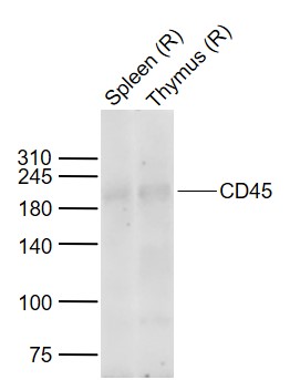 Sample:
Lane 1: Rat Spleen tissue lysates
Lane 2: Rat Thymus tissue lysates
Primary: Anti-CD45 (SL0522R) at 1/1000 dilution
Secondary: IRDye800CW Goat Anti-Rabbit IgG at 1/20000 dilution
Predicted band size: 220 kDa
Observed band size: 210 kDa
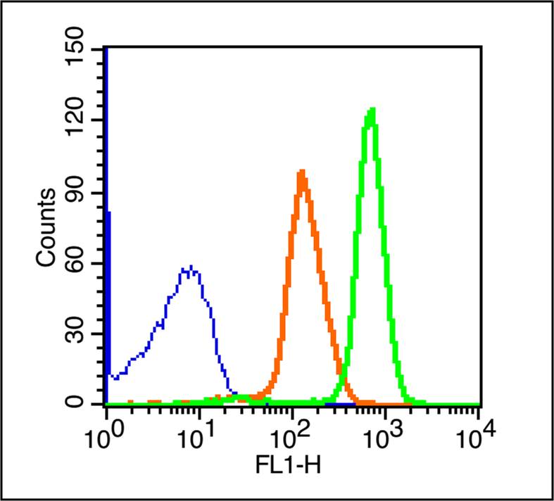 Blank control (blue line): Rabbit spleen cells(blue).
Primary Antibody (green line): Rabbit Anti-CD45/FITC Conjugated antibody (SL0522R-FITC)
Dilution: 1μg /10^6 cells;
Isotype Control Antibody (orange line): Rabbit IgG-FITC.
Protocol
The cells were fixed with 70% ice-cold methanol overnight at 4℃ . The cells were then incubated in 1 X PBS/2%BSA/10% goat serum to block non-specific protein-protein interactions followed by the antibody for 15 min at room temperature. Cells stained with Primary Antibody for 30 min at room temperature.Acquisition of 20,000 events was performed.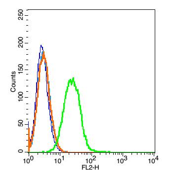 Blank control: Raji(blue).
Primary Antibody:Rabbit Anti- CD45 antibody(SL0522R), Dilution: 1μg in 100 μL 1X PBS containing 0.5% BSA;
Isotype Control Antibody: Rabbit IgG(orange) ,used under the same conditions );
Secondary Antibody: Goat anti-rabbit IgG-PE(white blue), Dilution: 1:200 in 1 X PBS containing 0.5% BSA.
Protocol
The cells were washed twice with phosphate-buffered saline (PBS).The cells were then incubated in 1 X PBS containing 0.5% BSA + 10% goat serum (15 min) to block non-specific protein-protein interactions followed by the antibody (SL0522R, 1μg /1x10^6 cells) for 30 min on ice. The secondary antibody used was Goat Anti-rabbit IgG/PE antibody at 1/200 dilution for 30 min on ice. Acquisition of 20,000 events was performed.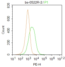 Blank control: Jurkat.
Primary Antibody (green line): Rabbit Anti-CD45 antibody (SL0522R)
Dilution: 2μg /10^6 cells;
Isotype Control Antibody (orange line): Rabbit IgG .
Secondary Antibody : Goat anti-rabbit IgG-PE
Dilution: 1μg /test.
Protocol
The cells were incubated in 5%BSA to block non-specific protein-protein interactions for 30 min at at room temperature .Cells stained with Primary Antibody for 30 min at room temperature. The secondary antibody used for 40 min at room temperature. Acquisition of 20,000 events was performed.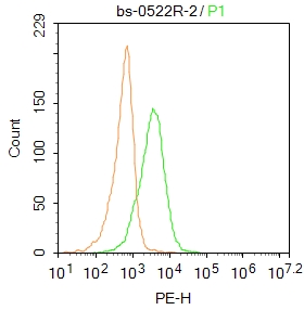 Blank control: Molt4.
Primary Antibody (green line): Rabbit Anti-CD45 antibody (SL0522R)
Dilution: 2μg /10^6 cells;
Isotype Control Antibody (orange line): Rabbit IgG .
Secondary Antibody : Goat anti-rabbit IgG-PE
Dilution: 1μg /test.
Protocol
The cells were incubated in 5%BSA to block non-specific protein-protein interactions for 30 min at at room temperature .Cells stained with Primary Antibody for 30 min at room temperature. The secondary antibody used for 40 min at room temperature. Acquisition of 20,000 events was performed.
Cartpieces
Totalgoods,subtotals:¥Checkout
Bought notes(bought amounts latest0)
No one bought this product
User Comment(Total0User Comment Num)
- No comment


 +86 571 56623320
 +86 18668110335

