
Rabbit Anti-CD2AP antibody
CD2-associated protein; Adapter protein CMS; AL024079; Cas ligand with multiple SH3 domains; C78928; Cd2ap; CMS; Mesenchyme to epithelium transition protein with SH3 domains 1; METS 1; Mets1; CD2AP_HUMAN.
View History [Clear]
Details
Product Name CD2AP Chinese Name 白Cell differentiation抗原CD2AP抗体 Alias CD2-associated protein; Adapter protein CMS; AL024079; Cas ligand with multiple SH3 domains; C78928; Cd2ap; CMS; Mesenchyme to epithelium transition protein with SH3 domains 1; METS 1; Mets1; CD2AP_HUMAN. literatures Research Area Signal transduction Transporter Binding protein Immunogen Species Rabbit Clonality Polyclonal React Species Mouse, Rat, (predicted: Human, Chicken, Dog, Pig, Cow, Horse, Rabbit, Sheep, Guinea Pig, ) Applications WB=1:500-2000 ELISA=1:5000-10000 IHC-P=1:100-500 IHC-F=1:100-500 ICC=1:100-500 IF=1:100-500 (Paraffin sections need antigen repair)
not yet tested in other applications.
optimal dilutions/concentrations should be determined by the end user.Theoretical molecular weight 71kDa Cellular localization cytoplasmic Form Liquid Concentration 1mg/ml immunogen KLH conjugated synthetic peptide derived from human CD2AP: 561-639/639 Lsotype IgG Purification affinity purified by Protein A Buffer Solution 0.01M TBS(pH7.4) with 1% BSA, 0.03% Proclin300 and 50% Glycerol. Storage Shipped at 4℃. Store at -20 °C for one year. Avoid repeated freeze/thaw cycles. Attention This product as supplied is intended for research use only, not for use in human, therapeutic or diagnostic applications. PubMed PubMed Product Detail This gene encodes a scaffolding molecule that regulates the actin cytoskeleton. The protein directly interacts with filamentous actin and a variety of cell membrane proteins through multiple actin binding sites, SH3 domains, and a proline-rich region containing binding sites for SH3 domains. The cytoplasmic protein localizes to membrane ruffles, lipid rafts, and the leading edges of cells. It is implicated in dynamic actin remodeling and membrane trafficking that occurs during receptor endocytosis and cytokinesis. Haploinsufficiency of this gene is implicated in susceptibility to glomerular disease. [provided by RefSeq, Jul 2008].
Function:
Seems to act as an adapter protein between membrane proteins and the actin cytoskeleton. May play a role in receptor clustering and cytoskeletal polarity in the junction between T-cell and antigen-presenting cell. May anchor the podocyte slit diaphragm to the actin cytoskeleton in renal glomerolus. Also required for cytokinesis.
Subunit:
Self-associates. Homodimer (Potential). Interacts with F-actin, PKD2, NPHS1 and NPHS2. Interacts with WTIP. Interacts with DDN; interaction is direct. Interacts (via SH3 2 domain) with CBL (via phosphorylated C-terminus). Interacts with BCAR1/p130Cas (via SH3 domain). Interacts with MVB12A and ARHGAP17. Interacts with ANLN, CD2 and CBLB. Interacts with PDCD6IP and TSG101. Interacts with RIN3.
Subcellular Location:
Cytoplasm, cytoskeleton. Cell projection, ruffle. Note=Colocalizes with F-actin and BCAR1/p130Cas in membrane ruffles. Located at podocyte slit diaphragm between podocyte foot processes. During late anaphase and telophase, concentrates in the vicinity of the midzone microtubules and in the midbody in late telophase.
Tissue Specificity:
Widely expressed in fetal and adult tissues.
Post-translational modifications:
Phosphorylated on tyrosine residues; probably by c-Abl, Fyn and c-Src.
DISEASE:
Focal segmental glomerulosclerosis 3 (FSGS3) [MIM:607832]: A renal pathology defined by the presence of segmental sclerosis in glomeruli and resulting in proteinuria, reduced glomerular filtration rate and progressive decline in renal function. Renal insufficiency often progresses to end-stage renal disease, a highly morbid state requiring either dialysis therapy or kidney transplantation. Note=Disease susceptibility is associated with variations affecting the gene represented in this entry.
Similarity:
Contains 3 SH3 domains.
SWISS:
Q9Y5K6
Gene ID:
23607
Database links:Entrez Gene: 23607 Human
Entrez Gene: 12488 Mouse
Omim: 604241 Human
SwissProt: Q9Y5K6 Human
SwissProt: Q9JLQ0 Mouse
Unigene: 485518 Human
Unigene: 218637 Mouse
Unigene: 212220 Rat
CD2AP可能作为细胞裂孔隔膜分子与Cytoskeleton的连接蛋白,在Cell differentiation即增殖的过程中发挥重要作用。
目前主用用于肾脏功能方面的研究,D2AP不仅参与T细胞的活化,而且对肾脏功能起着至关重要的作用,其异常表达可能是引起人类肾脏疾病的诱因之一。Product Picture 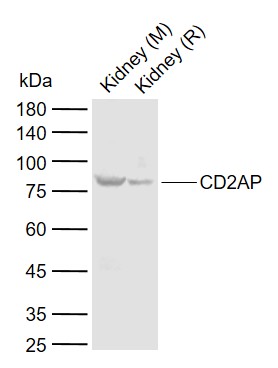 Sample:
Lane 1: Mouse Kidney tissue lysates
Lane 2: Rat Kidney tissue lysates
Primary: Anti-CD2AP (SL0512R) at 1/1000 dilution
Secondary: IRDye800CW Goat Anti-Rabbit IgG at 1/20000 dilution
Predicted band size: 71 kDa
Observed band size: 77 kDa
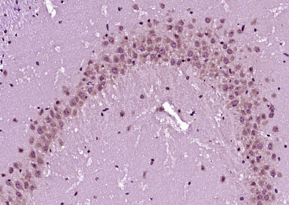 Paraformaldehyde-fixed, paraffin embedded (Mouse brain); Antigen retrieval by boiling in sodium citrate buffer (pH6.0) for 15min; Block endogenous peroxidase by 3% hydrogen peroxide for 20 minutes; Blocking buffer (normal goat serum) at 37°C for 30min; Antibody incubation with (CD2AP) Polyclonal Antibody, Unconjugated (SL0512R) at 1:400 overnight at 4°C, followed by operating according to SP Kit(Rabbit) (sp-0023) instructionsand DAB staining.
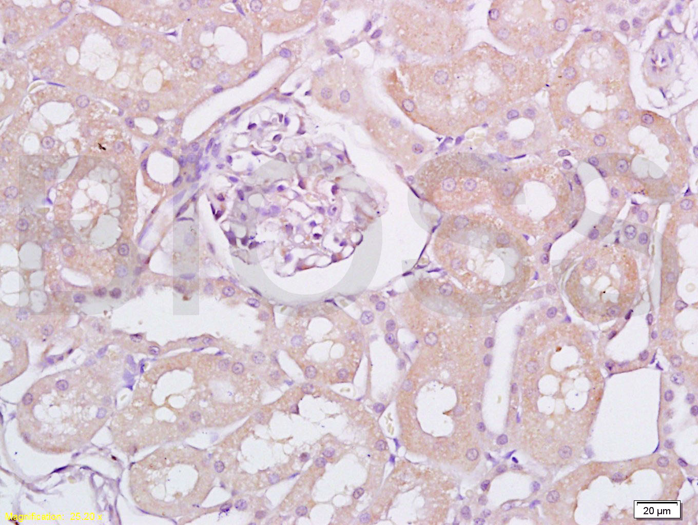 Tissue/cell: rat kidney tissue; 4% Paraformaldehyde-fixed and paraffin-embedded;
Antigen retrieval: citrate buffer ( 0.01M, pH 6.0 ), Boiling bathing for 15min; Block endogenous peroxidase by 3% Hydrogen peroxide for 30min; Blocking buffer (normal goat serum,C-0005) at 37℃ for 20 min;
Incubation: Anti-CD2AP Polyclonal Antibody, Unconjugated(SL0512R) 1:200, overnight at 4°C, followed by conjugation to the secondary antibody(SP-0023) and DAB(C-0010) staining
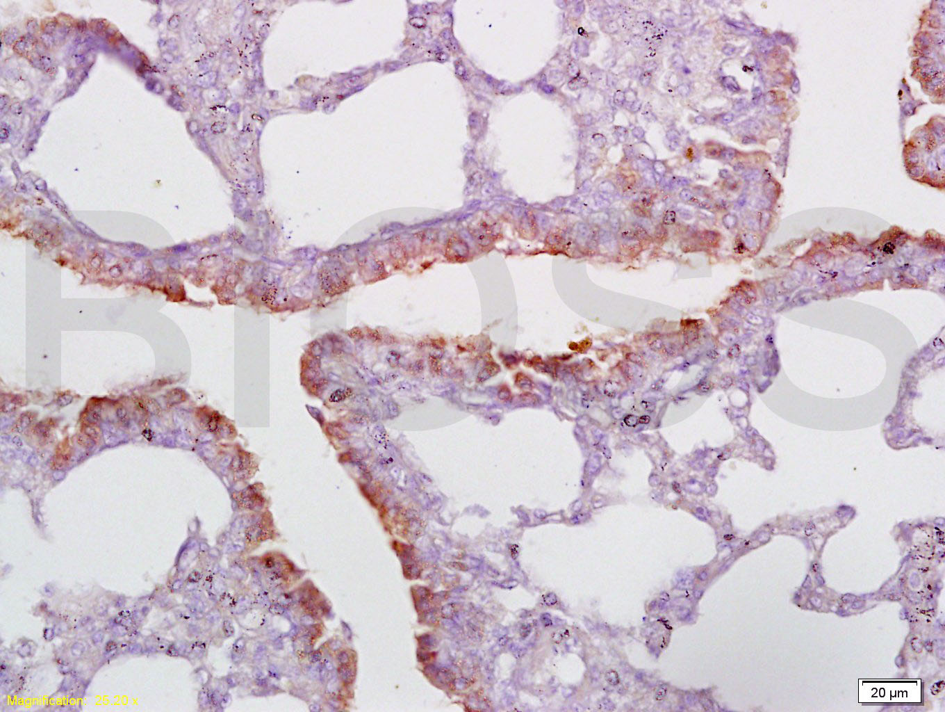 Tissue/cell: rat lung tissue; 4% Paraformaldehyde-fixed and paraffin-embedded;
Antigen retrieval: citrate buffer ( 0.01M, pH 6.0 ), Boiling bathing for 15min; Block endogenous peroxidase by 3% Hydrogen peroxide for 30min; Blocking buffer (normal goat serum,C-0005) at 37℃ for 20 min;
Incubation: Anti-CD2AP Polyclonal Antibody, Unconjugated(SL0512R) 1:200, overnight at 4°C, followed by conjugation to the secondary antibody(SP-0023) and DAB(C-0010) staining
Cartpieces
Totalgoods,subtotals:¥Checkout
Bought notes(bought amounts latest0)
No one bought this product
User Comment(Total0User Comment Num)
- No comment


 +86 571 56623320
 +86 18668110335

