
Rabbit Anti-JNK1 + JNK3 antibody
JNK1 + JNK3; JNK1 + 3; JNK1+JNK3; JNK1/3; c Jun N terminal kinase 1; JNK1; JNK3; JAK 1A; JAK1A; JNK 1; JNK 46; JNK; JNK1A2; JNK21B1/2; MAPK 8; MAPK8; Mitogen activated protein kinase 8; PRKM 8; PRKM8; Protein kinase JNK1; SAPK 1; SAPK gamma; SAPK1; c-Jun;
View History [Clear]
Details
Product Name JNK1 + JNK3 Chinese Name 氨基末端激酶1/3抗体 Alias JNK1 + JNK3; JNK1 + 3; JNK1+JNK3; JNK1/3; c Jun N terminal kinase 1; JNK1; JNK3; JAK 1A; JAK1A; JNK 1; JNK 46; JNK; JNK1A2; JNK21B1/2; MAPK 8; MAPK8; Mitogen activated protein kinase 8; PRKM 8; PRKM8; Protein kinase JNK1; SAPK 1; SAPK gamma; SAPK1; c-Jun; Stress activated protein kinase JNK1; Tyrosine protein kinase JAK1; MK08_HUMAN. literatures Research Area Tumour Cell biology immunology Signal transduction transcriptional regulatory factor Kinases and Phosphatases Immunogen Species Rabbit Clonality Polyclonal React Species Human, Mouse, Rat, (predicted: Chicken, Dog, Pig, Cow, Rabbit, ) Applications WB=1:500-2000 ELISA=1:5000-10000 IHC-P=1:100-500 IHC-F=1:100-500 Flow-Cyt=1μg/Test ICC=1:100 IF=1:100-500 (Paraffin sections need antigen repair)
not yet tested in other applications.
optimal dilutions/concentrations should be determined by the end user.Theoretical molecular weight 42kDa Cellular localization The nucleus cytoplasmic Form Liquid Concentration 1mg/ml immunogen KLH conjugated synthetic peptide derived from human JNK1: 201-300/427 Lsotype IgG Purification affinity purified by Protein A Buffer Solution 0.01M TBS(pH7.4) with 1% BSA, 0.03% Proclin300 and 50% Glycerol. Storage Shipped at 4℃. Store at -20 °C for one year. Avoid repeated freeze/thaw cycles. Attention This product as supplied is intended for research use only, not for use in human, therapeutic or diagnostic applications. PubMed PubMed Product Detail JNK1(MAPK8) is a member of the MAP kinase family. MAP kinases act as an integration point for multiple biochemical signals, and are involved in a wide variety of cellular processes such as proliferation, differentiation, transcription regulation and development. This kinase is activated by various cell stimuli, and targets specific transcription factors, and thus mediates immediate-early gene expression in response to cell stimuli. The activation of this kinase by tumor-necrosis factor alpha (TNF-alpha) is found to be required for TNF-alpha induced apoptosis. This kinase is also involved in UV radiation induced apoptosis, which is thought to be related to cytochrome c-mediated cell death pathway. Studies of the mouse counterpart of this gene suggested that this kinase play a key role in T cell proliferation, apoptosis and differentiation. Four alternatively spliced transcript variants encoding distinct isoforms have been reported. JNK1 is activated by threonine and tyrosine phosphorylation by either of two dual specificity kinases, MAP2K4 and MAP2K7. The JNK pathway is critically involved in diabetes and levels are abnormally elevated in obesity. The cell-permeable JNK inhibitory peptide may have promise as a therapeutic agent for diabetes.
Function:
Serine/threonine-protein kinase involved in various processes such as cell proliferation, differentiation, migration, transformation and programmed cell death. Extracellular stimuli such as proinflammatory cytokines or physical stress stimulate the stress-activated protein kinase/c-Jun N-terminal kinase (SAP/JNK) signaling pathway. In this cascade, two dual specificity kinases MAP2K4/MKK4 and MAP2K7/MKK7 phosphorylate and activate MAPK8/JNK1. In turn, MAPK8/JNK1 phosphorylates a number of transcription factors, primarily components of AP-1 such as JUN, JDP2 and ATF2 and thus regulates AP-1 transcriptional activity. Phosphorylates the replication licensing factor CDT1, inhibiting the interaction between CDT1 and the histone H4 acetylase HBO1 to replication origins. Loss of this interaction abrogates the acetylation required for replication initiation. Promotes stressed cell apoptosis by phosphorylating key regulatory factors including p53/TP53 and Yes-associates protein YAP1. In T-cells, MAPK8 and MAPK9 are required for polarized differentiation of T-helper cells into Th1 cells. Contributes to the survival of erythroid cells by phosphorylating the antagonist of cell death BAD upon EPO stimulation. Mediates starvation-induced BCL2 phosphorylation, BCL2 dissociation from BECN1, and thus activation of autophagy. Phosphorylates STMN2 and hence regulates microtubule dynamics, controlling neurite elongation in cortical neurons. In the developing brain, through its cytoplasmic activity on STMN2, negatively regulates the rate of exit from multipolar stage and of radial migration from the ventricular zone. Phosphorylates several other substrates including heat shock factor protein 4 (HSF4), the deacetylase SIRT1, ELK1, or the E3 ligase ITCH.
Subunit:
Binds to at least four scaffolding proteins, MAPK8IP1/JIP-1, MAPK8IP2/JIP-2, MAPK8IP3/JIP-3/JSAP1 and SPAG9/MAPK8IP4/JIP-4. These proteins also bind other components of the JNK signaling pathway. Forms a complex with MAPK8IP1 and RGNEF. Interacts with TP53 and WWOX. Interacts with JAMP. Interacts with NFATC4. Interacts with MECOM; regulates JNK signaling. Interacts with PIN1; this interaction mediates MAPK8 conformational changes leading to the binding of MAPK8 to its substrates. Interacts (phosphorylated form) with NFE2; the interaction phosphorylates NFE2 in undifferentiated cells.
Subcellular Location:
Cytoplasm. Nucleus.
Post-translational modifications:
Phosphorylated by TAOK2. Dually phosphorylated on Thr-183 and Tyr-185 by MAP2K7 and MAP2K4, which activates the enzyme.
Similarity:
Belongs to the protein kinase superfamily. CMGC Ser/Thr protein kinase family. MAP kinase subfamily.
Contains 1 protein kinase domain.
SWISS:
P45983
Gene ID:
5599
5601
Database links:Entrez Gene: 5599 Human
Entrez Gene: 5601 Human
Entrez Gene: 26419 Mouse
Entrez Gene: 26420 Mouse
Omim: 601158 Human
Omim: 602896 Human
SwissProt: P45983 Human
SwissProt: P45984 Human
SwissProt: Q91Y86 Mouse
SwissProt: Q9WTU6 Mouse
Unigene: 138211 Human
Unigene: 348446 Human
Unigene: 21495 Mouse
Unigene: 68933 Mouse
Product Picture 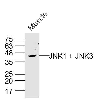 Sample:Muscle (Rat) Lysate at 40 ug
Primary: Anti-JNK1 + JNK3 (SL0501R) at 1/300 dilution
Secondary: IRDye800CW Goat Anti-Rabbit IgG at 1/20000 dilution
Predicted band size: 42 kD
Observed band size: 42 kD
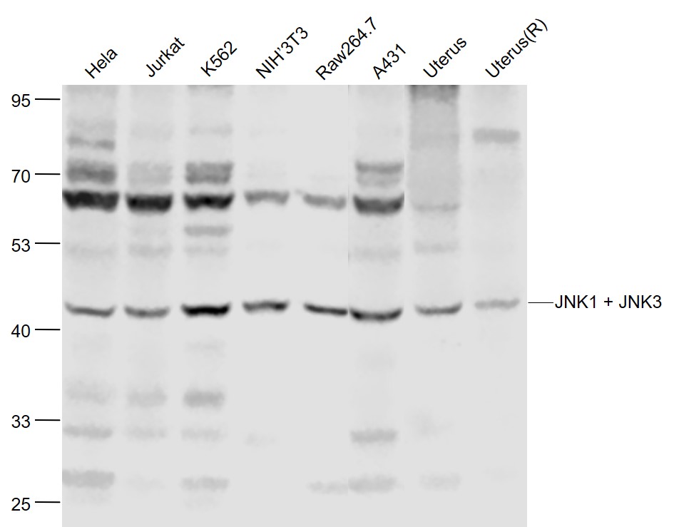 Sample:
Hela(Human) Cell Lysate at 30 ug
Jurkat(Human) Cell Lysate at 30 ug
K562(Human) Cell Lysate at 30 ug
NIH/3T3(Mouse) Cell Lysate at 30 ug
Raw264.7(Mouse) Cell Lysate at 30 ug
A431(Human) Cell Lysate at 30 ug
Uterus(Mouse) Lysate at 40 ug
Uterus(Rat) Lysate at 40 ug
Primary: Anti-JNK1 + JNK3 (SL0501R) at 1/1000 dilution
Secondary: IRDye800CW Goat Anti-Rabbit IgG at 1/20000 dilution
Predicted band size: 46'54 kD
Observed band size: 46 kD
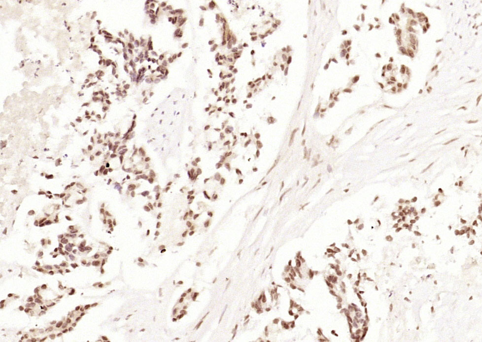 Paraformaldehyde-fixed, paraffin embedded (human gastric carcinoma); Antigen retrieval by boiling in sodium citrate buffer (pH6.0) for 15min; Block endogenous peroxidase by 3% hydrogen peroxide for 20 minutes; Blocking buffer (normal goat serum) at 37°C for 30min; Antibody incubation with (JNK1 + JNK3) Polyclonal Antibody, Unconjugated (SL0501R) at 1:200 overnight at 4°C, followed by operating according to SP Kit(Rabbit) (sp-0023) instructionsand DAB staining.
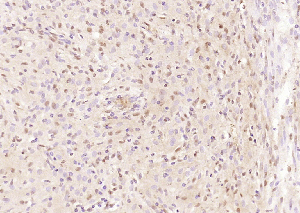 Paraformaldehyde-fixed, paraffin embedded (rat uterus); Antigen retrieval by boiling in sodium citrate buffer (pH6.0) for 15min; Block endogenous peroxidase by 3% hydrogen peroxide for 20 minutes; Blocking buffer (normal goat serum) at 37°C for 30min; Antibody incubation with (JNK1 + JNK3) Polyclonal Antibody, Unconjugated (SL0501R) at 1:200 overnight at 4°C, followed by operating according to SP Kit(Rabbit) (sp-0023) instructionsand DAB staining.
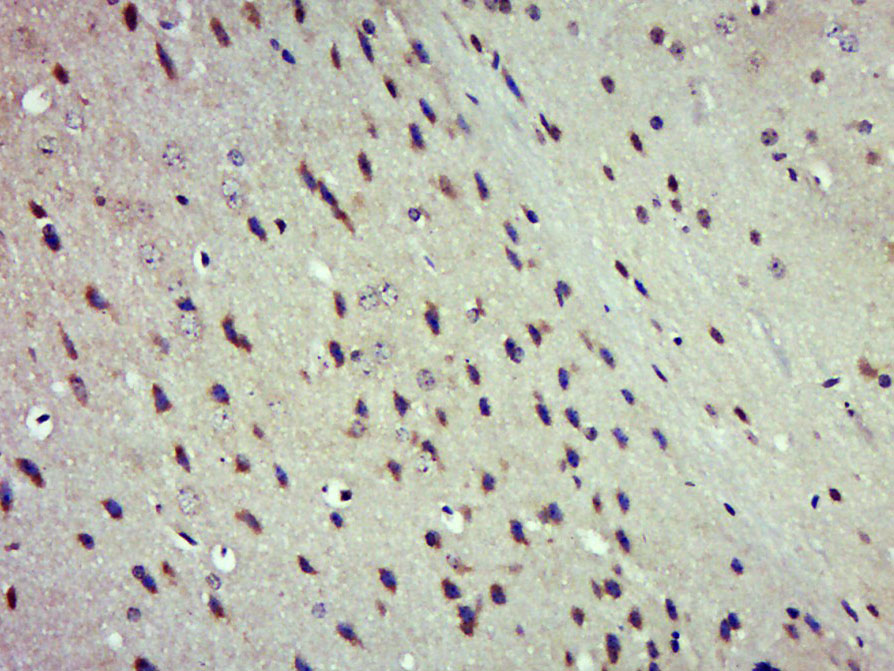 Paraformaldehyde-fixed, paraffin embedded (Mouse brain); Antigen retrieval by boiling in sodium citrate buffer (pH6.0) for 15min; Block endogenous peroxidase by 3% hydrogen peroxide for 20 minutes; Blocking buffer (normal goat serum) at 37°C for 30min; Antibody incubation with (JNK1 + JNK3) Polyclonal Antibody, Unconjugated (SL0501R) at 1:500 overnight at 4°C, followed by a conjugated secondary (sp-0023) for 20 minutes and DAB staining.
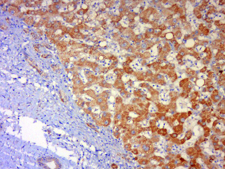 Tissue/cell: human liver carcinoma; 4% Paraformaldehyde-fixed and paraffin-embedded;
Antigen retrieval: citrate buffer ( 0.01M, pH 6.0 ), Boiling bathing for 15min; Block endogenous peroxidase by 3% Hydrogen peroxide for 30min; Blocking buffer (normal goat serum,C-0005) at 37℃ for 20 min;
Incubation: Anti-JNK1+ JNK3 Polyclonal Antibody, Unconjugated(SL0501R) 1:500, overnight at 4°C, followed by conjugation to the secondary antibody(SP-0023) and DAB(C-0010) staining
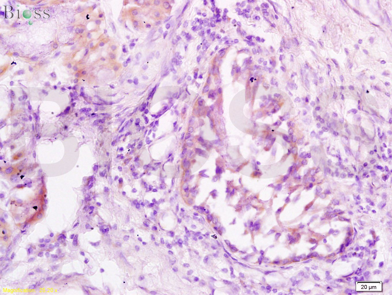 Tissue/cell: human lung carcinoma; 4% Paraformaldehyde-fixed and paraffin-embedded;
Antigen retrieval: citrate buffer ( 0.01M, pH 6.0 ), Boiling bathing for 15min; Block endogenous peroxidase by 3% Hydrogen peroxide for 30min; Blocking buffer (normal goat serum,C-0005) at 37℃ for 20 min;
Incubation: Anti-JNK1/3 Polyclonal Antibody, Unconjugated(SL0501R) 1:200, overnight at 4°C, followed by conjugation to the secondary antibody(SP-0023) and DAB(C-0010) staining
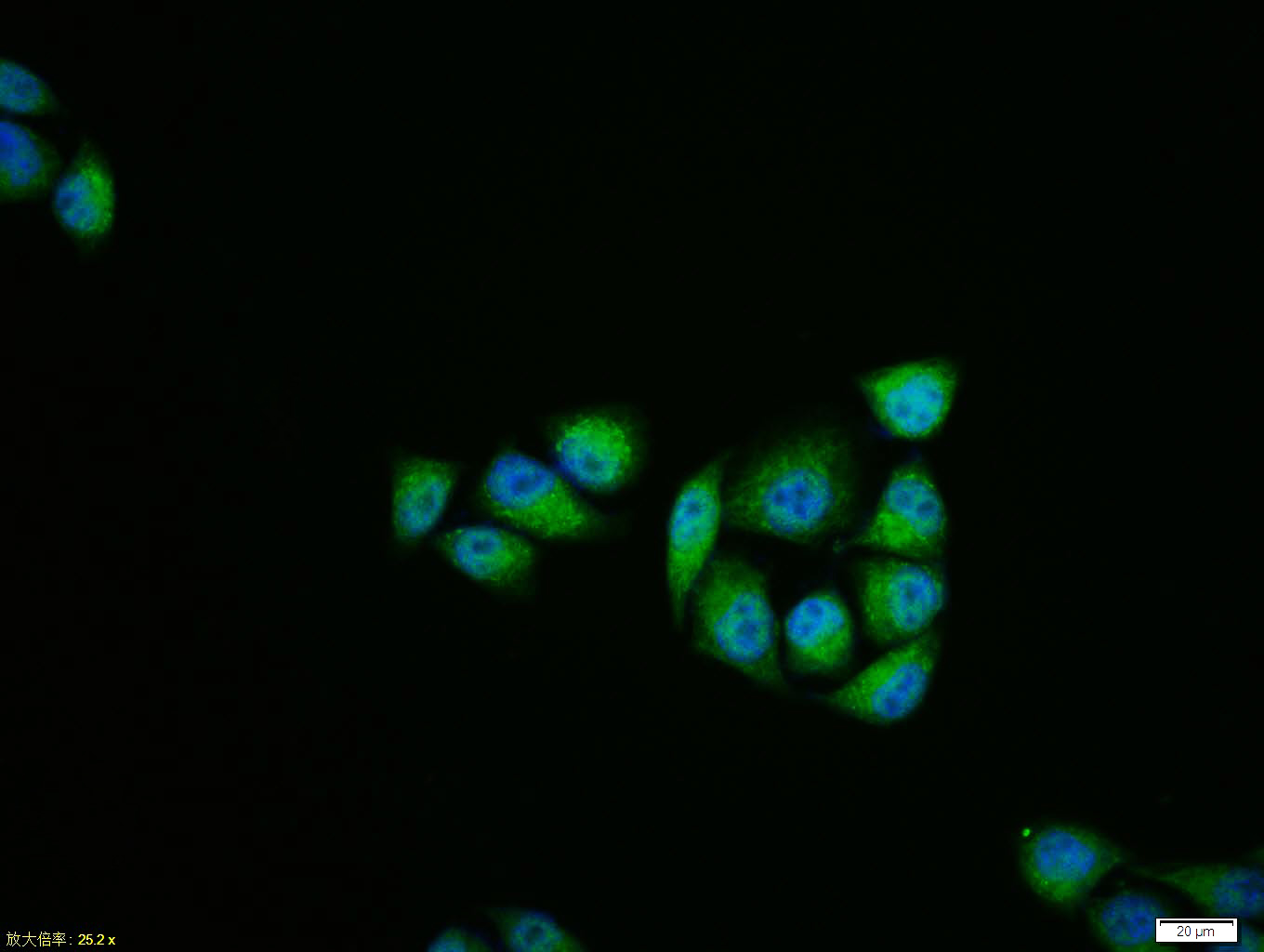 Hela cell; 4% Paraformaldehyde-fixed; Triton X-100 at room temperature for 20 min; Blocking buffer (normal goat serum, C-0005) at 37°C for 20 min; Antibody incubation with (JNK1 + JNK3) polyclonal Antibody, Unconjugated (SL0501R) 1:100, 90 minutes at 37°C; followed by a conjugated Goat Anti-Rabbit IgG antibody at 37°C for 90 minutes, DAPI (blue, C02-04002) was used to stain the cell nuclei.
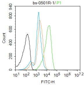 Blank control: K562.
Primary Antibody (green line): Rabbit Anti-JNK1 + JNK3 antibody (SL0501R)
Dilution: 1μg /10^6 cells;
Isotype Control Antibody (orange line): Rabbit IgG .
Secondary Antibody : Goat anti-rabbit IgG-FITC
Dilution: 1μg /test.
Protocol
The cells were fixed with 4% PFA (10min at room temperature)and then permeabilized with 90% ice-cold methanol for 20 min at-20℃. The cells were then incubated in 5%BSA to block non-specific protein-protein interactions for 30 min at room temperature .Cells stained with Primary Antibody for 30 min at room temperature. The secondary antibody used for 40 min at room temperature. Acquisition of 20,000 events was performed.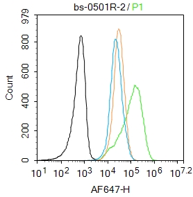 Blank control: Jurkat.
Primary Antibody (green line): Rabbit Anti-JNK1 + JNK3 antibody (SL0501R)
Dilution: 2μg /10^6 cells;
Isotype Control Antibody (orange line): Rabbit IgG .
Secondary Antibody : Goat anti-rabbit IgG-AF647
Dilution: 1μg /test.
Protocol
The cells were fixed with 4% PFA (10min at room temperature)and then permeabilized with 90% ice-cold methanol for 20 min at-20℃. The cells were then incubated in 5%BSA to block non-specific protein-protein interactions for 30 min at room temperature .Cells stained with Primary Antibody for 30 min at room temperature. The secondary antibody used for 40 min at room temperature. Acquisition of 20,000 events was performed.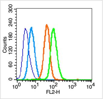 Blank control (blue line): Hep G2 (blue).
Primary Antibody (green line): Rabbit Anti-JNK1 + JNK3 antibody (SL0501R)
Dilution: 1μg /10^6 cells;
Isotype Control Antibody (orange line): Rabbit IgG .
Secondary Antibody (white blue line): Goat anti-rabbit IgG-PE
Dilution: 1μg /test.
Protocol
The cells were fixed with 70% ethanol (Overnight at 4℃) and then permeabilized with 90% methanol for 20 min at -20℃. Cells stained with Primary Antibody for 30 min at room temperature. The cells were then incubated in 1 X PBS/2%BSA/10% goat serum to block non-specific protein-protein interactions followed by the antibody for 15 min at room temperature. The secondary antibody used for 40 min at room temperature. Acquisition of 20,000 events was performed.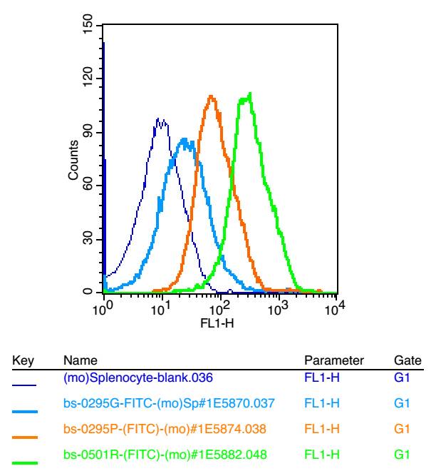 Blank control: mouse splenocytes(blue)
Isotype Control Antibody: Rabbit IgG(orange) ; Secondary Antibody: Goat anti-rabbit IgG-FITC(white blue), Dilution: 1:100 in 1 X PBS containing 0.5% BSA ; Primary Antibody Dilution: 1μl in 100 μL1X PBS containing 0.5% BSA(green).
Cartpieces
Totalgoods,subtotals:¥Checkout
Bought notes(bought amounts latest0)
No one bought this product
User Comment(Total0User Comment Num)
- No comment


 +86 571 56623320
 +86 18668110335

