
Rabbit Anti-MOG antibody
MOG_HUMAN; Myelin-oligodendrocyte glycoprotein; Myelin oligodendrocyte glycoprotein; BTN6; BTNL11; MOGIG2; NRCLP7;
View History [Clear]
Details
Product Name MOG Chinese Name 髓鞘少树突胶质细胞glycoprotein抗体 Alias MOG_HUMAN; Myelin-oligodendrocyte glycoprotein; Myelin oligodendrocyte glycoprotein; BTN6; BTNL11; MOGIG2; NRCLP7; literatures Research Area Cell biology Neurobiology Signal transduction Stem cells Apoptosis Cell adhesion molecule The cell membrane蛋白 Immunogen Species Rabbit Clonality Polyclonal React Species Mouse, Rat, (predicted: Human, Pig, Guinea Pig, ) Applications WB=1:500-2000 ELISA=1:5000-10000 IHC-P=1:100-500 IHC-F=1:100-500 ICC=1:100-500 IF=1:100-500 (Paraffin sections need antigen repair)
not yet tested in other applications.
optimal dilutions/concentrations should be determined by the end user.Theoretical molecular weight 24kDa Cellular localization The cell membrane Form Liquid Concentration 1mg/ml immunogen KLH conjugated synthetic peptide derived from mouse MOG: 35-55/247 <Extracellular> Lsotype IgG Purification affinity purified by Protein A Buffer Solution 0.01M TBS(pH7.4) with 1% BSA, 0.03% Proclin300 and 50% Glycerol. Storage Shipped at 4℃. Store at -20 °C for one year. Avoid repeated freeze/thaw cycles. Attention This product as supplied is intended for research use only, not for use in human, therapeutic or diagnostic applications. PubMed PubMed Product Detail The product of this gene is a membrane protein expressed on the oligodendrocyte cell surface and the outermost surface of myelin sheaths. Due to this localization, it is a primary target antigen involved in immune-mediated demyelination. This protein may be involved in completion and maintenance of the myelin sheath and in cell-cell communication. Alternatively spliced transcript variants encoding different isoforms have been identified. [provided by RefSeq, Jul 2008]
Function:
Mediates homophilic cell-cell adhesion. Minor component of the myelin sheath. May be involved in completion and/or maintenance of the myelin sheath and in cell-cell communication.
Subunit:
Homodimer. May form heterodimers between the different isoforms.
Subcellular Location:
Cell membrane; Multi-pass membrane protein (Potential).
Tissue Specificity:
Found exclusively in the CNS, where it is localized on the surface of myelin and oligodendrocyte cytoplasmic membranes.
DISEASE:
Defects in MOG are the cause of narcolepsy type 7 (NRCLP7) [MIM:614250]. Neurological disabling sleep disorder, characterized by excessive daytime sleepiness, sleep fragmentation, symptoms of abnormal rapid-eye-movement (REM) sleep, cataplexy, hypnagogic hallucinations, and sleep paralysis. Cataplexy is a sudden loss of muscle tone triggered by emotions, which is the most valuable clinical feature used to diagnose narcolepsy. Human narcolepsy is primarily a sporadically occurring disorder but familial clustering has been observed.
Similarity:
Belongs to the immunoglobulin superfamily. BTN/MOG family.
Contains 1 Ig-like V-type (immunoglobulin-like) domain.
SWISS:
Q61885
Gene ID:
4340
Database links:Entrez Gene: 4340 Human
Entrez Gene: 17441 Mouse
Omim: 159465 Human
SwissProt: Q16653 Human
SwissProt: Q61885 Mouse
Unigene: 141308 Human
Unigene: 210857 Mouse
Unigene: 9687 Rat
Product Picture
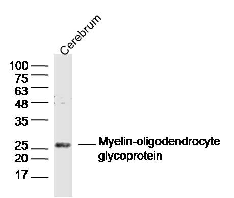
Cerebrum (Mouse) Lysate at 40 ug
Primary: Anti- Myelin-oligodendrocyte glycoprotein (SL0426R) at 1/300 dilution
Secondary: IRDye800CW Goat Anti-Rabbit IgG at 1/20000 dilution
Predicted band size: 24 kD
Observed band size: 26 kD
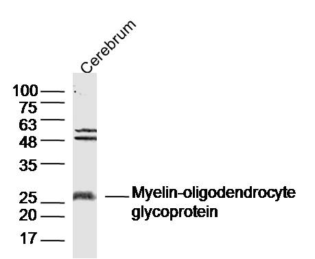
Sample:
Cerebrum (Rat) Lysate at 40 ug
Primary: Anti- Myelin-oligodendrocyte glycoprotein (SL0426R) at 1/300 dilution
Secondary: IRDye800CW Goat Anti-Rabbit IgG at 1/20000 dilution
Predicted band size: 24 kD
Observed band size: 26 kD
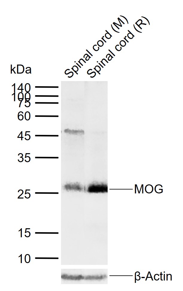
Sample:
Lane 1: Mouse Spinal cord tissue lysates
Lane 2: Rat Spinal cord tissue lysates
Primary: Anti-MOG (SL0426R) at 1/1000 dilution
Secondary: IRDye800CW Goat Anti-Rabbit IgG at 1/20000 dilution
Predicted band size: 24 kDa
Observed band size: 26 kDa
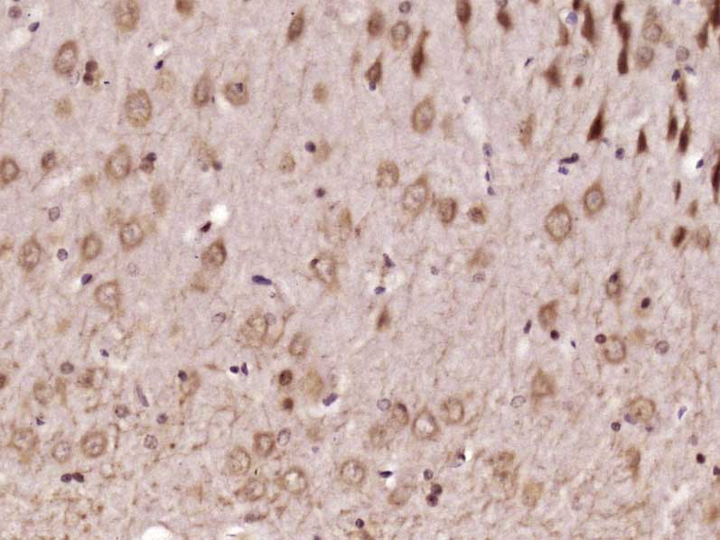
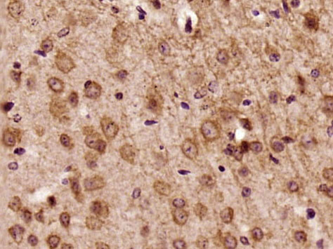
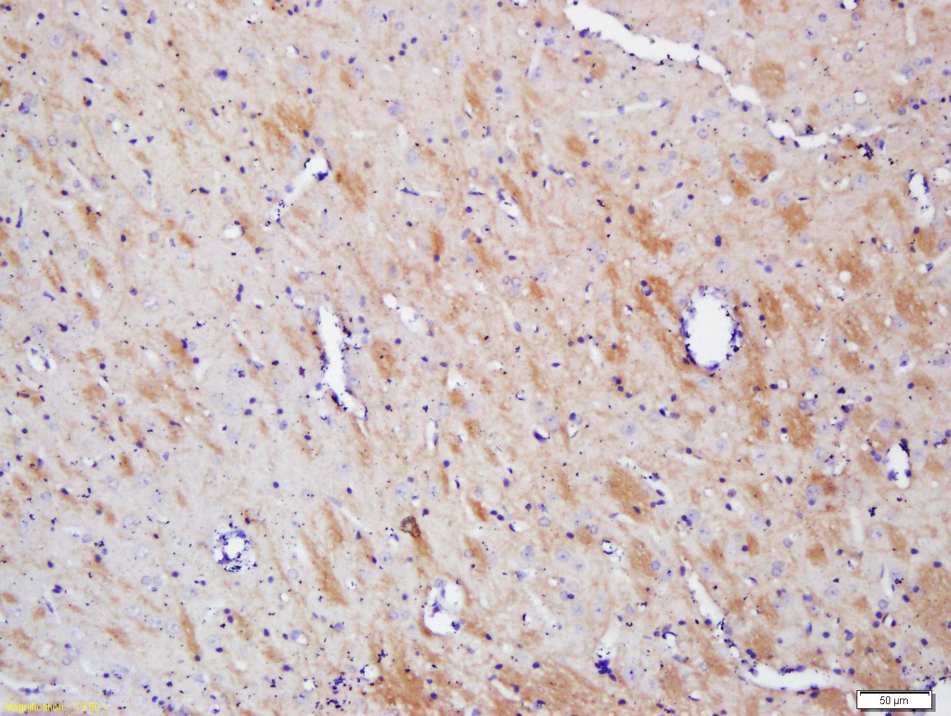
Paraformaldehyde-fixed, paraffin embedded (Rat brain); Antigen retrieval by boiling in sodium citrate buffer (pH6.0) for 15min; Block endogenous peroxidase by 3% hydrogen peroxide for 20 minutes; Blocking buffer (normal goat serum) at 37°C for 30min; Antibody incubation with (MOG) Polyclonal Antibody, Unconjugated (SL0426R) at 1:500 overnight at 4°C, followed by a conjugated secondary (sp-0023) for 20 minutes and DAB staining.Paraformaldehyde-fixed, paraffin embedded (Mouse brain); Antigen retrieval by boiling in sodium citrate buffer (pH6.0) for 15min; Block endogenous peroxidase by 3% hydrogen peroxide for 20 minutes; Blocking buffer (normal goat serum) at 37°C for 30min; Antibody incubation with (MOG) Polyclonal Antibody, Unconjugated (SL0426R) at 1:500 overnight at 4°C, followed by a conjugated secondary (sp-0023) for 20 minutes and DAB staining.Tissue/cell: rat brain tissue; 4% Paraformaldehyde-fixed and paraffin-embedded;
Antigen retrieval: citrate buffer ( 0.01M, pH 6.0 ), Boiling bathing for 15min; Block endogenous peroxidase by 3% Hydrogen peroxide for 30min; Blocking buffer (normal goat serum,C-0005) at 37← for 20 min;
Incubation: Anti- MOG Polyclonal Antibody, Unconjugated(SL0426R) 1:200, overnight at 4⒉C, followed by conjugation to the secondary antibody(SP-0023) and DAB(C-0010) staining
Partial purchase records(bought amounts latest0)
No one bought this product
User Comment(Total0User Comment Num)
- No comment


 +86 571 56623320


