
Rabbit Anti-DR3 antibody
Apo 3; Apo3; Apo-3; Apoptosis inducing receptor AIR; Apoptosis inducing receptor; Apoptosis mediating receptor; Apoptosis mediating receptor DR3; Apoptosis mediating receptor TRAMP; DDR 3; DDR3; Death domain receptor 3; Death Receptor 3; Death receptor be
View History [Clear]
Details
Product Name DR3 Chinese Name 死亡受体3抗体 Alias Apo 3; Apo3; Apo-3; Apoptosis inducing receptor AIR; Apoptosis inducing receptor; Apoptosis mediating receptor; Apoptosis mediating receptor DR3; Apoptosis mediating receptor TRAMP; DDR 3; DDR3; Death domain receptor 3; Death Receptor 3; Death receptor beta; DR 3; LARD; Lymphocyte associated receptor of death; TNFRSF 12; TNFRSF 25; TNFRSF12; TNFRSF25; TR 3; TR3; TRAMP; Translocating chain association membrane protein; Tumor necrosis factor receptor superfamily member 12; Tumor necrosis factor receptor superfamily member 25; WSL 1; WSL; WSL LR; WSL1; WSL1 protein; WSLLR; TNR25_HUMAN. literatures Research Area Tumour immunology Apoptosis Immunogen Species Rabbit Clonality Polyclonal React Species Human, Mouse, Rat, Applications WB=1:500-2000 ELISA=1:5000-10000 IHC-P=1:100-500 IHC-F=1:100-500 IF=1:100-500 (Paraffin sections need antigen repair)
not yet tested in other applications.
optimal dilutions/concentrations should be determined by the end user.Theoretical molecular weight 43kDa Cellular localization The cell membrane Secretory protein Form Liquid Concentration 1mg/ml immunogen KLH conjugated synthetic peptide derived from human DR3: 351-417/417 Lsotype IgG Purification affinity purified by Protein A Buffer Solution 0.01M TBS(pH7.4) with 1% BSA, 0.03% Proclin300 and 50% Glycerol. Storage Shipped at 4℃. Store at -20 °C for one year. Avoid repeated freeze/thaw cycles. Attention This product as supplied is intended for research use only, not for use in human, therapeutic or diagnostic applications. PubMed PubMed Product Detail The protein encoded by this gene is a member of the TNF-receptor superfamily. This receptor is expressed preferentially in the tissues enriched in lymphocytes, and it may play a role in regulating lymphocyte homeostasis. This receptor has been shown to stimulate NF-kappa B activity and regulate cell apoptosis. The signal transduction of this receptor is mediated by various death domain containing adaptor proteins. Knockout studies in mice suggested the role of this gene in the removal of self-reactive T cells in the thymus. Multiple alternatively spliced transcript variants of this gene encoding distinct isoforms have been reported, most of which are potentially secreted molecules. The alternative splicing of this gene in B and T cells encounters a programmed change upon T-cell activation, which predominantly produces full-length, membrane bound isoforms, and is thought to be involved in controlling lymphocyte proliferation induced by T-cell activation. [provided by RefSeq, Jul 2008]
Subunit:
Homodimer. Interacts strongly via the death domains with TNFRSF1 and TRADD to activate at least two distinct signaling cascades, apoptosis and NF-kappa-B signaling. Interacts with BAG4.
Subcellular Location:
Isoform 1: Cell membrane; Single-pass type I membrane protein.
Isoform 2: Cell membrane; Single-pass type I membrane protein.
Isoform 9: Cell membrane; Single-pass type I membrane protein.
Isoform 11: Cell membrane; Single-pass type I membrane protein.
Isoform 3: Secreted.
Isoform 4: Secreted.
Isoform 5: Secreted.
Isoform 6: Secreted.
Isoform 7: Secreted.
Isoform 8: Secreted.
Isoform 10: Secreted.
Isoform 12: Secreted.
Tissue Specificity:
Abundantly expressed in thymocytes and lymphocytes. Detected in lymphocyte-rich tissues such as thymus, colon, intestine, and spleen. Also found in the prostate.
Similarity:
Contains 1 death domain.
Contains 4 TNFR-Cys repeats.
SWISS:
Q93038
Gene ID:
8718
Database links:Entrez Gene: 8718 Human
Entrez Gene: 85030 Mouse
Omim: 603366 Human
SwissProt: Q93038 Human
Unigene: 462529 Human
Unigene: 101198 Mouse
Unigene: 106560 Rat
死亡受体(death receptor)已经鉴定的有DR3、DR4和DR5,DR3也称为WSL-1,Apo-3受体或TRAMP(TNFR介导的Apoptosis蛋白质TNF-receptor apoptosis-mediating protein,TRAMP)。
Tumour坏死因子-α受体(Tumour坏死因子受体超家TNF-α膜受体族成员)是近年来被鉴定发现的:主要在人的多lymphocyte组织中。它们同属TNF/NGF受体超家族成员。Product Picture 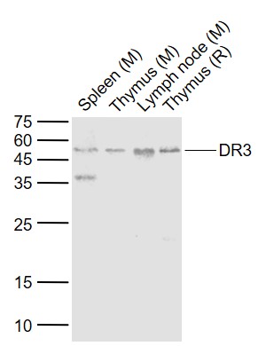 Sample:
Lane 1: Spleen (Mouse) Lysate at 40 ug
Lane 2: Thymus (Mouse) Lysate at 40 ug
Lane 3: Lymph node (Mouse) Lysate at 40 ug
Lane 4: Thymus (Rat) Lysate at 40 ug
Primary: Anti-DR3 (SL0421R) at 1/1000 dilution
Secondary: IRDye800CW Goat Anti-Rabbit IgG at 1/20000 dilution
Predicted band size: 50 kD
Observed band size: 50 kD
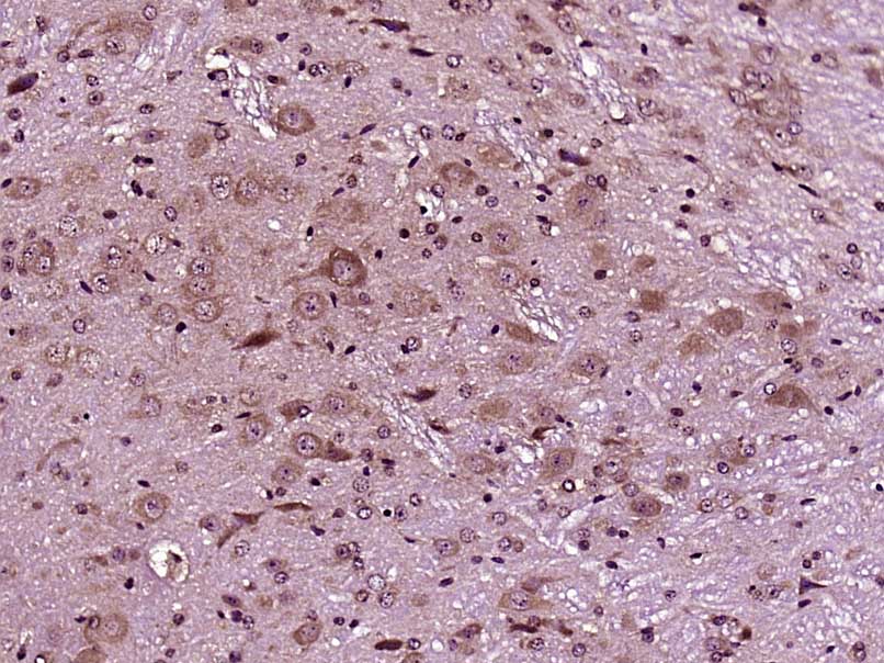 Paraformaldehyde-fixed, paraffin embedded (Mouse brain); Antigen retrieval by boiling in sodium citrate buffer (pH6.0) for 15min; Block endogenous peroxidase by 3% hydrogen peroxide for 20 minutes; Blocking buffer (normal goat serum) at 37°C for 30min; Antibody incubation with (DR3) Polyclonal Antibody, Unconjugated (SL0421R) at 1:400 overnight at 4°C, followed by operating according to SP Kit(Rabbit) (sp-0023) instructionsand DAB staining.
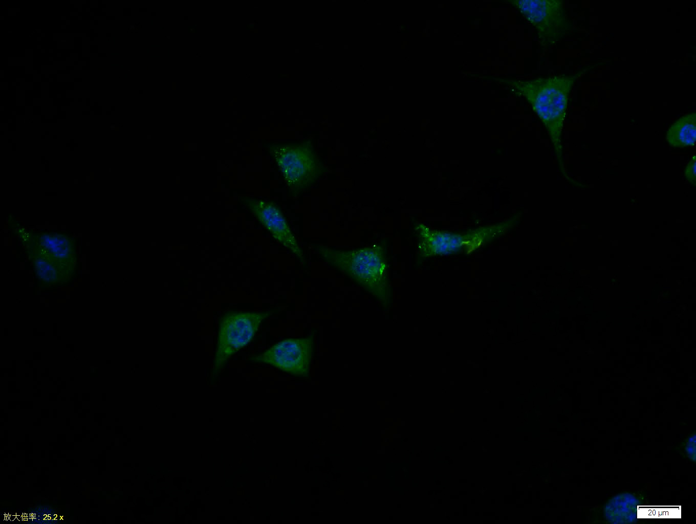 Tissue/cell:SH-SY5Y cell; 4% Paraformaldehyde-fixed; Triton X-100 at room temperature for 20 min; Blocking buffer (normal goat serum,C-0005) at 37°C for 20 min; Antibody incubation with (DR3) polyclonal Antibody, Unconjugated (SL0421R) 1:100, 90 minutes at 37°C; followed by a FITC conjugated Goat Anti-Rabbit IgG antibody at 37°C for 90 minutes, DAPI (blue, C02-04002) was used to stain the cell nuclei.
Cartpieces
Totalgoods,subtotals:¥Checkout
Partial purchase records(bought amounts latest0)
No one bought this product
User Comment(Total0User Comment Num)
- No comment


 +86 571 56623320


