
Rabbit Anti-GLUT2 antibody
GTR2_HUMAN; Solute carrier family 2, facilitated glucose transporter member 2; SLC2A2; Glucose transporter type 2, liver; GLUT-2; GLUT 2; solute carrier family 2 member 2;
View History [Clear]
Details
Product Name GLUT2 Chinese Name 葡萄糖Transporter2抗体 Alias GTR2_HUMAN; Solute carrier family 2, facilitated glucose transporter member 2; SLC2A2; Glucose transporter type 2, liver; GLUT-2; GLUT 2; solute carrier family 2 member 2; literatures Research Area Cell biology immunology Immunogen Species Rabbit Clonality Polyclonal React Species Human, Mouse, Rat, Goat, (predicted: Chicken, Dog, Pig, Cow, Sheep, ) Applications WB=1:500-2000 ELISA=1:5000-10000 IHC-P=1:100-500 IHC-F=1:100-500 Flow-Cyt=1μg /test IF=1:100-500 (Paraffin sections need antigen repair)
not yet tested in other applications.
optimal dilutions/concentrations should be determined by the end user.Theoretical molecular weight 54kDa Cellular localization The cell membrane Form Liquid Concentration 1mg/ml immunogen KLH conjugated synthetic peptide derived from human GLUT2: 431-524/524 <Cytoplasmic> Lsotype IgG Purification affinity purified by Protein A Buffer Solution 0.01M TBS(pH7.4) with 1% BSA, 0.03% Proclin300 and 50% Glycerol. Storage Shipped at 4℃. Store at -20 °C for one year. Avoid repeated freeze/thaw cycles. Attention This product as supplied is intended for research use only, not for use in human, therapeutic or diagnostic applications. PubMed PubMed Product Detail This gene encodes an integral plasma membrane glycoprotein of the liver, islet beta cells, intestine, and kidney epithelium. The encoded protein mediates facilitated bidirectional glucose transport. Because of its low affinity for glucose, it has been suggested as a glucose sensor. Mutations in this gene are associated with susceptibility to diseases, including Fanconi-Bickel syndrome and noninsulin-dependent diabetes mellitus (NIDDM). Alternative splicing results in multiple transcript variants of this gene. [provided by RefSeq, Jul 2013]
Function:
Facilitative glucose transporter. This isoform likely mediates the bidirectional transfer of glucose across the plasma membrane of hepatocytes and is responsible for uptake of glucose by the beta cells; may comprise part of the glucose-sensing mechanism of the beta cell. May also participate with the Na(+)/glucose cotransporter in the transcellular transport of glucose in the small intestine and kidney.
Subcellular Location:
Membrane; Multi-pass membrane protein.
Tissue Specificity:
Liver, insulin-producing beta cell, small intestine and kidney.
Post-translational modifications:
N-glycosylated; required for stability and retention at the cell surface of pancreatic beta cells.
DISEASE:
Defects in SLC2A2 are the cause of Fanconi-Bickel syndrome (FBS) [MIM:227810]. FBS is a rare, well-defined clinical entity, inherited in an autosomal recessive mode and characterized by hepatorenal glycogen accumulation, proximal renal tubular dysfunction, and impaired utilization of glucose and galactose.
Similarity:
Belongs to the major facilitator superfamily. Sugar transporter (TC 2.A.1.1) family. Glucose transporter subfamily.
SWISS:
P11168
Gene ID:
6514
Database links:Entrez Gene: 6514 Human
Entrez Gene: 20526 Mouse
Omim: 138160 Human
SwissProt: P11168 Human
SwissProt: P14246 Mouse
Unigene: 167584 Human
Unigene: 18443 Mouse
Unigene: 89295 Rat
Product Picture 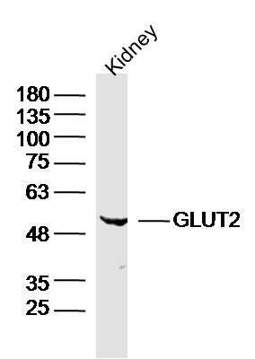 Sample:
Kidney (Mouse) Lysate at 40 ug
Primary: Anti- GLUT2 (SL0351R)at 1/300 dilution
Secondary: IRDye800CW Goat Anti-Rabbit IgG at 1/20000 dilution
Predicted band size: 54kD
Observed band size: 54kD
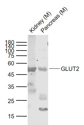 Sample:
Lane 1: Kidney (Mouse) Lysate at 40 ug
Lane 2: Pancreas (Mouse) Lysate at 40 ug
Primary: Anti-GLUT2 (SL0351R) at 1/1000 dilution
Secondary: IRDye800CW Goat Anti-Rabbit IgG at 1/20000 dilution
Predicted band size: 50-53 kD
Observed band size: 50 kD
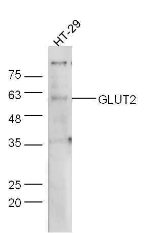 Sample:
HT-29 Cell (Human) Lysate at 30 ug
Primary: Anti-GLUT2 (SL0351R) at 1/300 dilution
Secondary: IRDye800CW Goat Anti-Rabbit IgG at 1/20000 dilution
Predicted band size: 54 kD
Observed band size: 59 kD
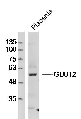 Sample:Placenta (Mouse)Lysate at 40 ug
Primary: Anti-GLUT2 (SL0351R)at 1/300 dilution
Secondary: IRDye800CW Goat Anti-RabbitIgG at 1/20000 dilution
Predicted band size: 54kD
Observed band size: 54kD
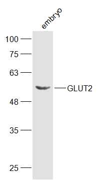 Sample:
embyo (Mouse) Lysate at 40 ug
Primary: Anti-GLUT2 (SL0351R) at 1/300 dilution
Secondary: IRDye800CW Goat Anti-Rabbit IgG at 1/20000 dilution
Predicted band size: 54 kD
Observed band size: 54 kD
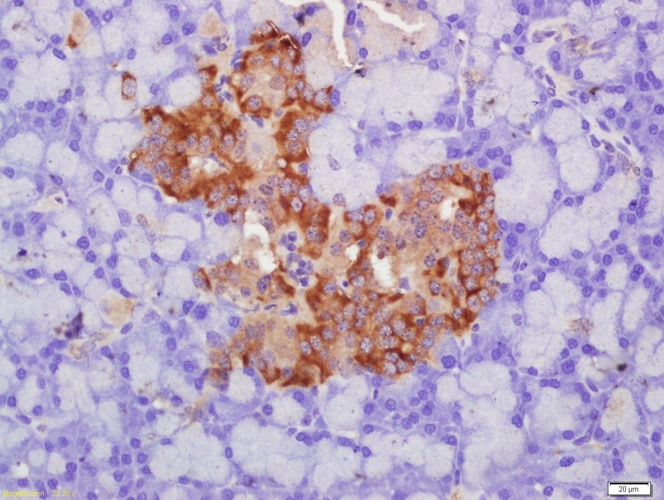 Paraformaldehyde-fixed, paraffin embedded (rat pancreas tissue); Antigen retrieval by boiling in sodium citrate buffer (pH6.0) for 15min; Block endogenous peroxidase by 3% hydrogen peroxide for 20 minutes; Blocking buffer (normal goat serum) at 37°C for 30min; Antibody incubation with (Glut2) Polyclonal Antibody, Unconjugated (SL0351R) at 1:400 overnight at 4°C, followed by a conjugated secondary (sp-0023) for 20 minutes and DAB staining.
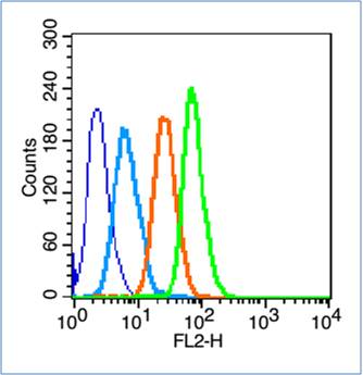 Blank control (blue line): Hep G2(blue).
Primary Antibody (green line): Rabbit Anti-GLUT2 antibody (SL0351R)
Dilution: 1μg /10^6 cells;
Isotype Control Antibody (orange line): Rabbit IgG .
Secondary Antibody (white blue line): Goat anti-rabbit IgG-PE
Dilution: 1μg /test.
Protocol
The cells were fixed with 70% ethanol Overnight at 4℃. Cells stained with Primary Antibody for 30 min at room temperature. The cells were then incubated in 1 X PBS/2%BSA/10% goat serum to block non-specific protein-protein interactions followed by the antibody for 15 min at room temperature. The secondary antibody used for 40 min at room temperature. Acquisition of 20,000 events was performed.
Cartpieces
Totalgoods,subtotals:¥Checkout
Bought notes(bought amounts latest0)
No one bought this product
User Comment(Total0User Comment Num)
- No comment


 +86 571 56623320
 +86 18668110335

