
Rabbit Anti-GNB1 antibody
G protein beta subunit GI/GS/GT; guanine nucleotide binding protein (G protein), beta polypeptide 1; guanine nucleotide-binding protein G; GBB1_HUMAN.
View History [Clear]
Details
Product Name GNB1 Chinese Name G蛋白/鸟苷酸Binding protein抗体 Alias G protein beta subunit GI/GS/GT; guanine nucleotide binding protein (G protein), beta polypeptide 1; guanine nucleotide-binding protein G; GBB1_HUMAN. Research Area Cell biology immunology Neurobiology Immunogen Species Rabbit Clonality Polyclonal React Species Mouse, Rat, (predicted: Human, Chicken, Dog, Pig, Cow, Horse, ) Applications WB=1:500-2000 ELISA=1:5000-10000 IHC-P=1:100-500 IHC-F=1:100-500 IF=1:100-500 (Paraffin sections need antigen repair)
not yet tested in other applications.
optimal dilutions/concentrations should be determined by the end user.Theoretical molecular weight 37kDa Cellular localization cytoplasmic The cell membrane Form Liquid Concentration 1mg/ml immunogen KLH conjugated synthetic peptide derived from human G protein beta subunit GI: 101-200/341 Lsotype IgG Purification affinity purified by Protein A Buffer Solution 0.01M TBS(pH7.4) with 1% BSA, 0.03% Proclin300 and 50% Glycerol. Storage Shipped at 4℃. Store at -20 °C for one year. Avoid repeated freeze/thaw cycles. Attention This product as supplied is intended for research use only, not for use in human, therapeutic or diagnostic applications. PubMed PubMed Product Detail G protein beta subunit GI/GS/GT (guanine nucleotide-binding protein G). WD40 domain, found in a number of eukaryotic proteins that cover a wide variety of functions including adaptor/regulatory modules in signal transduction, pre-mRNA processing and cytoskeleton assembly; typically contains a GH dipeptide 11-24 residues from its N-terminus and the WD dipeptide at its C-terminus and is 40 residues long, hence the name WD40; between GH and WD lies a conserved core; serves as a stable propeller-like platform to which proteins can bind either stably or reversibly; forms a propeller-like structure with several blades where each blade is composed of a four-stranded anti-parallel b-sheet; instances with few detectable copies are hypothesized to form larger structures by dimerization; each WD40 sequence repeat forms the first three strands of one blade and the last strand in the next blade; the last C-terminal WD40 repeat completes the blade structure of the first WD40 repeat to create the closed ring propeller-structure; residues on the top and bottom surface of the propeller are proposed to coordinate interactions with other proteins and/or small ligands; 7 copies of the repeat are present in this alignment.
Function:
Guanine nucleotide-binding proteins (G proteins) are involved as a modulator or transducer in various transmembrane signaling systems. The beta and gamma chains are required for the GTPase activity, for replacement of GDP by GTP, and for G protein-effector interaction.
Subunit:
G proteins are composed of 3 units, alpha, beta and gamma. Interacts with ARHGEF18 and RASD2. The heterodimer formed by GNB1 and GNG2 interacts with PTH1R (via C-terminus).
Post-translational modifications:
Phosphorylation at His-266 by NDKB contributes to G protein activation by increasing the high energetic phosphate transfer onto GDP.
Similarity:
Belongs to the WD repeat G protein beta family.
Contains 7 WD repeats.
SWISS:
P62879
Gene ID:
2782
Database links:Entrez Gene: 2782 Human
Entrez Gene: 14688 Mouse
Omim: 139380 Human
SwissProt: P62873 Human
SwissProt: P62874 Mouse
Unigene: 430425 Human
Unigene: 2344 Mouse
Unigene: 126047 Rat
G蛋白又称:鸟苷酸Binding protein,存在于全身各个组织和细胞中,G蛋白在信息传递中起到很重要的作用,参与细胞内外信息的相互传导。
激素受体与腺苷酸环化酶是The cell membrane上两类分开的蛋白质。激素受体结合的部分在The cell membrane的外表面,而腺苷环化酶在膜的胞浆面,在两者之间存在一种起耦联作用的调节蛋白——鸟苷酸Binding protein(guanine nucleotide-binding regulatory protein)细胞受到各种刺激信息,包括化学性的激素和神经递质及非化学性的声音、图像等。刺激信息通过G蛋白的激活作用,在胞浆中生成数目众多的第二信使分子,产生十万级放大效应,引起细胞兴奋,让后促动器官完成各自功能。
1977年,美国科学家阿尔弗雷德-戈德曼-吉尔曼(Alfred Goodman Gilman)发现了G蛋白及其在细胞兴奋传导方面的重要作用。1994年,吉尔曼因“G蛋白”方面的研究成果而荣获诺贝尔生理及医学奖。Product Picture 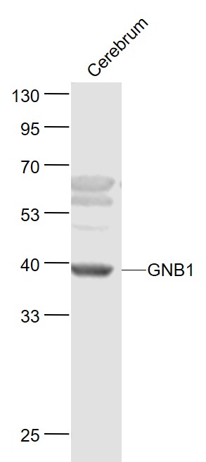 Sample:
Cerebrum (Mouse) Lysate at 40 ug
Primary: Anti-GNB1 (SL0348R) at 1/1000 dilution
Secondary: IRDye800CW Goat Anti-Rabbit IgG at 1/20000 dilution
Predicted band size: 37 kD
Observed band size: 37 kD
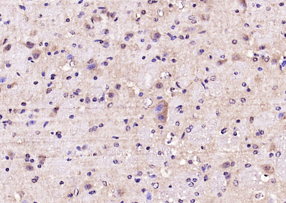 Paraformaldehyde-fixed, paraffin embedded (mouse brain); Antigen retrieval by boiling in sodium citrate buffer (pH6.0) for 15min; Block endogenous peroxidase by 3% hydrogen peroxide for 20 minutes; Blocking buffer (normal goat serum) at 37°C for 30min; Antibody incubation with (GNB1) Polyclonal Antibody, Unconjugated (SL0348R) at 1:200 overnight at 4°C, followed by operating according to SP Kit(Rabbit) (sp-0023) instructionsand DAB staining.
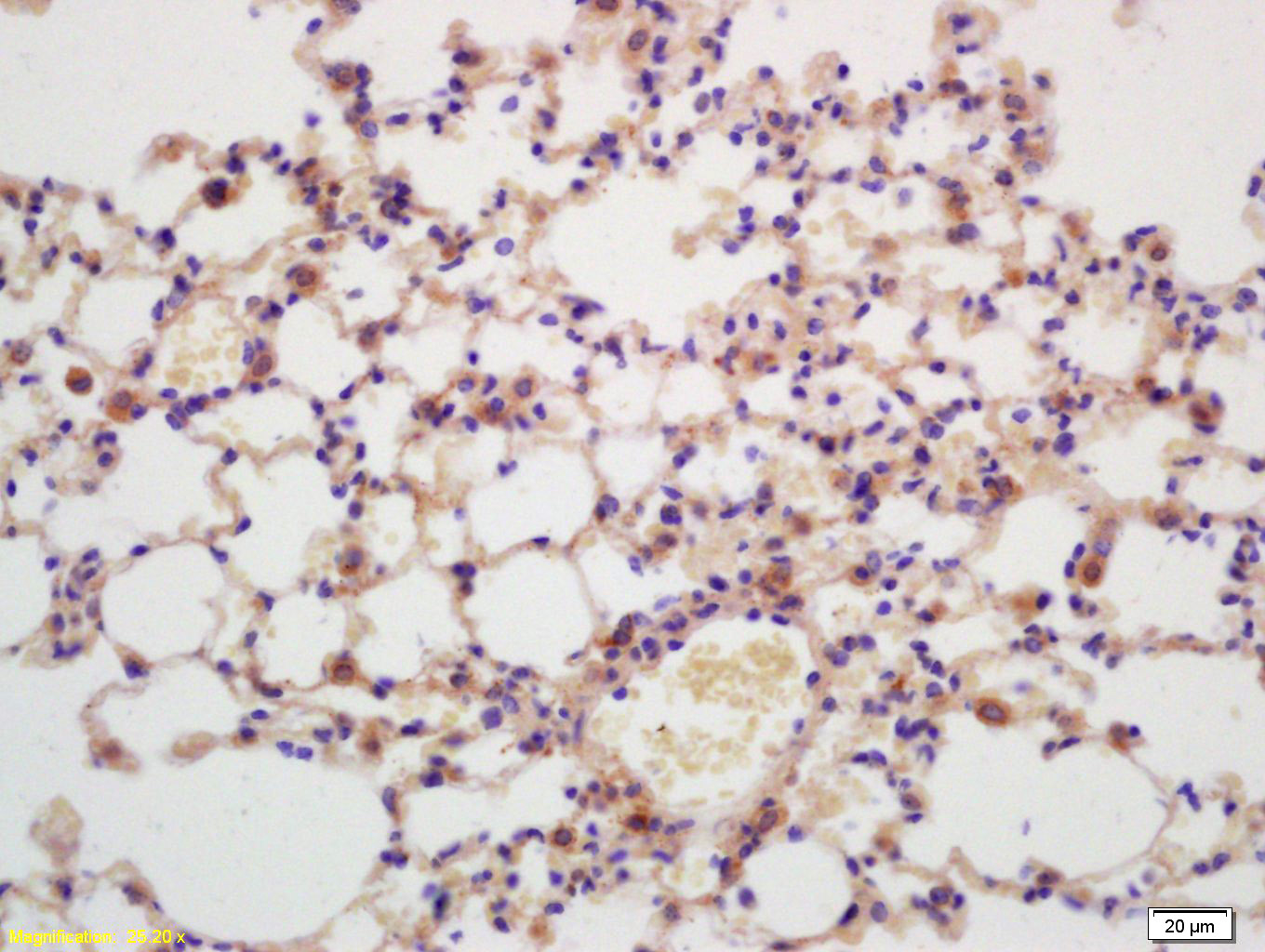 Tissue/cell: mouse lung tissue; 4% Paraformaldehyde-fixed and paraffin-embedded;
Antigen retrieval: citrate buffer ( 0.01M, pH 6.0 ), Boiling bathing for 15min; Block endogenous peroxidase by 3% Hydrogen peroxide for 30min; Blocking buffer (normal goat serum,C-0005) at 37℃ for 20 min;
Incubation: Anti-G protein beta subunit GI Polyclonal Antibody, Unconjugated(SL0348R) 1:200, overnight at 4°C, followed by conjugation to the secondary antibody(SP-0023) and DAB(C-0010) staining
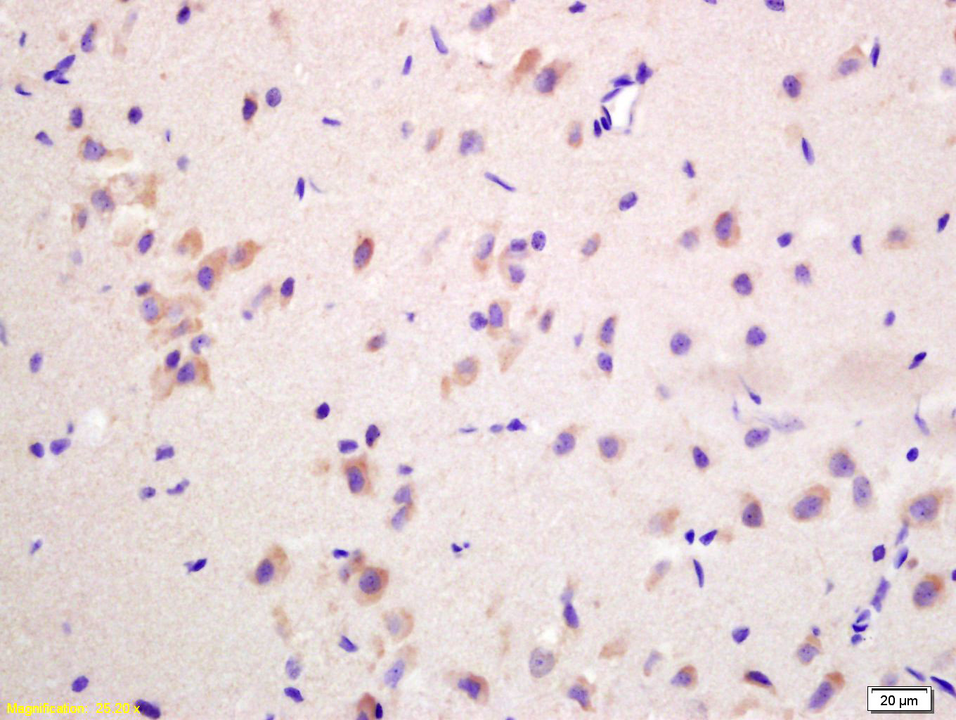 Tissue/cell: rat brain tissue; 4% Paraformaldehyde-fixed and paraffin-embedded;
Antigen retrieval: citrate buffer ( 0.01M, pH 6.0 ), Boiling bathing for 15min; Block endogenous peroxidase by 3% Hydrogen peroxide for 30min; Blocking buffer (normal goat serum,C-0005) at 37℃ for 20 min;
Incubation: Anti-G protein beta subunit GI Polyclonal Antibody, Unconjugated(SL0348R) 1:200, overnight at 4°C, followed by conjugation to the secondary antibody(SP-0023) and DAB(C-0010) staining
Cartpieces
Totalgoods,subtotals:¥Checkout
Bought notes(bought amounts latest0)
No one bought this product
User Comment(Total0User Comment Num)
- No comment


 +86 571 56623320
 +86 18668110335

