
Rabbit Anti-Insulin Receptor Alpha antibody
CD220; CD220 antigen; HHF5; HIR A; INSR alpha; INSR; Insulin receptor; Insulin receptor subunit alpha; IR; INSR_HUMAN.
View History [Clear]
Details
Product Name Insulin Receptor Alpha Chinese Name 胰岛素受体α抗体 Alias CD220; CD220 antigen; HHF5; HIR A; INSR alpha; INSR; Insulin receptor; Insulin receptor subunit alpha; IR; INSR_HUMAN. Research Area Cell biology Neurobiology Signal transduction Apoptosis Kinases and Phosphatases The cell membrane受体 Diabetes Immunogen Species Rabbit Clonality Polyclonal React Species Human, Rat, (predicted: Mouse, Dog, Pig, Cow, Horse, Rabbit, Sheep, Guinea Pig, ) Applications ELISA=1:5000-10000 IHC-P=1:100-500 IHC-F=1:100-500 IF=1:100-500 (Paraffin sections need antigen repair)
not yet tested in other applications.
optimal dilutions/concentrations should be determined by the end user.Theoretical molecular weight 80/152kDa Cellular localization The cell membrane Form Liquid Concentration 1mg/ml immunogen KLH conjugated synthetic peptide derived from human Insulin Receptor Alpha: 51-150/1382 Lsotype IgG Purification affinity purified by Protein A Buffer Solution 0.01M TBS(pH7.4) with 1% BSA, 0.03% Proclin300 and 50% Glycerol. Storage Shipped at 4℃. Store at -20 °C for one year. Avoid repeated freeze/thaw cycles. Attention This product as supplied is intended for research use only, not for use in human, therapeutic or diagnostic applications. PubMed PubMed Product Detail Insulin receptor binds insulin and has a tyrosine-protein kinase activity. It also has catalytic activity: ATP + a protein tyrosine = ADP + proteintyrosine phosphate. Tetramer of 2 alpha and 2 beta chains linked by disulfidebonds. The alpha chains contribute to the formation of the ligand-binding domain, while the beta chains carry the kinase domain. Interacts with SORBS1 but dissociates from it following insulin stimulation. [subcellular location] Type I membrane protein. Belongs to the Tyr protein kinase family. Insulin receptor subfamily.
Function:
Receptor tyrosine kinase which mediates the pleiotropic actions of insulin. Binding of insulin leads to phosphorylation of several intracellular substrates, including, insulin receptor substrates (IRS1, 2, 3, 4), SHC, GAB1, CBL and other signaling intermediates. Each of these phosphorylated proteins serve as docking proteins for other signaling proteins that contain Src-homology-2 domains (SH2 domain) that specifically recognize different phosphotyrosines residues, including the p85 regulatory subunit of PI3K and SHP2. Phosphorylation of IRSs proteins lead to the activation of two main signaling pathways: the PI3K-AKT/PKB pathway, which is responsible for most of the metabolic actions of insulin, and the Ras-MAPK pathway, which regulates expression of some genes and cooperates with the PI3K pathway to control cell growth and differentiation. Binding of the SH2 domains of PI3K to phosphotyrosines on IRS1 leads to the activation of PI3K and the generation of phosphatidylinositol-(3, 4, 5)-triphosphate (PIP3), a lipid second messenger, which activates several PIP3-dependent serine/threonine kinases, such as PDPK1 and subsequently AKT/PKB. The net effect of this pathway is to produce a translocation of the glucose transporter SLC2A4/GLUT4 from cytoplasmic vesicles to the cell membrane to facilitate glucose transport. Moreover, upon insulin stimulation, activated AKT/PKB is responsible for: anti-apoptotic effect of insulin by inducing phosphorylation of BAD; regulates the expression of gluconeogenic and lipogenic enzymes by controlling the activity of the winged helix or forkhead (FOX) class of transcription factors. Another pathway regulated by PI3K-AKT/PKB activation is mTORC1 signaling pathway which regulates cell growth and metabolism and integrates signals from insulin. AKT mediates insulin-stimulated protein synthesis by phosphorylating TSC2 thereby activating mTORC1 pathway. The Ras/RAF/MAP2K/MAPK pathway is mainly involved in mediating cell growth, survival and cellular differentiation of insulin. Phosphorylated IRS1 recruits GRB2/SOS complex, which triggers the activation of the Ras/RAF/MAP2K/MAPK pathway. In addition to binding insulin, the insulin receptor can bind insulin-like growth factors (IGFI and IGFII). Isoform Short has a higher affinity for IGFII binding. When present in a hybrid receptor with IGF1R, binds IGF1. PubMed:12138094 shows that hybrid receptors composed of IGF1R and INSR isoform Long are activated with a high affinity by IGF1, with low affinity by IGF2 and not significantly activated by insulin, and that hybrid receptors composed of IGF1R and INSR isoform Short are activated by IGF1, IGF2 and insulin. In contrast, PubMed:16831875 shows that hybrid receptors composed of IGF1R and INSR isoform Long and hybrid receptors composed of IGF1R and INSR isoform Short have similar binding characteristics, both bind IGF1 and have a low affinity for insulin.
Subunit:
Tetramer of 2 alpha and 2 beta chains linked by disulfide bonds. The alpha chains contribute to the formation of the ligand-binding domain, while the beta chains carry the kinase domain. Forms a hybrid receptor with IGF1R, the hybrid is a tetramer consisting of 1 alpha chain and 1 beta chain of INSR and 1 alpha chain and 1 beta chain of IGF1R. Interacts with SORBS1 but dissociates from it following insulin stimulation. Binds SH2B2. Activated form of INSR interacts (via Tyr-999) with the PTB/PID domains of IRS1 and SHC1. The sequences surrounding the phosphorylated NPXY motif contribute differentially to either IRS1 or SHC1 recognition. Interacts (via tyrosines in the C-terminus) with IRS2 (via PTB domain and 591-786 AA); the 591-786 would be the primary anchor of IRS2 to INSR while the PTB domain would have a stabilizing action on the interaction with INSR. Interacts with the SH2 domains of the 85 kDa regulatory subunit of PI3K (PIK3R1) in vitro, when autophosphorylated on tyrosine residues. Interacts with SOCS7. Interacts (via the phosphorylated Tyr-999), with SOCS3. Interacts (via the phosphorylated Tyr-1185, Tyr-1189, Tyr-1190) with SOCS1. Interacts with CAV2 (tyrosine-phosphorylated form); the interaction is increased with 'Tyr-27'phosphorylation of CAV2 (By similarity). Interacts with ARRB2 (By similarity). Interacts with GRB10; this interaction blocks the association between IRS1/IRS2 and INSR, significantly reduces insulin-stimulated tyrosine phosphorylation of IRS1 and IRS2 and thus decreases insulin signaling. Interacts with GRB7 (By similarity). Interacts with PDPK1. Interacts (via Tyr-1190) with GRB14 (via BPS domain); this interaction protects the tyrosines in the activation loop from dephosphorylation, but promotes dephosphorylation of Tyr-999, this results in decreased interaction with, and phosphorylation of, IRS1. Interacts (via subunit alpha) with ENPP1 (via 485-599 AA); this interaction blocks autophosphorylation. Interacts with PTPRE; this interaction is dependent of Tyr-1185, Tyr-1189 and Tyr-1190 of the INSR. Interacts with STAT5B (via SH2 domain). Interacts with PTPRF.
Subcellular Location:
Membrane; Single-pass type I membrane protein.
Tissue Specificity:
Isoform Long and isoform Short are predominantly expressed in tissue targets of insulin metabolic effects: liver, adipose tissue and skeletal muscle but are also expressed in the peripheral nerve, kidney, pulmonary alveoli, pancreatic acini, placenta vascular endothelium, fibroblasts, monocytes, granulocytes, erythrocytes and skin. Isoform Short is preferentially expressed in fetal cells such as fetal fibroblasts, muscle, liver and kidney. Found as a hybrid receptor with IGF1R in muscle, heart, kidney, adipose tissue, skeletal muscle, hepatoma, fibroblasts, spleen and placenta (at protein level). Overexpressed in several tumors, including breast, colon, lung, ovary, and thyroid carcinomas.
Post-translational modifications:
After being transported from the endoplasmic reticulum to the Golgi apparatus, the single glycosylated precursor is further glycosylated and then cleaved, followed by its transport to the plasma membrane.
Autophosphorylated on tyrosine residues in response to insulin. Phosphorylation of Tyr-999 is required for IRS1-, SHC1-, and STAT5B-binding. Dephosphorylated by PTPRE on Tyr-999, Tyr-1185, Tyr-1189 and Tyr-1190 residues. Dephosphorylated by PTPRF.
DISEASE:
Defects in INSR are the cause of Rabson-Mendenhall syndrome (RMS) [MIM:262190]; also known as Mendenhall syndrome. RMS is a severe insulin resistance syndrome characterized by insulin-resistant diabetes mellitus with pineal hyperplasia and somatic abnormalities. Typical features include coarse, senile-appearing facies, dental and skin abnormalities, abdominal distension, and phallic enlargement. Inheritance is autosomal recessive.
Defects in INSR are the cause of leprechaunism (LEPRCH) [MIM:246200]; also known as Donohue syndrome. Leprechaunism represents the most severe form of insulin resistance syndrome, characterized by intrauterine and postnatal growth retardation and death in early infancy. Inheritance is autosomal recessive.
Defects in INSR may be associated with noninsulin-dependent diabetes mellitus (NIDDM) [MIM:125853]; also known as diabetes mellitus type 2.
Defects in INSR are the cause of familial hyperinsulinemic hypoglycemia type 5 (HHF5) [MIM:609968]. Familial hyperinsulinemic hypoglycemia [MIM:256450], also referred to as congenital hyperinsulinism, nesidioblastosis, or persistent hyperinsulinemic hypoglycemia of infancy (PPHI), is the most common cause of persistent hypoglycemia in infancy and is due to defective negative feedback regulation of insulin secretion by low glucose levels.
Defects in INSR are the cause of insulin-resistant diabetes mellitus with acanthosis nigricans type A (IRAN type A) [MIM:610549]. This syndrome is characterized by the association of severe insulin resistance (manifested by marked hyperinsulinemia and a failure to respond to exogenous insulin) with the skin lesion acanthosis nigricans and ovarian hyperandrogenism in adolescent female subjects. Women frequently present with hirsutism, acne, amenorrhea or oligomenorrhea, and virilization. This syndrome is different from the type B that has been demonstrated to be secondary to the presence of circulating autoantibodies against the insulin receptor.
Similarity:
Belongs to the protein kinase superfamily. Tyr protein kinase family. Insulin receptor subfamily.
Contains 3 fibronectin type-III domains.
Contains 1 protein kinase domain.
SWISS:
P06213
Gene ID:
3643
Database links:Entrez Gene: 3643 Human
Entrez Gene: 16337 Mouse
Omim: 147670 Human
SwissProt: P06213 Human
SwissProt: P15208 Mouse
Unigene: 465744 Human
Unigene: 9876 Rat
胰岛素受体是一个四聚体,由两个α亚基和两个β亚基通过二硫键连接。两个α亚基位于细胞质膜的外侧,其上有胰岛素的结合位点;两个β亚基是Transmembrane protein,起Signal transduction作用。无胰岛素结 合时,受体的酪氨酸蛋白激酶没有活性。当胰岛素与受体的α亚基结合并改变了β亚基的构型后,酪氨酸蛋白激酶才被激活,激活后可催化两个反应: ①使四聚体复合物中β亚基特异位点的酪氨酸残基磷酸化,这种过程称为自我磷酸化(autophosphorylation); ②将胰岛素受体底物(insulin receptor substrate,IRSs)上具有重要作用的十几个酪氨酸残基磷酸化,磷酸化的IRSs能够结合并激活下游效应物。Product Picture 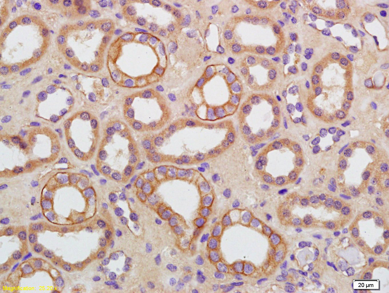 Tissue/cell: human kidney tissue; 4% Paraformaldehyde-fixed and paraffin-embedded;
Antigen retrieval: citrate buffer ( 0.01M, pH 6.0 ), Boiling bathing for 15min; Block endogenous peroxidase by 3% Hydrogen peroxide for 30min; Blocking buffer (normal goat serum,C-0005) at 37℃ for 20 min;
Incubation: Anti-AQP-9 Polyclonal Antibody, Unconjugated(SL2060R) 1:200, overnight at 4°C, followed by conjugation to the secondary antibody(SP-0023) and DAB(C-0010) staining
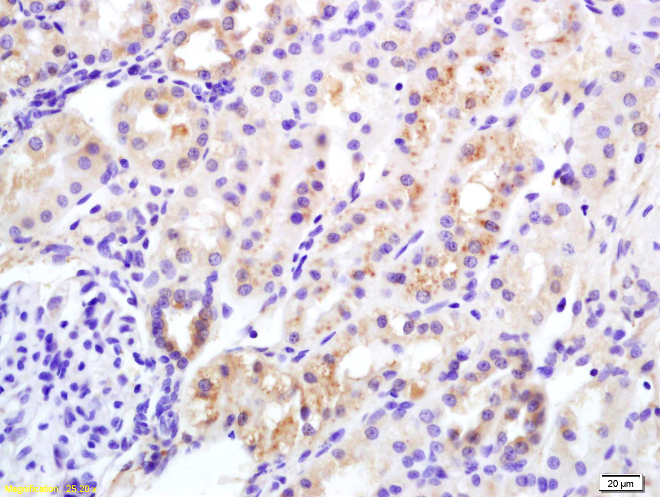 Tissue/cell: rat kidney tissue; 4% Paraformaldehyde-fixed and paraffin-embedded;
Antigen retrieval: citrate buffer ( 0.01M, pH 6.0 ), Boiling bathing for 15min; Block endogenous peroxidase by 3% Hydrogen peroxide for 30min; Blocking buffer (normal goat serum,C-0005) at 37℃ for 20 min;
Incubation: Anti-Insulin Receptor Alpha Polyclonal Antibody, Unconjugated(SL0260R) 1:200, overnight at 4°C, followed by conjugation to the secondary antibody(SP-0023) and DAB(C-0010) staining
Cartpieces
Totalgoods,subtotals:¥Checkout
Bought notes(bought amounts latest0)
No one bought this product
User Comment(Total0User Comment Num)
- No comment


 +86 571 56623320
 +86 18668110335

