
Rabbit Anti-Bax antibody
apoptosis regulator BAX; Apoptosis regulator BAX cytoplasmic isoform beta; Apoptosis regulator BAX membrane isoform alpha; Bax isoform psi; BAX protein cytoplasmic isoform delta; Bax protein cytoplasmic isoform delta. antibody Bax protein cytoplasmic isof
View History [Clear]
Details
Product Name Bax Chinese Name Bax抗体 Alias apoptosis regulator BAX; Apoptosis regulator BAX cytoplasmic isoform beta; Apoptosis regulator BAX membrane isoform alpha; Bax isoform psi; BAX protein cytoplasmic isoform delta; Bax protein cytoplasmic isoform delta. antibody Bax protein cytoplasmic isoform gamma; Bax zeta; Bax-protein; Bcl-2-like protein 4; BCL2 associated X protein; BCL2L4; BAX_HUMAN; Bcl2-L-4. literatures Research Area Cell biology Signal transduction Apoptosis Mitochondrion Immunogen Species Rabbit Clonality Polyclonal React Species Human, Mouse, Rat, Rabbit, (predicted: Dog, Pig, Cow, Sheep, ) Applications WB=1:500-2000 ELISA=1:5000-10000 IHC-P=1:100-500 IHC-F=1:100-500 Flow-Cyt=1μg /test ICC=1:100 IF=1:100-500 (Paraffin sections need antigen repair)
not yet tested in other applications.
optimal dilutions/concentrations should be determined by the end user.Theoretical molecular weight 21kDa Cellular localization cytoplasmic The cell membrane Mitochondrion Form Liquid Concentration 1mg/ml immunogen KLH conjugated synthetic peptide derived from human Bax: 84-175/192 Lsotype IgG Purification affinity purified by Protein A Buffer Solution 0.01M TBS(pH7.4) with 1% BSA, 0.03% Proclin300 and 50% Glycerol. Storage Shipped at 4℃. Store at -20 °C for one year. Avoid repeated freeze/thaw cycles. Attention This product as supplied is intended for research use only, not for use in human, therapeutic or diagnostic applications. PubMed PubMed Product Detail The protein encoded by this gene belongs to the BCL2 protein family. BCL2 family members form hetero- or homodimers and act as anti- or pro-apoptotic regulators that are involved in a wide variety of cellular activities. This protein forms a heterodimer with BCL2, and functions as an apoptotic activator. This protein is reported to interact with, and increase the opening of, the mitochondrial voltage-dependent anion channel (VDAC), which leads to the loss in membrane potential and the release of cytochrome c. The expression of this gene is regulated by the tumor suppressor P53 and has been shown to be involved in P53-mediated apoptosis. Multiple alternatively spliced transcript variants, which encode different isoforms, have been reported for this gene. [provided by RefSeq, Jul 2008].
Function:
Accelerates programmed cell death by binding to, and antagonizing the apoptosis repressor BCL2 or its adenovirus homolog E1B 19k protein. Under stress conditions, undergoes a conformation change that causes translocation to the mitochondrion membrane, leading to the release of cytochrome c that then triggers apoptosis. Promotes activation of CASP3, and thereby apoptosis.
Subunit:
Homodimer. Forms higher oligomers under stress conditions. Interacts with BCL2L11. Interaction with BCL2L11 promotes BAX oligomerization and association with mitochondrial membranes, with subsequent release of cytochrome c. Forms heterodimers with BCL2, E1B 19K protein, BCL2L1 isoform Bcl-X(L), BCL2L2, MCL1 and A1. Interacts with SH3GLB1 and HN. Interacts with SFN and YWHAZ; the interaction occurs in the cytoplasm. Under stress conditions, JNK-mediated phosphorylation of SFN and YWHAZ, releases BAX to mitochondria. Isoform Sigma interacts with BCL2A1 and BCL2L1 isoform Bcl-X(L). Interacts with RNF144B, which regulates the ubiquitin-dependent stability of BAX. Interacts with CLU under stress conditions that cause a conformation change leading to BAX oligomerization and association with mitochondria. Does not interact with CLU in unstressed cells. Interacts with FAIM2/LFG2.
Subcellular Location:
Isoform Alpha: Mitochondrion membrane; Single-pass membrane protein. Cytoplasm. Note=Colocalizes with 14-3-3 proteins in the cytoplasm. Under stress conditions, undergoes a conformation change that causes release from JNK-phosphorylated 14-3-3 proteins and translocation to the mitochondrion membrane.
Isoform Beta: Cytoplasm.
Isoform Gamma: Cytoplasm.
Isoform Delta: Cytoplasm (Potential).
Tissue Specificity:
Expressed in a wide variety of tissues. Isoform Psi is found in glial tumors. Isoform Alpha is expressed in spleen, breast, ovary, testis, colon and brain, and at low levels in skin and lung. Isoform Sigma is expressed in spleen, breast, ovary, testis, lung, colon, brain and at low levels in skin. Isoform Alpha and isoform Sigma are expressed in pro-myelocytic leukemia, histiocytic lymphoma, Burkitt's lymphoma, T-cell lymphoma, lymphoblastic leukemia, breast adenocarcinoma, ovary adenocarcinoma, prostate carcinoma, prostate adenocarcinoma, lung carcinoma, epidermoid carcinoma, small cell lung carcinoma and colon adenocarcinoma cell lines.
Similarity:
Belongs to the Bcl-2 family.
SWISS:
Q07812
Gene ID:
581
Database links:Entrez Gene: 581 Human
Entrez Gene: 12028 Mouse
Omim: 600040 Human
SwissProt: Q07812 Human
SwissProt: Q07813 Mouse
Unigene: 624291 Human
Unigene: 19904 Mouse
Unigene: 10668 Rat
可识别分子量为21KDa的Bax蛋白,此抗体与Bax有较高特异性,且与Bc1-2及Bc1-X蛋白无React Species,Bax、Bc1-2和Bc1-X蛋白是凋亡调节蛋白家庭成员。与Bc1-2和Bc1-X相反,Bax蛋白的过量表达加速Apoptosis。Bax在组织中广泛表达。
Bax与Bc1-2比值的高低可用于判断恶性Tumour耐药及复发。此抗体用于Tumour及Apoptosis等方面的研究。最新的研究表明:Bax可能具有Tumour抑制作用。
Product Picture 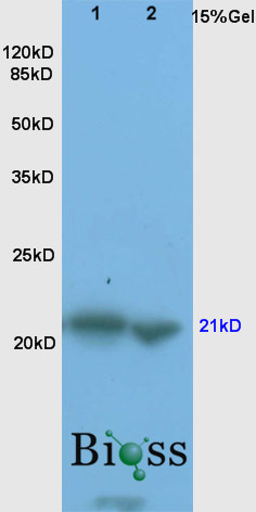 Sample: Brain(Rat)lysate 30ug;
Liver(Mouse) lysates, 30ug;
Primary: Anti-Bax (SL0127R) at 1:200;
Secondary: HRP conjugated Goat-Anti-Rabbit IgG(bse-0295G) at 1: 3000;
ECL excitated the fluorescence;
Predicted band size : 21kD
Observed band size : 21kD
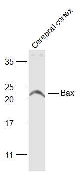 Sample:
Cerebral cortex (Rat) Lysate at 40 ug
Primary: Anti-Bax (SL0127R) at 1/1000 dilution
Secondary: IRDye800CW Goat Anti-Rabbit IgG at 1/20000 dilution
Predicted band size: 21 kD
Observed band size: 21 kD
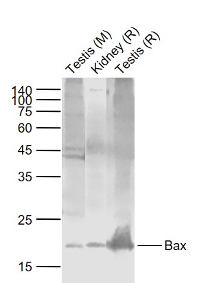 Sample:
Lane 1: Testis (Mouse) Lysate at 40 ug
Lane 2: Kidney (Rat) Lysate at 40 ug
Lane 3: Testis (Rat) Lysate at 40 ug
Primary:
Anti-Bax (SL0127R) at 1/1000 dilution
Secondary: IRDye800CW Goat Anti-Rabbit IgG at 1/20000 dilution
Predicted band size: 21 kD
Observed band size: 21 kD
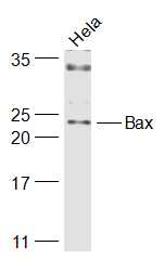
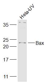 Sample:
Hela-UV(Human) Cell Lysate at 30 ug
Primary: Anti-Bax (SL0127R) at 1/1000 dilution
Secondary: IRDye800CW Goat Anti-Rabbit IgG at 1/20000 dilution
Predicted band size: 21 kD
Observed band size: 23 kD
 Sample:
Hela(Human) Cell Lysate at 30 ug
Primary: Anti-Bax (SL0127R) at 1/1000 dilution
Secondary: IRDye800CW Goat Anti-Rabbit IgG at 1/20000 dilution
Predicted band size: 21 kD
Observed band size: 23 kD
 Paraformaldehyde-fixed, paraffin embedded (Rat spinal cord); Antigen retrieval by boiling in sodium citrate buffer (pH6.0) for 15min; Block endogenous peroxidase by 3% hydrogen peroxide for 20 minutes; Blocking buffer (normal goat serum) at 37°C for 30min; Antibody incubation with (Bax) Polyclonal Antibody, Unconjugated (SL0127R) at 1:400 overnight at 4°C, followed by operating according to SP Kit(Rabbit) (sp-0023) instructionsand DAB staining.
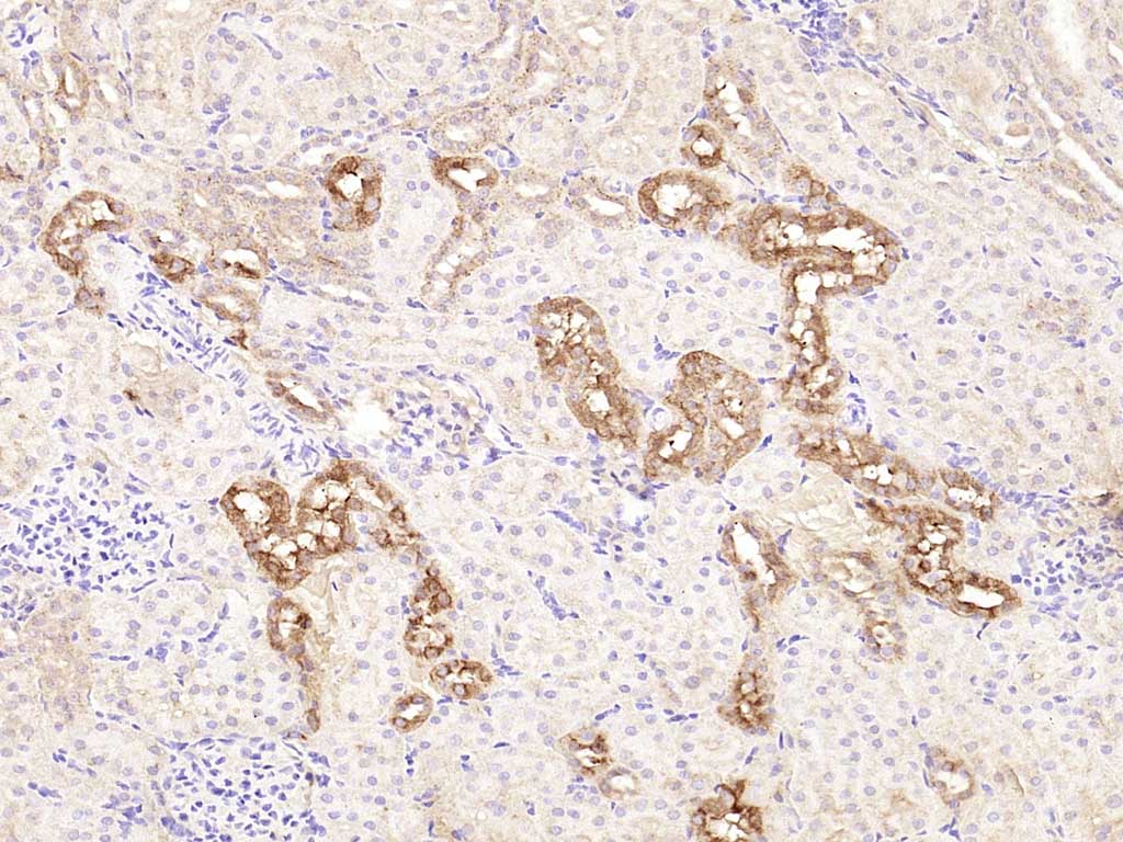 Paraformaldehyde-fixed, paraffin embedded (rat kidney); Antigen retrieval by boiling in sodium citrate buffer (pH6.0) for 15min; Block endogenous peroxidase by 3% hydrogen peroxide for 20 minutes; Blocking buffer (normal goat serum) at 37°C for 30min; Antibody incubation with (Bax) Polyclonal Antibody, Unconjugated (SL0127R) at 1:2000 overnight at 4°C, followed by operating according to SP Kit(Rabbit) (sp-0023) instructionsand DAB staining.
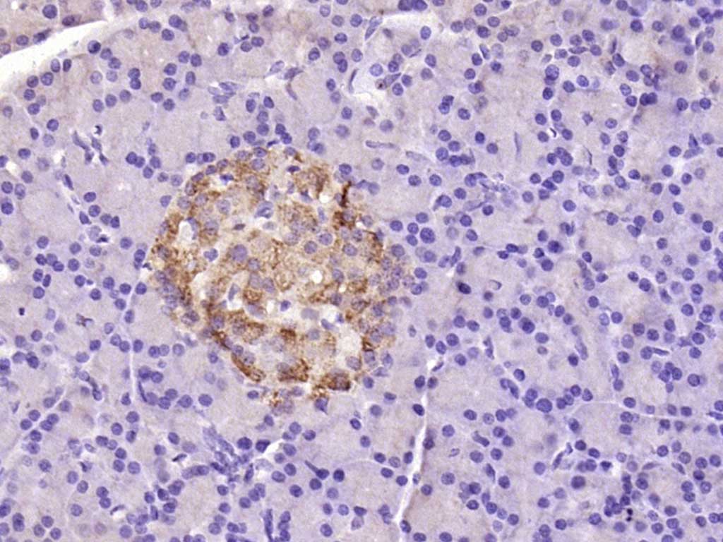 Paraformaldehyde-fixed, paraffin embedded (mouse pancreas); Antigen retrieval by boiling in sodium citrate buffer (pH6.0) for 15min; Block endogenous peroxidase by 3% hydrogen peroxide for 20 minutes; Blocking buffer (normal goat serum) at 37°C for 30min; Antibody incubation with (Bax) Polyclonal Antibody, Unconjugated (SL0127R) at 1:2000 overnight at 4°C, followed by operating according to SP Kit(Rabbit) (sp-0023) instructionsand DAB staining.
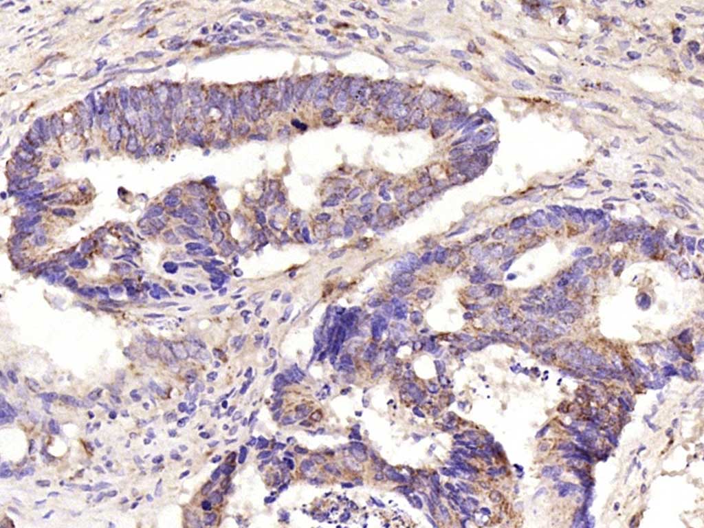 Paraformaldehyde-fixed, paraffin embedded (human colon carcinoma); Antigen retrieval by boiling in sodium citrate buffer (pH6.0) for 15min; Block endogenous peroxidase by 3% hydrogen peroxide for 20 minutes; Blocking buffer (normal goat serum) at 37°C for 30min; Antibody incubation with (Bax) Polyclonal Antibody, Unconjugated (SL0127R) at 1:2000 overnight at 4°C, followed by operating according to SP Kit(Rabbit) (sp-0023) instructionsand DAB staining.
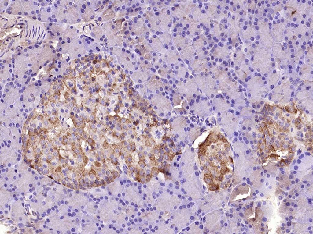 Paraformaldehyde-fixed, paraffin embedded (rat pancreas); Antigen retrieval by boiling in sodium citrate buffer (pH6.0) for 15min; Block endogenous peroxidase by 3% hydrogen peroxide for 20 minutes; Blocking buffer (normal goat serum) at 37°C for 30min; Antibody incubation with (Bax) Polyclonal Antibody, Unconjugated (SL0127R) at 1:2000 overnight at 4°C, followed by operating according to SP Kit(Rabbit) (sp-0023) instructionsand DAB staining.
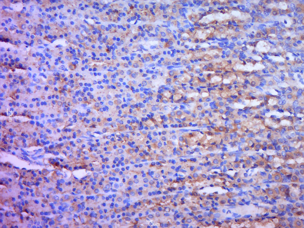 Tissue/cell: rat stomach tissue; 4% Paraformaldehyde-fixed and paraffin-embedded;
Antigen retrieval: citrate buffer ( 0.01M, pH 6.0 ), Boiling bathing for 15min; Block endogenous peroxidase by 3% Hydrogen peroxide for 30min; Blocking buffer (normal goat serum,C-0005) at 37℃ for 20 min;
Incubation: Anti-Bax Polyclonal Antibody, Unconjugated(SL0127R) 1:200, overnight at 4癈, followed by conjugation to the secondary antibody(SP-0023) and DAB(C-0010) staining
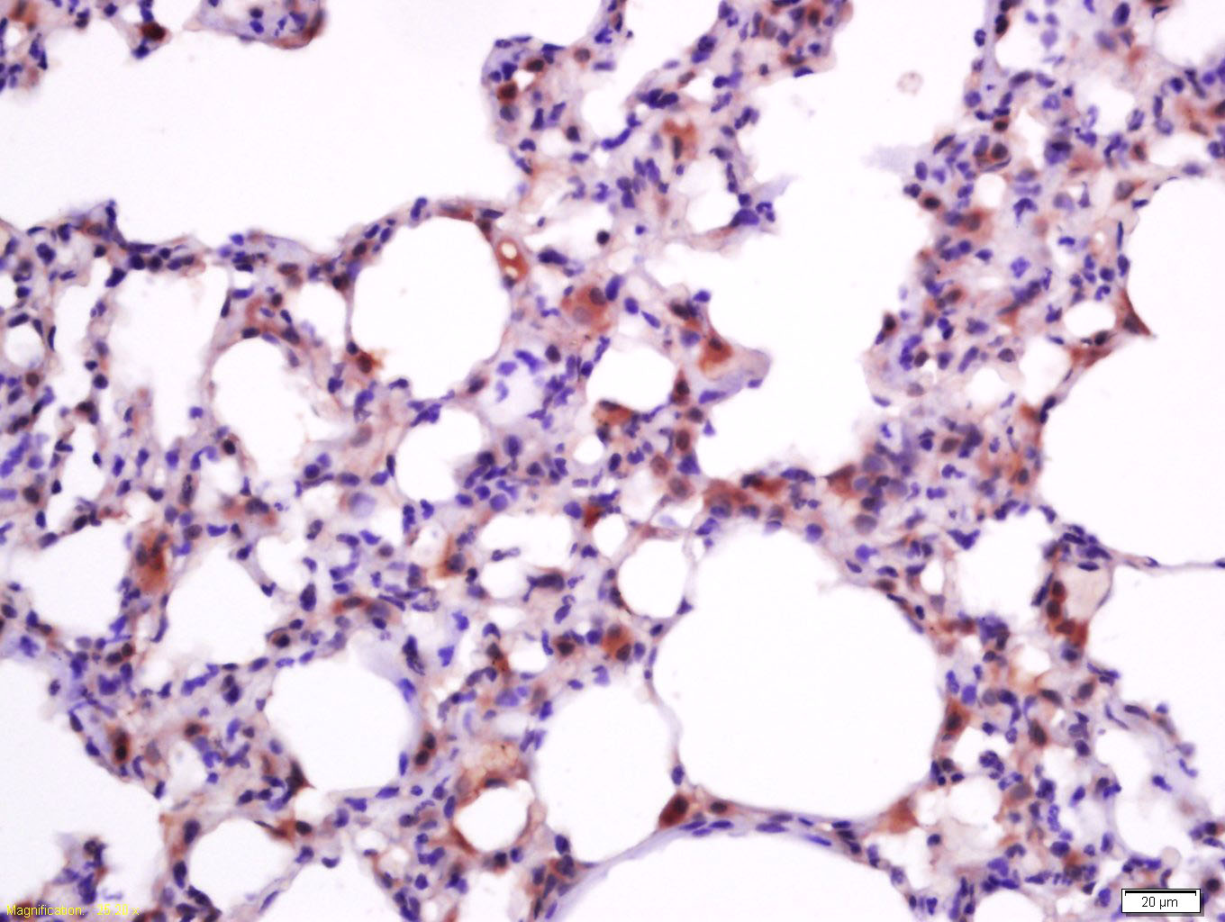 Tissue/cell: rat lung tissue; 4% Paraformaldehyde-fixed and paraffin-embedded;
Antigen retrieval: citrate buffer ( 0.01M, pH 6.0 ), Boiling bathing for 15min; Block endogenous peroxidase by 3% Hydrogen peroxide for 30min; Blocking buffer (normal goat serum,C-0005) at 37℃ for 20 min;
Incubation: Anti-Bax Polyclonal Antibody, Unconjugated(SL0127R) 1:200, overnight at 4癈, followed by conjugation to the secondary antibody(SP-0023) and DAB(C-0010) staining
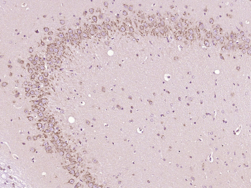
 Tissue/cell: rat brain tissue; 4% Paraformaldehyde-fixed and paraffin-embedded;
Antigen retrieval: citrate buffer ( 0.01M, pH 6.0 ), Boiling bathing for 15min; Block endogenous peroxidase by 3% Hydrogen peroxide for 30min; Blocking buffer (normal goat serum,C-0005) at 37℃ for 20 min;
Incubation: Anti-Bax Polyclonal Antibody, Unconjugated(SL0127R) 1:800, overnight at 4癈, followed by conjugation to the secondary antibody(SP-0023) and DAB(C-0010) staining
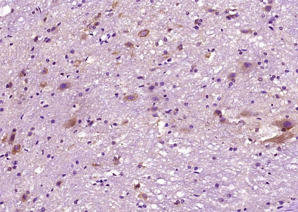
 Paraformaldehyde-fixed, paraffin embedded (Rat brain); Antigen retrieval by boiling in sodium citrate buffer (pH6.0) for 15min; Block endogenous peroxidase by 3% hydrogen peroxide for 20 minutes; Blocking buffer (normal goat serum) at 37°C for 30min; Antibody incubation with (Bax) Polyclonal Antibody, Unconjugated (SL0127R) at 1:400 overnight at 4°C, followed by operating according to SP Kit(Rabbit) (sp-0023) instructionsand DAB staining.
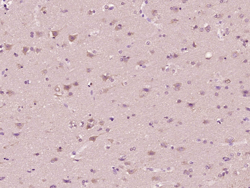 Paraformaldehyde-fixed, paraffin embedded (Human brain glioma); Antigen retrieval by boiling in sodium citrate buffer (pH6.0) for 15min; Block endogenous peroxidase by 3% hydrogen peroxide for 20 minutes; Blocking buffer (normal goat serum) at 37°C for 30min; Antibody incubation with (Bax) Polyclonal Antibody, Unconjugated (SL0127R) at 1:400 overnight at 4°C, followed by operating according to SP Kit(Rabbit) (sp-0023) instructionsand DAB staining.
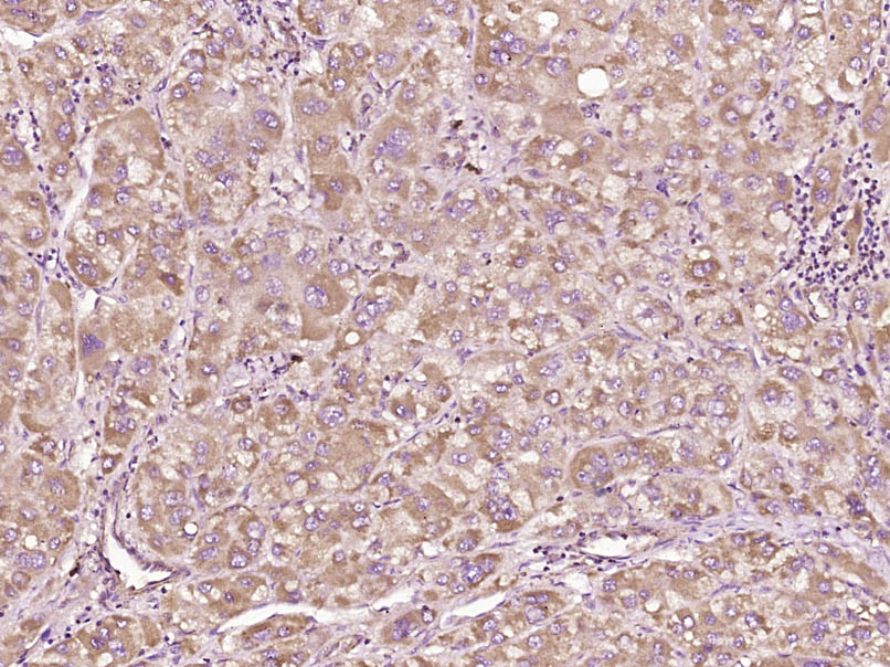 Paraformaldehyde-fixed, paraffin embedded (Human liver carcinoma); Antigen retrieval by boiling in sodium citrate buffer (pH6.0) for 15min; Block endogenous peroxidase by 3% hydrogen peroxide for 20 minutes; Blocking buffer (normal goat serum) at 37°C for 30min; Antibody incubation with (Bax) Polyclonal Antibody, Unconjugated (SL0127R) at 1:400 overnight at 4°C, followed by operating according to SP Kit(Rabbit) (sp-0023) instructionsand DAB staining.
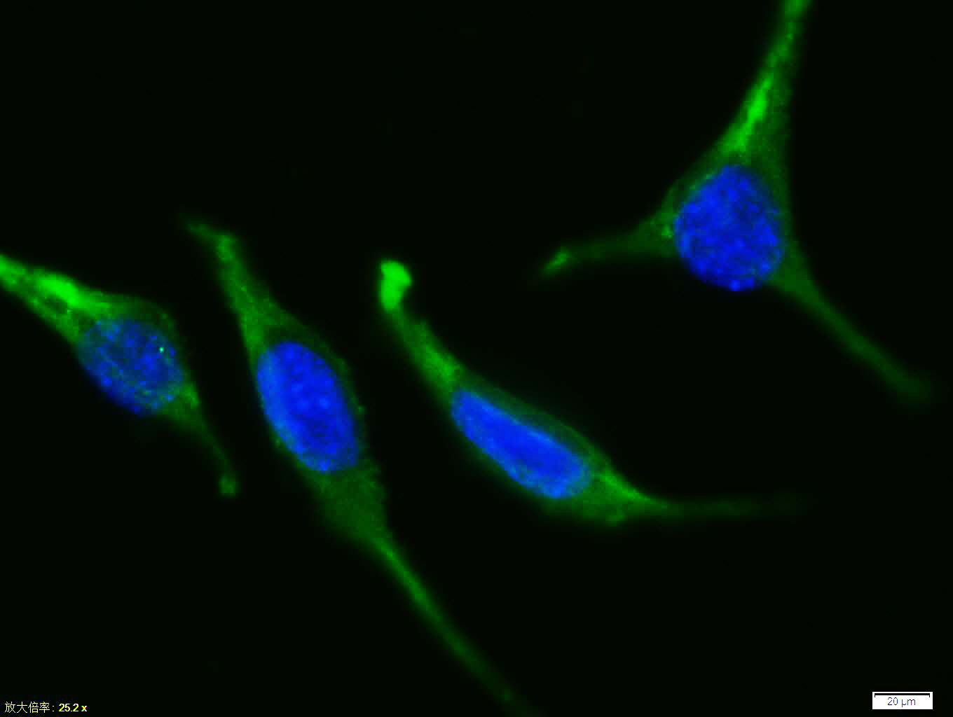 Tissue/cell:Sh-sy5y cell; 4% Paraformaldehyde-fixed; Triton X-100 at room temperature for 20 min; Blocking buffer (normal goat serum, C-0005) at 37°C for 20 min; Antibody incubation with (Bax) polyclonal Antibody, Unconjugated (SL0127R) 1:100, 90 minutes at 37°C; followed by a FITC conjugated Goat Anti-Rabbit IgG antibody at 37°C for 90 minutes, DAPI (blue, C02-04002) was used to stain the cell nuclei.Tissue/cell:Sh-sy5y cell; 4% Paraformaldehyde
Cartpieces
Totalgoods,subtotals:¥CheckoutBought notes(bought amounts latest0)
No one bought this productUser Comment(Total0User Comment Num)
- No comment
+86 571 56623320
[email protected]
+86 18668110335
Scan Wechat Qrcode


