
Mouse Anti-PKM2 antibody
PK 1; PK 2; PK 3; PK Muscle type; PK1; PK2; Pk3; PKL; PKLR; PKM 2; PKM; PYKM; Pyruvate kinase 1; Pyruvate kinase 2/3; Pyruvate kinase 3; Pyruvate kinase isozyme R/L; Pyruvate kinase isozymes M1/M2; Pyruvate kinase liver and blood cell; Pyruvate kinase liv
View History [Clear]
Details
Product Name PKM2 Chinese Name 丙酮酸激酶-M2抗体 Alias PK 1; PK 2; PK 3; PK Muscle type; PK1; PK2; Pk3; PKL; PKLR; PKM 2; PKM; PYKM; Pyruvate kinase 1; Pyruvate kinase 2/3; Pyruvate kinase 3; Pyruvate kinase isozyme R/L; Pyruvate kinase isozymes M1/M2; Pyruvate kinase liver and blood cell; Pyruvate kinase liver and RBC; Pyruvate kinase liver and RBC type; Pyruvate kinase M2; Pyruvate kinase muscle; Pyruvate kinase muscle isozyme; Pyruvate kinase type L; R type/L type pyruvate kinase; Red cell/liver pyruvate kinase; RPK; TCB; THBP 1; THBP1; Thyroid hormone binding protein cytosolic; CTHBP; Cytosolic thyroid hormone binding protein; MGC3932; OIP 3; Oip3; Tumor M2-PK; p58; OIP-3; KPYM_HUMAN. literatures Research Area Tumour immunology Signal transduction Cyclin Kinases and Phosphatases TumourCell biologyMaker The new supersedes the old Immunogen Species Mouse Clonality Polyclonal React Species Human, Mouse, Rat, (predicted: Pig, Cow, Horse, Rabbit, ) Applications WB=1:500-2000 ELISA=1:5000-10000 IHC-P=1:100-500 IHC-F=1:100-500 ICC=1:100 IF=1:100-500 (Paraffin sections need antigen repair)
not yet tested in other applications.
optimal dilutions/concentrations should be determined by the end user.Theoretical molecular weight 58kDa Cellular localization The nucleus cytoplasmic Form Liquid Concentration 1mg/ml immunogen KLH conjugated synthetic peptide derived from human PKM2: 51-150/531 Lsotype IgG Purification affinity purified by Protein A Buffer Solution 0.01M TBS(pH7.4) with 1% BSA, 0.03% Proclin300 and 50% Glycerol. Storage Shipped at 4℃. Store at -20 °C for one year. Avoid repeated freeze/thaw cycles. Attention This product as supplied is intended for research use only, not for use in human, therapeutic or diagnostic applications. PubMed PubMed Product Detail The protein encoded by this gene is a pyruvate kinase that catalyzes the production of phosphoenolpyruvate from pyruvate and ATP. This protein has been shown to interact with thyroid hormone, and thus may mediate cellular metabolic effects induced by thyroid hormones. This protein has been found to bind Opa protein, a bacterial outer membrane protein involved in gonococcal adherence to and invasion of human cells, suggesting a role of this protein in bacterial pathogenesis. Three alternatively spliced transcript variants encoding two distinct isoforms have been reported.
Function:
Glycolytic enzyme that catalyzes the transfer of a phosphoryl group from phosphoenolpyruvate (PEP) to ADP, generating ATP. Stimulates POU5F1-mediated transcriptional activation. Plays a general role in caspase independent cell death of tumor cells. The ratio betwween the highly active tetrameric form and nearly inactive dimeric form determines whether glucose carbons are channeled to biosynthetic processes or used for glycolytic ATP production. The transition between the 2 forms contributes to the control of glycolysis and is important for tumor cell proliferation and survival.
Subunit:
Monomer and homotetramer. Exists as a monomer in the absence of FBP, and reversibly associates to form a homotetramer in the presence of FBP. The monomeric form binds T3. Tetramer formation induces pyruvate kinase activity. The tetrameric form has high affinity for the substrate and is associated within the glycolytic enzyme complex. Exists in a nearly inactive dimeric form in tumor cells and the dimeric form has less affinity for the substrate. Binding to certain oncoproteins such as HPV-16 E7 oncoprotein can trigger dimerization. FBP stimulates the formation of tetramers from dimers. Interacts with HERC1, POU5F1 and PML. Interacts (isoform M2) with EGLN3; the interaction hydroxylates PKM under hypoxia and enhances binding to HIF1A. Interacts (isoform M2) with HIF1A; the interaction is enhanced by binding of EGLN3, promoting enhanced transcription activity under hypoxia.
Subcellular Location:
Cytoplasm. Nucleus. Note=Translocates to the nucleus in response to different apoptotic stimuli. Nuclear translocation is sufficient to induce cell death that is caspase independent, isoform-specific and independent of its enzymatic activity.
Tissue Specificity:
Specifically expressed in proliferating cells, such as embryonic stem cells, embryonic carcinoma cells, as well as cancer cells.
Post-translational modifications:
ISGylated.
Under hypoxia, hydroxylated by EGLN3.
Acetylation at Lys-305 is stimulated by high glucose concentration, it decreases enzyme activity and promotes its lysosomal-dependent degradation via chaperone-mediated autophagy.
Similarity:
Belongs to the pyruvate kinase family.
SWISS:
P14618
Gene ID:
5315
Database links:Entrez Gene: 5315 Human
Entrez Gene: 18746 Mouse
Omim: 179050 Human
SwissProt: P14618 Human
SwissProt: P52480 Mouse
Unigene: 534770 Human
Unigene: 326167 Mouse
Unigene: 405069 Mouse
Unigene: 1556 Rat
丙酮酸激酶(PK)有L、R、M1、M2四种同功酶,均为四聚体。 L型主要分布于肝脏,R型存在于红细胞,在结构、immunology和动力学上十分相似,由同一基因调控;M1存在于肌肉中,M2分布于除上述以外的其他组织(尤以肾脏最多)以及胎儿各组织。PK也是一种癌胚蛋白,在恶性Tumour中,增高的都是M2型。Product Picture 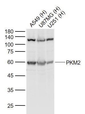 Sample:
Lane 1: A549 (Human) Cell Lysate at 30 ug
Lane 2: U87MG (Human) Cell Lysate at 30 ug
Lane 3: U251 (Human) Cell Lysate at 30 ug
Primary: Anti-PKM2 (SL0102M) at 1/1000 dilution
Secondary: IRDye800CW Goat Anti-Mouse IgG at 1/20000 dilution
Predicted band size: 60 kD
Observed band size: 60 kD
 Sample:
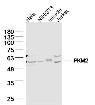
Hela Cell (Human) Lysate at 40 ug
NIH/3T3 Cell (Mouse) Lysate at 40 ug
Muscle (Mouse) Lysate at 40 ug
Jurkat Cell (Human) Lysate at 40 ug
Primary: Anti-PKM2 (SL0102M) at 1/300 dilution
Secondary: IRDye800CW Goat Anti-Mouse IgG at 1/20000 dilution
Predicted band size: 58 kD
Observed band size: 58 kD
 Sample:
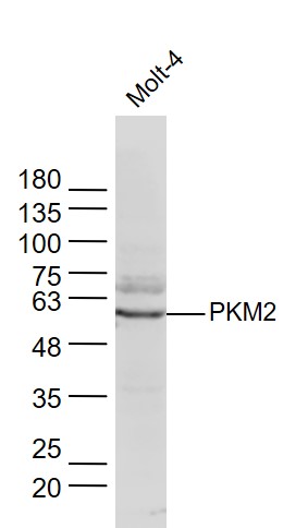
MOLT-4(Human) Cell Lysate at 30 ug
Primary: Anti-PKM2 (SL0102M) at 1/300 dilution
Secondary: IRDye800CW Goat Anti-Mouse IgG at 1/20000 dilution
Predicted band size: 58 kD
Observed band size: 58 kD
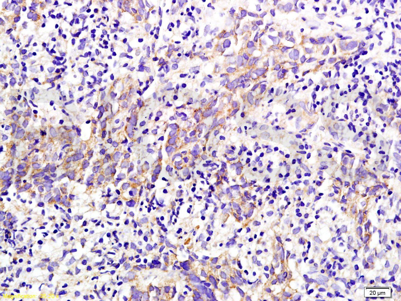 Tissue/cell: Human nasopharyngeal carcinoma; 4% Paraformaldehyde-fixed and paraffin-embedded;
Antigen retrieval: citrate buffer ( 0.01M, pH 6.0 ), Boiling bathing for 15min; Block endogenous peroxidase by 3% Hydrogen peroxide for 30min; Blocking buffer (normal goat serum,C-0005) at 37℃ for 20 min;
Incubation: Anti-M2-PK Polyclonal Antibody, Unconjugated(SL0102M) 1:200, overnight at 4°C, followed by conjugation to the secondary antibody(SP-0024) and DAB(C-0010) staining
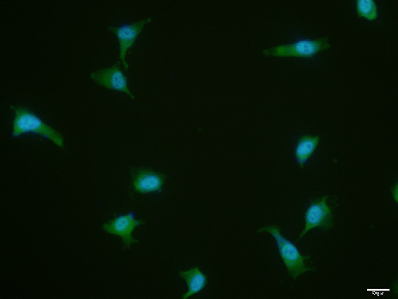 U-87MG cell; 4% Paraformaldehyde-fixed; Triton X-100 at room temperature for 20 min; Blocking buffer (normal goat serum, C-0005) at 37°C for 20 min; Antibody incubation with (PKM2) polyclonal Antibody, Unconjugated (SL0102M) 1:100, 90 minutes at 37°C; followed by a conjugated Goat Anti-Mouse IgG antibody at 37°C for 90 minutes, DAPI (blue, C02-04002) was used to stain the cell nuclei.
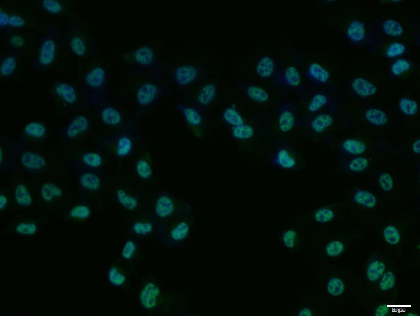 Hela cell; 4% Paraformaldehyde-fixed; Triton X-100 at room temperature for 20 min; Blocking buffer (normal goat serum, C-0005) at 37°C for 20 min; Antibody incubation with (PKM2) polyclonal Antibody, Unconjugated (SL0102M) 1:100, 90 minutes at 37°C; followed by a conjugated Goat Anti-Mouse IgG antibody at 37°C for 90 minutes, DAPI (blue, C02-04002) was used to stain the cell nuclei.
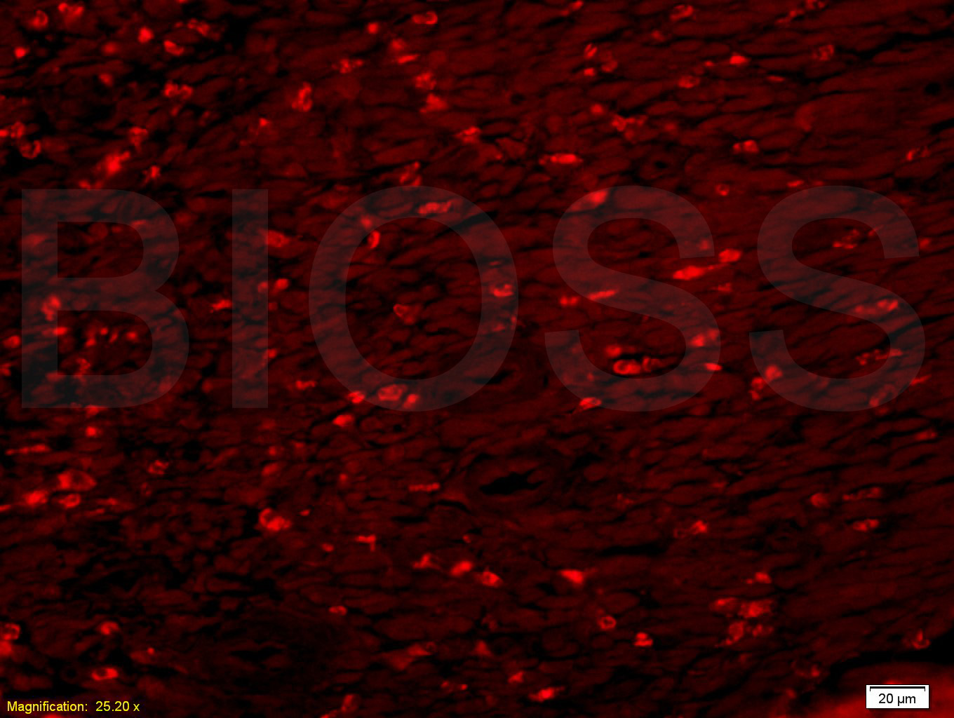 Tissue/cell: rat colitis tissue;4% Paraformaldehyde-fixed and paraffin-embedded;
Antigen retrieval: citrate buffer ( 0.01M, pH 6.0 ), Boiling bathing for 15min; Blocking buffer (normal goat serum,C-0005) at 37℃ for 20 min;
Incubation: Anti-M2-PK Polyclonal Antibody, Unconjugated(SL0102M) 1:200, overnight at 4°C; The secondary antibody was Goat Anti-Mouse IgG, Cy5 conjugated(SL0296G-Cy5)used at 1:200 dilution for 40 minutes at 37°C.
Cartpieces
Totalgoods,subtotals:¥Checkout
Bought notes(bought amounts latest0)
No one bought this product
User Comment(Total0User Comment Num)
- No comment


 +86 571 56623320
 +86 18668110335

