
Rabbit Anti-TGF alpha antibody
EGF like TGF; ETGF; TFGA; TGF A; TGF type 1; TGFA; Transforming growth factor alpha; Transforming growth factor alpha precursor; Wa1; Waved 1; TGFA_HUMAN; TGFα; TGF-α; TGF α.
View History [Clear]
Details
Product Name TGF alpha Chinese Name 转移生长因子α抗体 Alias EGF like TGF; ETGF; TFGA; TGF A; TGF type 1; TGFA; Transforming growth factor alpha; Transforming growth factor alpha precursor; Wa1; Waved 1; TGFA_HUMAN; TGFα; TGF-α; TGF α. literatures Research Area Tumour Cardiovascular Signal transduction Growth factors and hormones cell factor Immunogen Species Rabbit Clonality Polyclonal React Species Human, Mouse, Rat, Applications WB=1:500-2000 ELISA=1:5000-10000
not yet tested in other applications.
optimal dilutions/concentrations should be determined by the end user.Theoretical molecular weight 17kDa Cellular localization The cell membrane Extracellular matrix Secretory protein Form Liquid Concentration 1mg/ml immunogen KLH conjugated synthetic peptide derived from human TGF-alpha : 40-89/160 Lsotype IgG Purification affinity purified by Protein A Buffer Solution 0.01M TBS(pH7.4) with 1% BSA, 0.03% Proclin300 and 50% Glycerol. Storage Shipped at 4℃. Store at -20 °C for one year. Avoid repeated freeze/thaw cycles. Attention This product as supplied is intended for research use only, not for use in human, therapeutic or diagnostic applications. PubMed PubMed Product Detail This gene encodes a growth factor that is a ligand for the epidermal growth factor receptor, which activates a signaling pathway for cell proliferation, differentiation and development. This protein may act as either a transmembrane-bound ligand or a soluble ligand. This gene has been associated with many types of cancers, and it may also be involved in some cases of cleft lip/palate. Alternatively spliced transcript variants encoding different isoforms have been found for this gene. [provided by RefSeq, Sep 2011].
Function:
TGF alpha is a mitogenic polypeptide that is able to bind to the EGF receptor/EGFR and to act synergistically with TGF beta to promote anchorage-independent cell proliferation in soft agar (By similarity). Inhibitor of acid secretion. Inhibitor of aminopyrine uptake in parietal cells (in vitro).
Subunit:
Interacts with the PDZ domains of MAGI3, SDCBP and SNTA1. The interaction with SDCBP, is required for the targeting to the cell surface. In the endoplasmic reticulum, in its immature form (i.e. with a prosegment and lacking full N-glycosylation), interacts with CNIH. In the Golgi apparatus, may form a complex with CNIH and GORASP2. Interacts (via cytoplasmic C-terminal domain) with NKD2.
Subcellular Location:
Transforming growth factor alpha: Secreted, extracellular space. Protransforming growth factor alpha: Cell membrane; Single-pass type I membrane protein.
Tissue Specificity:
Isoform 1, isoform 3 and isoform 4 are expressed in keratinocytes and tumor-derived cell lines.
Similarity:
Contains 1 EGF-like domain.
SWISS:
P01135
Gene ID:
7039
Database links:
Entrez Gene: 7039 Human
Entrez Gene: 21802 Mouse
Omim: 190170 Human
SwissProt: P01135 Human
SwissProt: P48030 Mouse
Unigene: 170009 Human
Unigene: 137222 Mouse
Unigene: 9952 Rat
Growth factors and hormones
TGFα是一个强有力的只有丝分裂多肽,主要由恶性Tumour产生。Product Picture
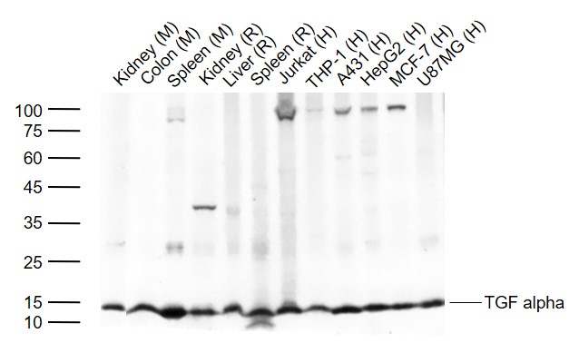
Lane 1: Mouse Kidney tissue lysates
Lane 2: Mouse Colon tissue lysates
Lane 3: Mouse Spleen tissue lysates
Lane 4: Rat Kidney tissue lysates
Lane 5: Rat Liver tissue lysates
Lane 6: Rat Spleen tissue lysates
Lane 7: Human Jurkat cell lysates
Lane 8: Human THP-1 cell lysates
Lane 9: Human A431 cell lysates
Lane 10: Human HepG2 cell lysates
Lane 11: Human MCF-7 cell lysates
Lane 12: Human U87MG cell lysates
Primary: Anti- TGF alpha (SL0066R) at 1/1000 dilution
Secondary: IRDye800CW Goat Anti-Rabbit IgG at 1/20000 dilution
Predicted band size: 17 kDa
Observed band size: 15 kDa
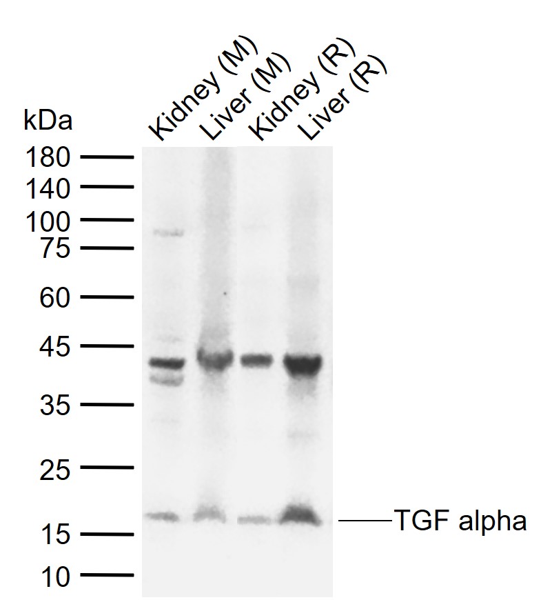
Sample:
Lane 1: Mouse Kidney tissue lysates
Lane 2: Mouse Liver tissue lysates
Lane 3: Rat Kidney tissue lysates
Lane 4: Rat Liver tissue lysates
Primary: Anti-TGF alpha (SL0066R) at 1/1000 dilution
Secondary: IRDye800CW Goat Anti-Rabbit IgG at 1/20000 dilution
Predicted band size: 17 kDa
Observed band size: 17 kDa
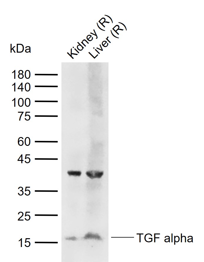
Sample:
Lane 1: Rat Kidney tissue lysates
Lane 2: Rat Liver tissue lysates
Primary: Anti-TGF alpha (SL0066R) at 1/1000 dilution
Secondary: IRDye800CW Goat Anti-Rabbit IgG at 1/20000 dilution
Predicted band size: 17 kDa
Observed band size: 17 kDa
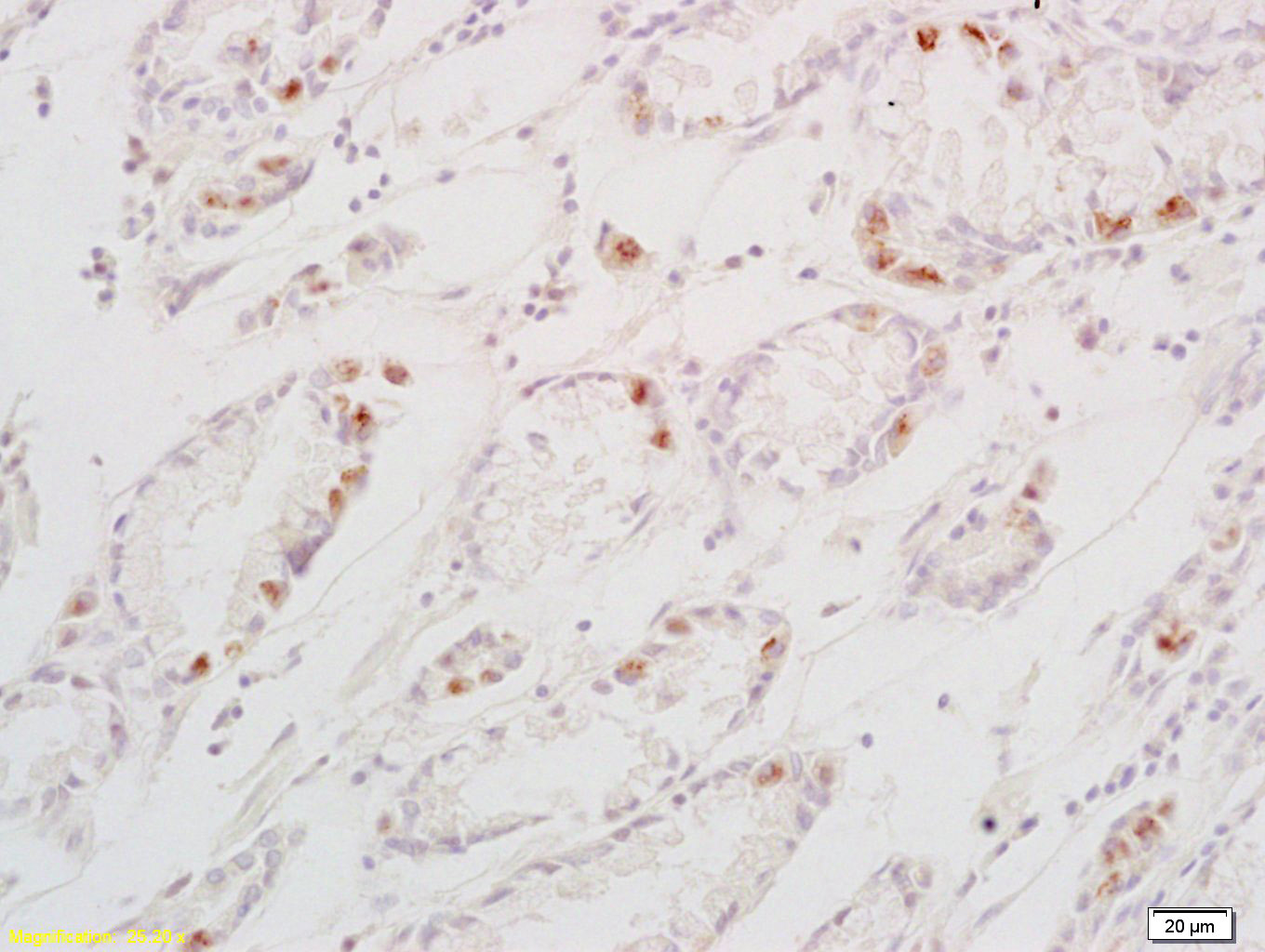
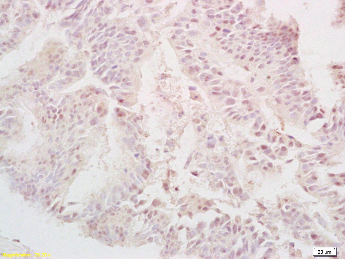
Paraformaldehyde-fixed, paraffin embedded (rat stomach tissue); Antigen retrieval by boiling in sodium citrate buffer (pH6.0) for 15min; Block endogenous peroxidase by 3% hydrogen peroxide for 20 minutes; Blocking buffer (normal goat serum) at 37°C for 30min; Antibody incubation with (TGF alpha) Polyclonal Antibody, Unconjugated (SL0066R) at 1:400 overnight at 4°C, followed by a conjugated secondary (sp-0023) for 20 minutes and DAB staining.Tissue/cell: human gastric carcinoma; 4% Paraformaldehyde-fixed and paraffin-embedded;
Antigen retrieval: citrate buffer ( 0.01M, pH 6.0 ), Boiling bathing for 15min; Block endogenous peroxidase by 3% Hydrogen peroxide for 30min; Blocking buffer (normal goat serum,C-0005) at 37℃ for 20 min;
Incubation: Anti-TGF alpha Polyclonal Antibody, Unconjugated(SL0066R) 1:200, overnight at 4°C, followed by conjugation to the secondary antibody(SP-0023) and DAB(C-0010) staining
Tissue/cell: human colon carcinoma; 4% Paraformaldehyde-fixed and paraffin-embedded;
Antigen retrieval: citrate buffer ( 0.01M, pH 6.0 ), Boiling bathing for 15min; Block endogenous peroxidase by 3% Hydrogen peroxide for 30min; Blocking buffer (normal goat serum,C-0005) at 37℃ for 20 min;
Incubation: Anti-TGF alpha Polyclonal Antibody, Unconjugated(SL0066R) 1:200, overnight at 4°C, followed by conjugation to the secondary antibody(SP-0023) and DAB(C-0010) staining
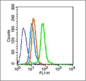
Blank control (blue line): Hela (blue).
Primary Antibody (green line): Rabbit Anti-TGF alpha antibody (SL0066R)
Dilution: 1μg /10^6 cells;
Isotype Control Antibody (orange line): Rabbit IgG .
Secondary Antibody (white blue line): Goat anti-rabbit IgG-FITC
Dilution: 1μg /test.
Protocol
The cells were fixed with 70% methanol (Overnight at -20℃) and then permeabilized with ice-cold 90% methanol for 30 min on ice. Cells stained with Primary Antibody for 30 min at room temperature. The cells were then incubated in 1 X PBS/2%BSA/10% goat serum to block non-specific protein-protein interactions followed by the antibody for 15 min at room temperature. The secondary antibody used for 40 min at room temperature. Acquisition of 20,000 events was performed.
Partial purchase records(bought amounts latest0)
No one bought this product
User Comment(Total0User Comment Num)
- No comment



 +86 571 56623320


