Rabbit Anti-Calretinin antibody
29 kDa calbindin; CAB 29; CAB29; CAL 2; CAL2; CALB 2; CALB2; Calbindin 2 29kDa; Calbindin 2; Calbindin D29K; Calbindin2; CR; CALB2_HUMAN; Calretinin.
View History [Clear]
Details
Product Name Calretinin Chinese Name 钙Binding protein抗体 Alias 29 kDa calbindin; CAB 29; CAB29; CAL 2; CAL2; CALB 2; CALB2; Calbindin 2 29kDa; Calbindin 2; Calbindin D29K; Calbindin2; CR; CALB2_HUMAN; Calretinin. literatures Research Area Neurobiology Binding protein Immunogen Species Rabbit Clonality Polyclonal React Species Human, Rat, (predicted: Mouse, Dog, Pig, Cow, Horse, ) Applications WB=1:500-2000 ELISA=1:5000-10000 IHC-P=1:100-500 IHC-F=1:100-500 Flow-Cyt=2ug/Test IF=1:100-500 (Paraffin sections need antigen repair)
not yet tested in other applications.
optimal dilutions/concentrations should be determined by the end user.Theoretical molecular weight 29kDa Cellular localization cytoplasmic The cell membrane Form Liquid Concentration 1mg/ml immunogen KLH conjugated synthetic peptide derived from human Calretinin: 211-271/271 Lsotype IgG Purification affinity purified by Protein A Buffer Solution 0.01M TBS(pH7.4) with 1% BSA, 0.03% Proclin300 and 50% Glycerol. Storage Shipped at 4℃. Store at -20 °C for one year. Avoid repeated freeze/thaw cycles. Attention This product as supplied is intended for research use only, not for use in human, therapeutic or diagnostic applications. PubMed PubMed Product Detail Calretinin is a calcium-binding protein which is abundant in auditory neurons. It belongs to the calbindin family. Calbindin 2 (calretinin), closely related to calbindin 1, is an intracellular calcium-binding protein belonging to the troponin C superfamily. Calbindin 1 is known to be involved in the vitamin-D-dependent calcium absorption through intestinal and renal epithelia, while the function of neuronal calbindin 1 and calbindin2 is poorly understood. The sequence of the calbindin 2 cDNA reveals an open reading frame of 271 codons coding for a protein of 31,520 Da, and shares 58% identical residues with human calbindin1.
Function:
Calretinin is a calcium-binding protein which is abundant in auditory neurons.
Tissue Specificity:
Brain.
Similarity:
Belongs to the calbindin family.
Contains 6 EF-hand domains.
SWISS:
P22676
Gene ID:
794
Database links:Entrez Gene: 396255 Chicken
Entrez Gene: 794 Human
Entrez Gene: 12308 Mouse
Entrez Gene: 393684 Zebrafish
Omim: 114051 Human
SwissProt: P07090 Chicken
SwissProt: P22676 Human
SwissProt: Q08331 Mouse
Unigene: 106857 Human
Unigene: 2755 Mouse
Unigene: 10321 Rat
钙Binding protein(Calretinin,CR)主要存在于神经组织中,近10%的腺癌可以表达此蛋白的局部染色阳性,而绝大多数的间皮瘤为阳性表达,因此它是诊断间皮瘤的一个有用的标记物 。
钙Binding protein是平滑肌和非肌性调控蛋白。它可以与肌动蛋白、肌球蛋白、原肌球蛋白和钙调素相互作用。
Product Picture
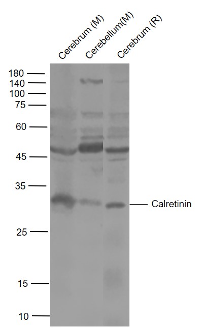
Lane 1: Mouse Cerebrum Lysates
Lane 2: Mouse Cerebellum Lysates
Lane 3: Rat Cerebrum Lysates
Primary: Anti- Calretinin (SL0062R) at 1/1000 dilution
Secondary: IRDye800CW Goat Anti-Rabbit IgG at 1/20000 dilution
Predicted band size: 29 kDa
Observed band size: 29 kDa
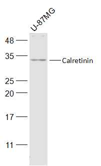
Sample:
U-87MG(Human) Cell Lysate at 30 ug
Primary: Anti-Calretinin (SL0062R) at 1/500 dilution
Secondary: IRDye800CW Goat Anti-Rabbit IgG at 1/20000 dilution
Predicted band size: 29 kD
Observed band size: 29 kD
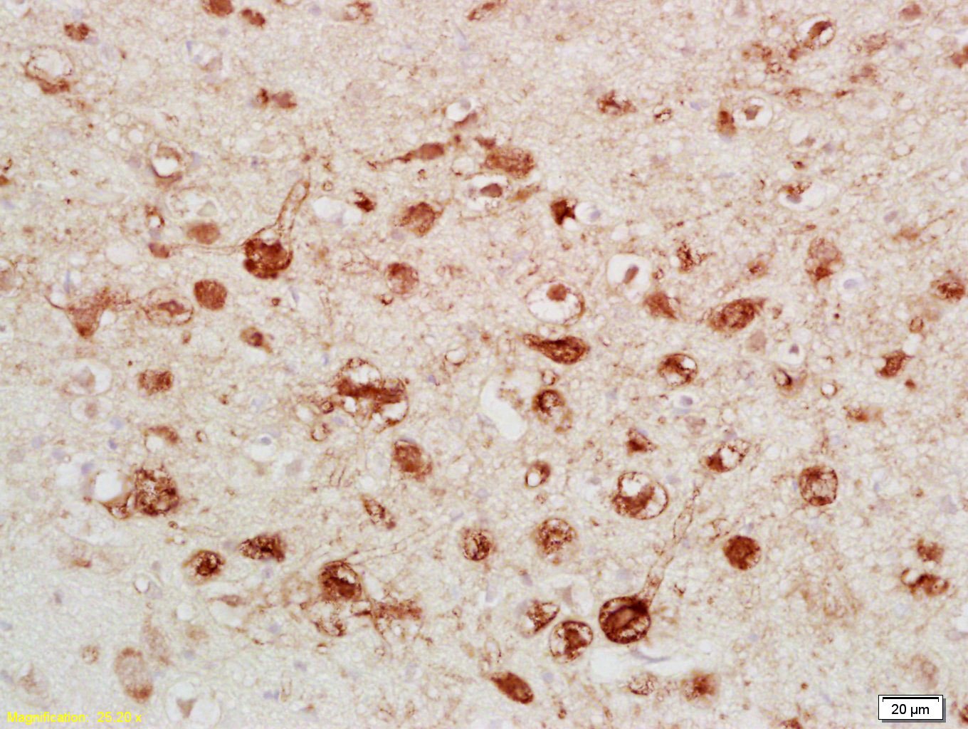
Tissue/cell: rat brain tissue; 4% Paraformaldehyde-fixed and paraffin-embedded;
Antigen retrieval: citrate buffer ( 0.01M, pH 6.0 ), Boiling bathing for 15min; Block endogenous peroxidase by 3% Hydrogen peroxide for 30min; Blocking buffer (normal goat serum,C-0005) at 37℃ for 20 min;
Incubation: Anti-Calretinin/CA Polyclonal Antibody, Unconjugated(SL0062R) 1:200, overnight at 4°C, followed by conjugation to the secondary antibody(SP-0023) and DAB(C-0010) staining
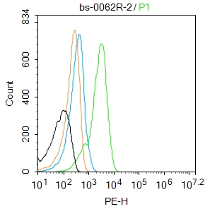
Blank control:A431.
Primary Antibody (green line): Rabbit Anti-S100A13 antibody (SL2617R)
Dilution: 2μg /10^6 cells;
Isotype Control Antibody (orange line): Rabbit IgG .
Secondary Antibody : Goat anti-rabbit IgG-AF488
Dilution: 1μg /test.
Protocol
The cells were fixed with 4% PFA (10min at room temperature)and then permeabilized with 0.1% PBST for 20 min at room temperature.The cells were then incubated in 5%BSA to block non-specific protein-protein interactions for 30 min at room temperature .Cells stained with Primary Antibody for 30 min at room temperature. The secondary antibody used for 40 min at room temperature. Acquisition of 20,000 events was performed.
Partial purchase records(bought amounts latest0)
No one bought this product
User Comment(Total0User Comment Num)
- No comment


 +86 571 56623320


